. 1998 Nov;18(11):6178-90.
doi: 10.1128/MCB.18.11.6178.

Two independent internal ribosome entry sites are involved in translation initiation of vascular endothelial growth factor mRNA

Affiliations

Two independent internal ribosome entry sites are involved in translation initiation of vascular endothelial growth factor mRNA

I Huez et al. Mol Cell Biol. 1998 Nov.

Abstract

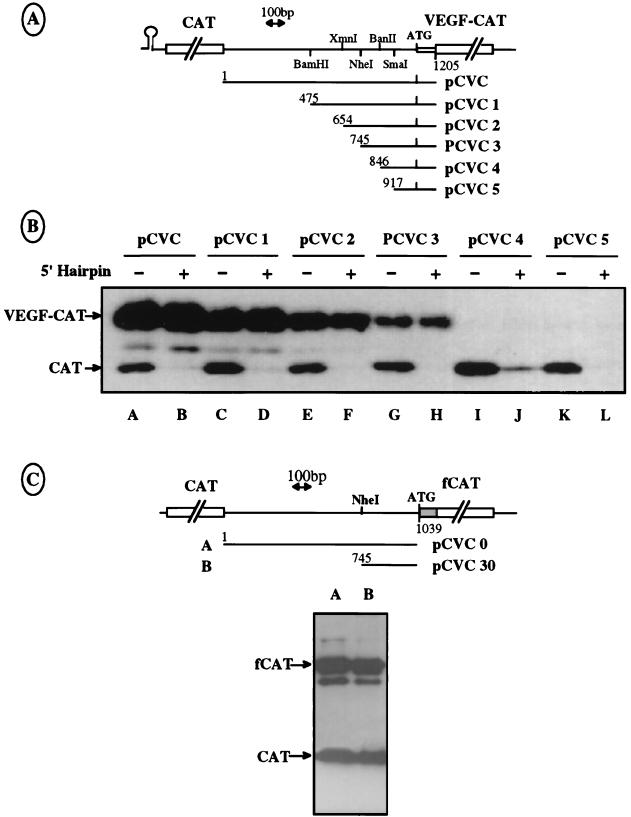
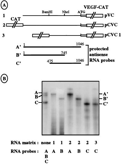
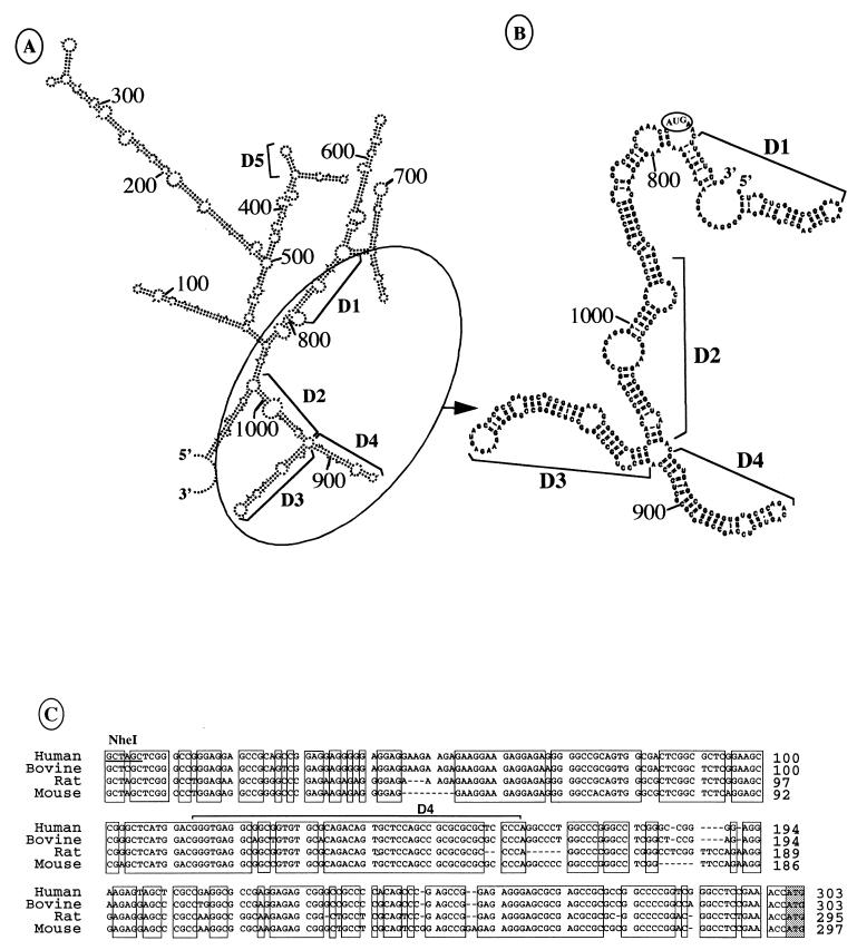
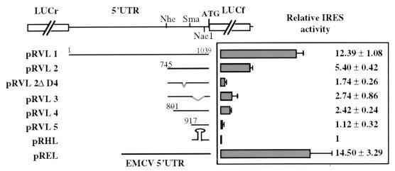
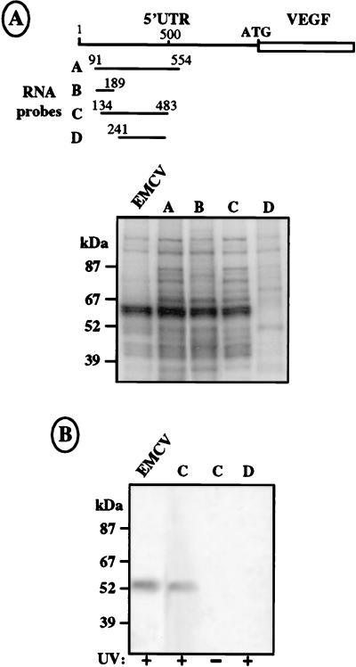
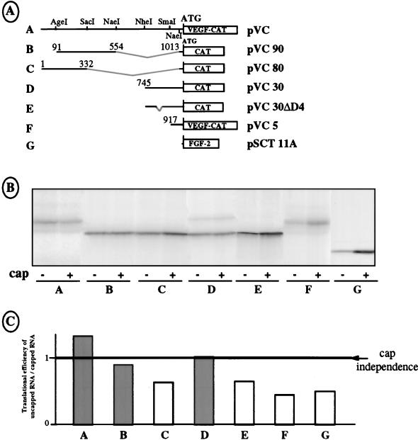
The mRNA of vascular endothelial growth factor (VEGF), the major angiogenic growth factor, contains an unusually long (1,038 nucleotides) and structured 5' untranslated region (UTR). According to the classical translation initiation model of ribosome scanning, such a 5' UTR is expected to be a strong translation inhibitor. In vitro and bicistronic strategies were used to show that the VEGF mRNA translation was cap independent and occurred by an internal ribosome entry process. For the first time, we demonstrate that two independent internal ribosome entry sites (IRESs) are present in this 5' UTR. IRES A is located within the 300 nucleotides upstream from the AUG start codon. RNA secondary structure prediction and site-directed mutagenesis allowed the identification of a 49-nucleotide structural domain (D4) essential to IRES A activity. UV cross-linking experiments revealed that IRES A activity was correlated with binding of a 100-kDa protein to the D4 domain. IRES B is located in the first half of the 5' UTR. An element between nucleotides 379 and 483 is required for its activity. Immunoprecipitation experiments demonstrated that a main IRES B-bound protein was the polypyrimidine tract binding protein (PTB), a well-known regulator of picornavirus IRESs. However, we showed that binding of the PTB on IRES B does not seem to be correlated with its activity. Evidence is provided of an original cumulative effect of two IRESs, probably controlled by different factors, to promote an efficient initiation of translation at the same AUG codon.


Figures

FIG. 1
FIG. 1
Identification of an IRES in the VEGF mRNA 5′ UTR. (A) Schematic representation of the chimeric constructs used for transfection experiments. pVC (construct A) corresponds to the VEGF-CAT fusion in which nt 1 to 1205 of the VEGF cDNA were fused to the CAT coding sequence (see Materials and Methods). This fusion gives rise to a chimeric VEGF-CAT protein of 32 kDa. pHVC (construct B) is derived from pVC and carries an additional 5′ hairpin (ΔG = −40 kcal/mol) downstream from the CMV promoter. pCVC (construct C) is a bicistronic vector containing the CAT gene as a first cistron upstream from the VEGF-CAT fusion in the pVC construct. pHCVC (construct D), derived from pCVC, contains a 5′ hairpin (ΔG = −40 kcal/mol) upstream from the first CAT cistron. (B) The constructs depicted in panel A were transiently transfected in COS-7 cells, and their expression was analyzed by Western immunoblotting using an anti-CAT antibody. The amount of transfected cell protein extract loaded on each lane was adjusted to the quantity of the mono- and bicistronic mRNAs present in each extract. The control (Ct) lane corresponds to untransfected COS-7 cells. The positions of CAT and VEGF-CAT proteins are indicated by arrows.
FIG. 2
FIG. 2
Mapping of the IRES by progressive deletions in the VEGF mRNA 5′ UTR. (A) Schematic representation of the different deletions of the 5′ leader performed in the bicistronic vectors pCVC and pHCVC. Only the pHCVC series is represented here. (B) Western immunoblotting was performed as described for Fig. 1B after transfection of COS-7 cells with the constructs detailed in panel A. The presence or absence of a hairpin in the vector, as well as the name of the vector, is indicated above each lane. The same quantity of COS-7 cell protein extract was loaded in all lanes. Positions of the CAT and VEGF-CAT proteins are indicated with arrows. (C) Representation of two bicistronic vectors containing another VEGF-CAT fusion in which nt 1 to 1046 (including the AUG codon) of the VEGF 5′ leader are fused to the chimeric fCAT gene resulting from the translational fusion of part of the nucleolin gene with the CAT gene (9). These two plasmids were transfected in COS-7 cells, and the extracts were analyzed as described for Fig. 1B. The positions of the CAT and fCAT proteins are indicated with arrows.
FIG. 3
FIG. 3
Verification of the integrity of the bicistronic mRNA by RNase protection assay. (A) Schematic representation of monocistronic vector pVC (construct 1) and bicistronic vectors pCVC and pCVC1 (constructs 2 and 3) used to generate RNA templates. The regions A′, B′, and C′, protected by the three antisense RNA probes A, B, and C, are indicated. The RNA probes A, B, and C are slightly longer than the protected fragments because of the presence of additional nucleotides in the polylinker regions of the plasmids used as templates for the probes (see Materials and Methods). (B) Vectors shown in panel A were transfected in COS-7 cells. Total mRNAs were purified and analyzed by RNase A and T1 protection (see Materials and Methods), using the RNA probes A (1054 nt), B (748 nt), and C (572 nt), complementary to nt 1 to 1046, 1 to 745, and 475 to 1046, respectively. The first lane corresponds to a mix of the three probes alone, without RNase treatment. The RNA templates and probes used are indicated at the bottom. The fragments protected by the probes A, B, and C are notated as A′ (1,017 nt), B′ (690 nt), and C′ (535 nt), respectively.
FIG. 4
FIG. 4
IRES A secondary predicted structures and sequence conservation in mammals. (A) Secondary structure of the complete VEGF mRNA 5′ UTR predicted by the ESSA folding program (6). The 5′ and 3′ ends of the sequence corresponding to nt 1 and 1047, respectively, are indicated. Nucleotide positions and the IRES A predicted domains D1 to D4 are also indicated. D5 corresponds to a stem-loop structure bearing an unpaired loop-located GNRA sequence. (B) Secondary predicted structure of the IRES A. Nucleotide positions and the four domains D1 to D4 are indicated. The 5′ and 3′ ends of the region analyzed correspond to nt 745 to 1052, respectively. (C) Alignment of the cDNA sequence of the region corresponding to human IRES A with bovine, rat, and mouse VEGF cDNA sequences. The conserved regions are boxed. The D4 domain is indicated. Relative positions of the nucleotides aligned from the transcription start point of rat, human, and mouse cDNAs are indicated. The complete 5′ UTR of bovine VEGF mRNA is not known.
FIG. 5
FIG. 5
Characterization of IRES A cis-acting elements. Schematic drawing of the bicistronic vectors containing the LUCr gene as the first cistron, all or part of the VEGF mRNA leader sequence in the intercistronic region, and the LUCf gene as the second cistron. Construct pREL contains the EMCV IRES in the intercistronic region (positive control); construct pRHL contains a hairpin (ΔG = −40 kcal/mol) in the intercistronic region (negative control). These plasmids were transfected in COS-7 cells, and luciferase activities were measured as described in Materials and Methods. On the right, the histogram and corresponding values represent the ratio between the LUCf/LUCr activities obtained with each construct and that obtained with pRHL. Each value represents the average of at least four independent transfection experiments.
FIG. 6
FIG. 6
UV cross-linking of COS-7 cell proteins on the VEGF mRNA 5′ UTR. (A) Drawing of the different 32P-labeled RNA probes, obtained from T7 in vitro transcription and corresponding to the complete or parts of the VEGF 5′ UTR mRNA. Relative positions of the 5′ and 3′ ends of each probe are indicated. (B) UV cross-linking experiments performed with probes A to E. S10 COS-7 cell extracts were incubated with 106 cpm of the different probes followed by UV irradiation and treatment with RNases A and ONE (see Materials and Methods). The control (Ct) lane corresponds to proteinase K treatment of sample loaded in the first lane. Size markers are indicated. (C) 32P-labeled probes corresponding to VEGF probe A (complete 5′ UTR) and to EMCV IRES were cross-linked with proteins extracted from S10 COS-7 cells, and the complex was immunoprecipitated with an anti-PTB antibody. The samples were analyzed before (CL) and after (I) immunoprecipitation. The use of a VEGF or EMCV probe is indicated above the lanes. PTB migration is indicated with an arrow.
FIG. 7
FIG. 7
Mapping of IRES B. (A) Schematic drawing of the bicistronic constructs containing the complete or truncated VEGF 5′ UTR between the first CAT cistron and the second chimeric fCAT cistron (depicted in Fig. 2C). The 5′ and 3′ boundaries of the deletions are indicated. Western immunoblotting was performed (right) as described for Fig. 1B after transfection of COS-7 cells with plasmids A to E. The CAT and fCAT proteins are shown by arrows. The control (Ct) lane corresponds to untransfected cells. (B) Representation of bicistronic vectors containing a stable hairpin structure upstream from the LUCr gene (first cistron), fragments of the VEGF mRNA leader sequence in the intercistronic region, and the LUCf gene as the second cistron. The 5′ and 3′ boundaries of the deletions are indicated. These plasmids were transfected into COS-7 cells, and luciferase activities measured as described in Materials and Methods. On the right, the histogram and corresponding values represent the LUCf/LUCr activity ratio obtained with each construct and that obtained with pRHL. Each value represents the average of at least four independent transfection experiments.
FIG. 8
FIG. 8
UV cross-linking of COS-7 cell proteins on IRES B. (A) Top, drawing of the different 32P-labeled RNA probes, obtained from T7 in vitro transcription and corresponding to the complete or parts of the IRES B sequence. Relative positions of the 5′ and 3′ ends of each probe are indicated. Bottom, UV cross-linking experiments performed with probes A to D and a probe corresponding to the EMCV IRES (first lane). S10 COS-7 cell extracts were incubated with 1.5 × 10⁶ cpm of the different probes followed by UV irradiation and treatment with RNases A and ONE (see Materials and Methods). Size markers are indicated. (B) Immunoprecipitation with an anti-PTB antibody of COS-7 cell protein extract cross-linked to EMCV and VEGF probes C and D (lane 1, 2, and 4). Lane 3 corresponds to a control immunoprecipitation of cell extract and probe C without previous cross-linking. UV cross-linking or absence of cross-linking before immunoprecipitation is indicated by a plus or a minus sign, respectively, below each lane.
FIG. 9
FIG. 9
Activities of IRES A and IRES B in vitro. (A) Schematic drawing of the monocistronic vectors used as T7 polymerase templates. The in vitro transcriptions were performed in the presence or absence of a cap structure. In constructs A and F, the translation product is the chimeric VEGF-CAT protein depicted in Fig. 1A. In constructs B to E, the translation product is the CAT protein. In construct G, the translation product is the FGF-2 protein. (B) Identical quantities of the different capped or noncapped mRNAs were translated in RRL. The presence or absence of a cap structure in the mRNA is indicated by a plus or minus sign, respectively, below each lane, together with the construct used. (C) The lanes in panel B were quantified with PhosphorImager. The histogram indicates the ratio of the translation efficiency observed in uncapped RNA versus capped mRNA. The line corresponds to cap-independent translation initiation (ratio of 1). The experiment reported here is representative of five independent experiments.

Similar articles

Cited by

References

    1. Belsham G J, Sonenberg N. RNA-protein interactions in regulation of picornavirus RNA translation. Microbiol Rev. 1996;60:499–511. - PMC - PubMed
    1. Bernstein J, Sella O, Le S Y, Elroy-Stein O. PDGF2/c-sis mRNA leader contains a differentiation-linked internal ribosomal entry site (D-IRES) J Biol Chem. 1997;272:9356–9362. - PubMed
    1. Borman A, Howell M T, Patton J G, Jackson R J. The involvement of a spliceosome component in internal initiation of human rhinovirus RNA translation. J Gen Virol. 1993;74:1775–1788. - PubMed
    1. Borovjagin A, Pestova T, Shatsky I. Pyrimidine tract binding protein strongly stimulates in vitro encephalomyocarditis virus RNA translation at the level of preinitiation complex formation. FEBS Lett. 1994;351:299–302. - PubMed
    1. Brogi E, Wu T, Namiki A, Isner J M. Indirect angiogenic cytokines upregulate VEGF and bFGF gene expression in vascular smooth muscle cells, whereas hypoxia upregulates VEGF expression only. Circulation. 1994;90:649–652. - PubMed

Publication types

MeSH terms