. 1998 Nov;18(11):6213-23.
doi: 10.1128/MCB.18.11.6213.

MFR, a putative receptor mediating the fusion of macrophages

Affiliations

MFR, a putative receptor mediating the fusion of macrophages

C Saginario et al. Mol Cell Biol. 1998 Nov.

Abstract

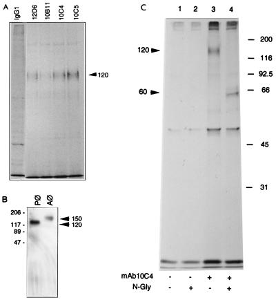
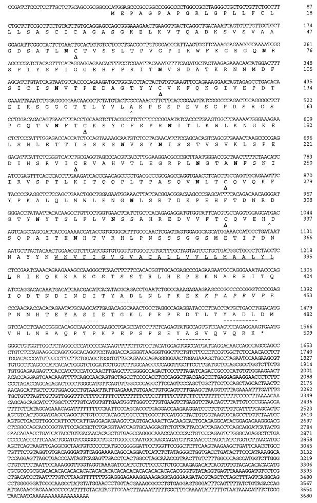
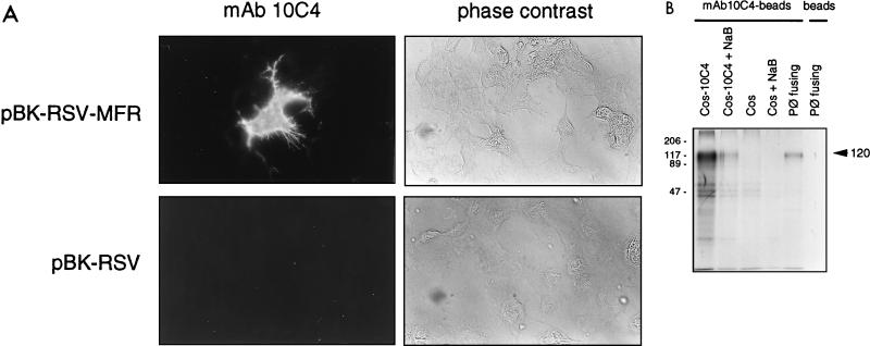
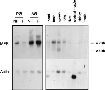
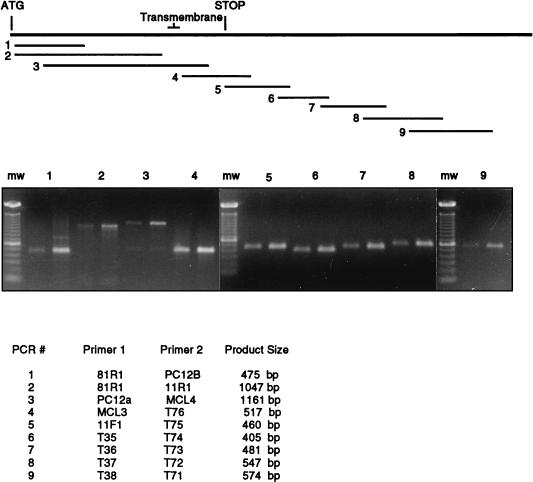
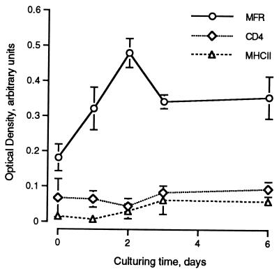
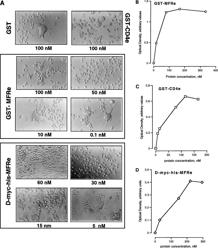
We had previously identified a macrophage surface protein whose expression is highly induced, transient, and specific, as it is restricted to actively fusing macrophages in vitro and in vivo. This protein is recognized by monoclonal antibodies that block macrophage fusion. We have now purified this protein and cloned its corresponding cDNA. This protein belongs to the superfamily of immunoglobulins and is similar to immune antigen receptors such as the T-cell receptor, B-cell receptor, and viral receptors such as CD4. We have therefore named this protein macrophage fusion receptor (MFR). We show that the extracellular domain of MFR prevents fusion of macrophages in vitro and therefore propose that MFR belongs to the fusion machinery of macrophages. MFR is identical to SHPS-1 and BIT and is a homologue of P84, SIRPalpha, and MyD-1, all of which have been recently cloned and implicated in cell signaling and cell-cell interaction events.


Figures

FIG. 1
FIG. 1
(A) Antifusion MAbs recognize a 120-kDa protein from fusing rat peritoneal macrophages. Macrophages were cultured for 3 days in fusogenic conditions, metabolically labeled with [35S]methionine for 17 h, and then subjected to immunoprecipitation with either mouse IgG1 or MAb 12D6, 10B11, 10C4, or 10C5. Immunoprecipitates were analyzed by SDS-PAGE. All four MAbs precipitate a 120-kDa protein from peritoneal macrophages. Each lane represents immunoprecipitates from extracts prepared from 5 × 10⁶ plated peritoneal macrophages. In all panels, molecular masses of markers are indicated in kilodaltons. (B) MAb 10C4 recognizes 120- and 150-kDa proteins from fusing peritoneal and alveolar macrophages (Pφ and Aφ), respectively. Macrophages were cultured for 3 days in fusogenic conditions prior to being subjected to Western blot analysis. Total cell lysates were run on a 10% polyacrylamide gel in the absence of both SDS and β-mercaptoethanol and blotted with MAb 12D6 followed by HRP-conjugated goat anti-mouse IgG. The ECL substrate reaction was developed with XAR film. (C) 10C4 antigen is heavily glycosylated. Rat peritoneal macrophages were cultured in fusogenic conditions for 3 days prior to being metabolically labeled with [35S]methionine for 17 h. Immunoprecipitates were subjected to N-glycanase (N-Gly) digestion.
FIG. 2
FIG. 2
Nucleotide and deduced amino acid sequences of rat MFR. The putative transmembrane domain is underlined. Possible N-glycosylation sites (Asn-X-Ser/Thr) are indicated in boldface. Cysteines likely to form disulfide bonds creating the three Ig-like domains (▵) and putative binding sites for SH2 domains of PTPases (–––) are indicated. The putative binding site for SH3 domains is in italics.
FIG. 3
FIG. 3
MAb 10C4 recognizes recombinant MFR. (A) COS-7 cells were transiently transfected with either pBK-RSV-MFR or pBK-RSV, and the recombinant protein was detected by incubating unfixed cells with either MAb 10C4 or PBS, followed by LRSC-conjugated F(ab′)2 goat anti-mouse IgG (H+L). Magnification, ×200. (B) COS-7 cells transiently transfected with pBK-RSV-MFR, nontransfected COS-7 cells, and fusing peritoneal macrophages (Pφ) were metabolically labeled with [35S]methionine and subjected to immunoprecipitation with protein A-Sepharose-MAb 10C4-conjugated beads. Peritoneal macrophage lysates were incubated with protein A-Sepharose beads as a control. Immunoprecipitates were analyzed by SDS-PAGE. The mobilities of molecular mass standards are indicated in kilodaltons.
FIG. 4
FIG. 4
Northern blot analysis of MFR. Each lane contained approximately 2 μg of mRNA from the tissues indicated and 8 μg of total RNA from freshly isolated nonfusing (NF) and fusing (F) alveolar and peritoneal macrophages (Aφ and Pφ). Hybridization was performed with 32P-labeled PCR-generated mouse brain-based sequence (Table 1) and β-actin probes.
FIG. 5
FIG. 5
Comparison of 10C4 transcripts in alveolar (left product of each pair) and peritoneal (right product of each pair) macrophages by RT-PCR. PCRs were performed with the primer combinations listed. Primers were designed on the basis of the sequence of cloned MFR cDNA. The corresponding locations of the PCR products with respect to MFR cDNA are shown. PCR primer combinations were chosen such that the products overlap, and the majority of the cDNA sequence was analyzed. PCR products were separated by agarose gel electrophoresis and stained with ethidium bromide. The approximate sizes of the PCR products are indicated. mw, molecular weight markers.
FIG. 6
FIG. 6
Cell surface expression of MFR is transiently induced at the onset of fusion. MFR, CD4, and MHCII cell surface expression was quantitated by ELISA on alveolar macrophages cultured in fusogenic conditions. Expression was recorded at the indicated times.
FIG. 7
FIG. 7
(A) MFRe blocks macrophage fusion in vitro. GST-CD4e, GST-MFRe, and D-Myc-His-MFRe were added at the indicated concentrations to fusing macrophages. Cells were cultured for 4 days prior to being fixed and examined for multinucleation. Magnification, ×100. (B to D) Specific binding of GST-MFRe (B), GST-CD4e (C), and D-Myc-His-MFRe (D) to fusing macrophages in vitro. Fusing macrophages were cultured for 24 h in fusogenic conditions prior to being subjected to binding analysis as described in Materials and Methods. Binding was detected by ELISA.

Similar articles

Cited by

References

    1. Alkhatib G, Combadiere C, Broder C, Feng Y, Kennedy P, Murphy P, Berger E. CC CKR5: a RANTES, MIP-1α, MIP-1β receptor as a fusion cofactor for macrophage-tropic HIV-1. Science. 1996;272:1955–1958. - PubMed
    1. Bergelson J, Cunningham J, Droguett G, Kurt-Jones E, Krithivas A, Hong J, Horwitz M, Crowell R, Finberg R. Isolation of a common receptor for coxsackie B viruses and adenoviruses 2 and 5. Science. 1997;275:1320–1323. - PubMed
    1. Blobel C, Wolfsberg T, Turck C, Myles D, Primakoff P, White J. A potential fusion peptide and an integrin ligand domain in a protein active in sperm-egg fusion. Nature. 1992;356:248–252. - PubMed
    1. Brooke G, Parsons K, Howard C. Cloning of two members of the SIRPα family of protein tyrosine phosphatase binding proteins in cattle that are expressed on monocytes and a subpopulation of dendritic cells and which mediate binding to CD4 T cells. Eur J Immunol. 1998;28:1–11. - PubMed
    1. Chiozzi P, Sanz J, Ferrari D, Falzoni S, Aleotti A, Buell G, Collo G, DiVirgilio F. Spontaneous cell fusion in macrophage cultures expressing high levels of the P2Z/P2X7 receptor. J Cell Biol. 1997;138:697–706. - PMC - PubMed

Publication types

MeSH terms

Associated data

LinkOut - more resources