. 1998 Nov;18(11):6224-37.
doi: 10.1128/MCB.18.11.6224.

Ectopic expression of cdc2/cdc28 kinase subunit Homo sapiens 1 uncouples cyclin B metabolism from the mitotic spindle cell cycle checkpoint

Affiliations

Ectopic expression of cdc2/cdc28 kinase subunit Homo sapiens 1 uncouples cyclin B metabolism from the mitotic spindle cell cycle checkpoint

M L Hixon et al. Mol Cell Biol. 1998 Nov.

Abstract

Primary human fibroblasts arrest growth in response to the inhibition of mitosis by mitotic spindle-depolymerizing drugs. We show that the mechanism of mitotic arrest is transient and implicates a decrease in the expression of cdc2/cdc28 kinase subunit Homo sapiens 1 (CKsHs1) and a delay in the metabolism of cyclin B. Primary human fibroblasts infected with a retroviral vector that drives the expression of a mutant p53 protein failed to downregulate CKsHs1 expression, degraded cyclin B despite the absence of chromosomal segregation, and underwent DNA endoreduplication. In addition, ectopic expression of CKsHs1 interfered with the control of cyclin B metabolism by the mitotic spindle cell cycle checkpoint and resulted in a higher tendency to undergo DNA endoreduplication. These results demonstrate that an altered regulation of CKsHs1 and cyclin B in cells that carry mutant p53 undermines the mitotic spindle cell cycle checkpoint and facilitates the development of aneuploidy. These data may contribute to the understanding of the origin of heteroploidy in mutant p53 cells.


Figures

FIG. 1
FIG. 1
(A) Immunoprecipitation of mutant p53 with antibody P Ab 240 in primary NHF infected with vector pBabe (NHF-pBabe) or vector pBabe p53 143A (NHF-pBabe p53 143A). The cells were incubated with radiolabeled methionine, harvested, and lysed, P Ab 240-reactive p53 was immunoprecipitated, and the immunoprecipitates were resolved by SDS-PAGE and exposed to a PhosphorImager screen. (B) Analysis of the cell cycle distribution of the DNA content in NHF-pBabe and NHF-pBabe p53 143A cells. Cells were incubated in the absence or presence of 200 ng of colcemid per ml for two to four population doubling times (PDL). The population doubling times for NHF-pBabe and NHF-pBabe p53 143A cells were 48 and 42 h, respectively. Following incubations, the cells were harvested and processed for flow cytometric determination of DNA content as indicated in Materials and Methods. Polyclonal populations at passage 2 were assayed. The data are representative of at least three independent experiments.
FIG. 2
FIG. 2
Western analysis of cyclin B and β-actin in NHF carrying the control retroviral vector (NHF-pBabe) or a retroviral vector containing mutant 143A p53 sequences (NHF-pBabe p53 143A). Confluent cell cultures (4 × 10⁴ to 5 × 10⁴ cells/cm2) were synchronized by a 2-day incubation in low-serum medium (0.5% calf serum), incubated at low density (1 × 10⁴ to 2 × 10⁴ cells/cm2) in 10% FBS in the absence (A and B) or presence (C and D) of 200 ng of colcemid per ml, and harvested at the indicated intervals. Colcemid was added at 12 h after cell passage. Western blotting was carried out as indicated in Materials and Methods. Data are representative of three independent experiments.
FIG. 3
FIG. 3
(A) Western analysis of p53 and β-actin in NHF carrying the retroviral vectors pBabe (puromycin resistance, no insert) and LXSN (neomycin resistance, no insert) or LXSN-E6. For the induction of p53, the cells were incubated for 2 days in 10 μM mycophenolic acid (a GMP biosynthesis inhibitor). (B) Immunoprecipitation of mutant p53 and β-actin in primary NHF following double infection with the retroviral vector pBabe and LXSN or LXSN-E6 or with the vector pBabe p53 143A and LXSN or LXSN-E6. The cells were metabolically labeled and the proteins were analyzed as in Fig. 1. (C to E) Western analysis of cyclin B and β-actin in NHF carrying the retroviral vectors pBabe and LXSN (C), pBabe and LXSN-E6 (D), or pBabe p53 143A and LXSN-E6 (E). The cells were incubated and Western blotting was carried out as in Fig. 2. Data are representative of three independent experiments.
FIG. 4
FIG. 4
(A) Northern analysis of CKsHs1 and GAPDH expression in NHF-pBabe (pB), NHF-pBabe p53 143A (pB-143A), and HT1080 cells. HT1080 is a fibrosarcoma cell line that carries two mutated p53 alleles. The cells were incubated in DMEM with 10% FBS until confluent (approximately 4 × 10⁴ to 5 × 10⁴ cells/cm2) and processed for Northern analysis. A CKsHs1 sequence obtained by RT-PCR from NHF was subcloned into Bluescribe (Stratagene), sequenced, and used as a probe. RNA integrity was verified by reprobing with a GAPDH sequence (American Type Culture Collection). Other experimental details were as indicated in Materials and Methods. (B) Northern analysis of the expression of a murine homolog of CKsHs1 (CKsMm1) and GAPDH in C2C12-pBabe (pB) and C2C12-pBabe p53 143A (pB-143A) cells. The cells were incubated as in panel A. (C) Northern analysis of CKsHs1 and GAPDH expression in quiescent or exponentially growing NHF-pBabe (pB) and NHF-pBabe p53 143A (pB-143A) cells. The cells were incubated for 2 days at confluence in DMEM with 0.5% calf serum (No Serum) or for 2 days at low density (1 × 10⁴ to 2 × 10⁴ cells/cm2) in DMEM with 10% FBS (Exp. Growth). (D) Flow-cytometric analysis of the cell cycle distribution of the DNA content in NHF-pBabe (pB) and NHF-pBabe p53 143A (pB-143A) cells at confluence. The cells were incubated for 2 days at confluence (4 × 10⁴ to 5 × 10⁴ cells/cm2) in DMEM with 10% FBS, 100 μM BrdU was added, and the cells were incubated for an additional 4 h. The cultures were harvested, fixed, and processed for flow cytometry as indicated in Materials and Methods.
FIG. 5
FIG. 5
Northern analysis of CKsHs1 expression in NHF-pBabe (A) and NHF-pBabe p53 143A (B) cells incubated in the presence of increasing concentrations of colcemid. The cells were synchronized at G0 by incubation in low-serum medium as in Fig. 2 and then incubated for 40 h in 10% FBS in the presence of 0 (lanes a), 100 (lanes b), 200 (lanes c), or 1,000 (lanes d) ng of colcemid per ml. Colcemid was added 12 h after cell passage. (C and D) Northern analysis of CKsHs1 expression in NHF-pBabe cells incubated in the absence (C) or presence (D) of 200 ng of colcemid per ml. The cells were synchronized and incubated as indicated above. Northern analysis was carried out as in Fig. 4. Data are representative of three independent experiments. (E and F) Northern analysis of CKsHs1 expression in NHF-pBabe 143A cells incubated in the absence (E) or presence (F) of 200 ng of colcemid per ml. The cells were synchronized and incubated as indicated above. Northern analysis was carried out as described in Fig. 4. Data are representative of two independent experiments.
FIG. 6
FIG. 6
(A) Western analysis of CKsHs1 and β-actin levels in control NHF (NHF-pBabe) (A) and in NHF expressing mutant p53 proteins (NHF-pBabe p53 143A) (B). The cells were synchronized by a 2-day incubation in low-serum medium (0.5% calf serum) and then incubated in 10% FBS in the absence (No Colcemid) or presence (Colcemid) of 200 ng of colcemid per ml and harvested at the indicated intervals. Colcemid was added 12 h after cell passage. Western blotting was carried out as indicated in Materials and Methods, except for the colcemid-treated NHF-pBabe blot, which was probed with a 1:25 CKsHs1 antibody dilution. Data are representative of three independent experiments.
FIG. 7
FIG. 7
(A) Western analysis of CKsHs1 and β-actin levels in control NHF (NHF-pBabe) and in NHF expressing mutant p53 proteins (NHF-pBabe p53 143A). Exponentially growing cells were incubated in the presence (+) or absence (−) of 200 ng of colcemid per ml for 40 h. (B) Western analysis of CKsHs1 and β-actin in primary NHF stably transfected with murine sarcoma virus long terminal repeat neo-based expression plasmids containing no insert (NHF-LTR), mutant p53 175H (NHF-LTR p53 175H), or mutant p53 273H (NHF-LTR p53 273H) cDNA sequences. The cells were incubated as in panel A. (C) Western analysis of CKsHs1 and β-actin in NHF pBabe cells. Confluent cell cultures (4 × 10⁴ to 5 × 10⁴ cells/cm2) were synchronized by a 2-day incubation in low-serum medium (0.5% calf serum) and then incubated at low density (1 × 10⁴ to 2 × 10⁴ cells/cm2) in 10% FBS. Colcemid (200 ng/ml) was added to the cells at the indicated times, and the cultures were harvested at 48 h. Western analysis was performed as indicated in Materials and Methods. Data are representative of three independent experiments.
FIG. 8
FIG. 8
(A) Immunoprecipitation of cdc2-associated CKsHs1. NHF pBabe cells were synchronized and incubated as in Fig. 2 and metabolically labeled for 2 h before being harvested at the indicated times. Preparation of extracts and cdc2 immunoprecipitations (CDC2 IP) were carried out as indicated in Materials and Methods. CKsHs1 was independently immunoprecipitated (CKsHs1 IP) at the 48-h timepoint. As a control, 5 μg of the CKsHs1 peptide epitope was added to half of the CKsHs1 immunoprecipitation extract. Immunoprecipitates were resolved by PAGE (15% polyacrylamide), and the gels were dried and exposed to a PhosphorImager screen. (B) Immunoprecipitation of total and cdc2-associated CKsHs1. Exponentially growing cells (48 h after synchronization) were incubated in the presence (+) or absence (−) of 200 ng of colcemid per ml. Extracts were immunoprecipitated with the indicated antibodies. Other experimental details are as in panel A. Data are representative of three independent experiments.
FIG. 9
FIG. 9
Western analysis of CKsHs1 and β-actin in C2C12 cells stably transfected with CMVneo-bcl2 and pBabe or pBabe-CKsHs1 expression vectors. The cells were synchronized as in Fig. 2 and then incubated for the indicated times in 10% FBS in the absence (A, No colcemid) or presence (B and C, Colcemid) of 200 ng of colcemid per ml. Colcemid was added 12 h after cell passage. Western blotting was carried out as indicated in Materials and Methods. Data are representative of two independent experiments.
FIG. 10
FIG. 10
Western analysis of cyclin B levels in C2C12 cells ectopically expressing bcl2 or expressing bcl2 and CKsHs1. The cells were synchronized as in Fig. 2, incubated in 10% FBS in the absence (A and B) or presence (C and D) of 200 ng of colcemid per ml, and harvested at the indicated intervals. Colcemid was added 12 h after cell passage. Western blotting was carried out as indicated in Materials and Methods. Data are representative of two independent experiments.
FIG. 11
FIG. 11
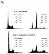
(A) Flow-cytometric analysis of the cell cycle distribution of the DNA content in C2C12 cells ectopically expressing CKsHs1 and/or bcl-2. Cells were incubated in the absence or presence of 200 ng of colcemid per ml for two population doubling times (the population doubling times of C2C12 bcl2/pBabe and C2C12 bcl2/pBabe CKsHs1 cells were 38 and 32 h, respectively). Following incubations, the cells were harvested and processed for flow cytometry of DNA content as indicated in Materials and Methods. Polyclonal populations at passage 2 were assayed. (B) Flow-cytometric analysis of the cell cycle distribution of the DNA content in 10T1/2 cells stably transfected with pBabe, pBabe CKsHs1, or pBabe CKsHs2. The cells were incubated in the absence or presence of 200 ng of colcemid per ml for two population doubling times (72, 66, and 62 h, respectively). Other experimental details are as in panel A. Data are representative of three independent experiments.
FIG. 11
FIG. 11
(A) Flow-cytometric analysis of the cell cycle distribution of the DNA content in C2C12 cells ectopically expressing CKsHs1 and/or bcl-2. Cells were incubated in the absence or presence of 200 ng of colcemid per ml for two population doubling times (the population doubling times of C2C12 bcl2/pBabe and C2C12 bcl2/pBabe CKsHs1 cells were 38 and 32 h, respectively). Following incubations, the cells were harvested and processed for flow cytometry of DNA content as indicated in Materials and Methods. Polyclonal populations at passage 2 were assayed. (B) Flow-cytometric analysis of the cell cycle distribution of the DNA content in 10T1/2 cells stably transfected with pBabe, pBabe CKsHs1, or pBabe CKsHs2. The cells were incubated in the absence or presence of 200 ng of colcemid per ml for two population doubling times (72, 66, and 62 h, respectively). Other experimental details are as in panel A. Data are representative of three independent experiments.

Similar articles

Cited by

References

    1. Agapova L S, Ilyinskaya G V, Turovets N A, Ivanov A V, Chumakov P M, Kopnin B P. Chromosome changes caused by alterations of p53 expression. Mutat Res. 1996;354:129–138. - PubMed
    1. Andreassen P R, Margolis R L. Microtubule dependency of p34cdc2 inactivation and mitotic exit in mammalian cells. J Cell Biol. 1994;127:789–802. - PMC - PubMed
    1. Basi G, Draetta G. The cdc2 kinase: structure, activation, and its role at mitosis in vertebrate cells. In: Hutchison C, Glover D M, editors. Cell cycle control. Vol. 10. Oxford, United Kingdom: Oxford University Press; 1995. pp. 106–134.
    1. Basi G, Draetta G. p13suc1 of Schizosaccharomyces pombe regulates two distinct forms of the mitotic cdc2 kinase. Mol Cell Biol. 1995;15:2028–2036. - PMC - PubMed
    1. Bischoff F Z, Yim S O, Pathak S, Grant G, Siciliano M J, Giovanella B C, Strong L C, Tainsky M A. Spontaneous abnormalities in normal fibroblasts from patients with Li-Fraumeni cancer syndrome: aneuploidy and immortalization. Cancer Res. 1990;50:7979–7984. - PubMed

MeSH terms