. 1998 Nov;18(11):6374-86.
doi: 10.1128/MCB.18.11.6374.

Yeast Los1p has properties of an exportin-like nucleocytoplasmic transport factor for tRNA

Affiliations

Yeast Los1p has properties of an exportin-like nucleocytoplasmic transport factor for tRNA

K Hellmuth et al. Mol Cell Biol. 1998 Nov.

Abstract

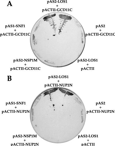
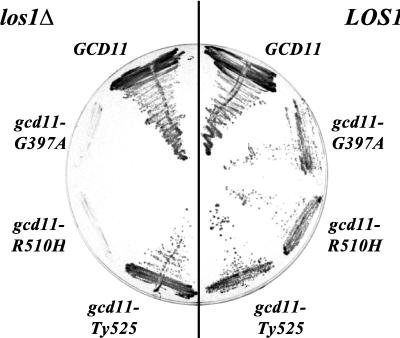
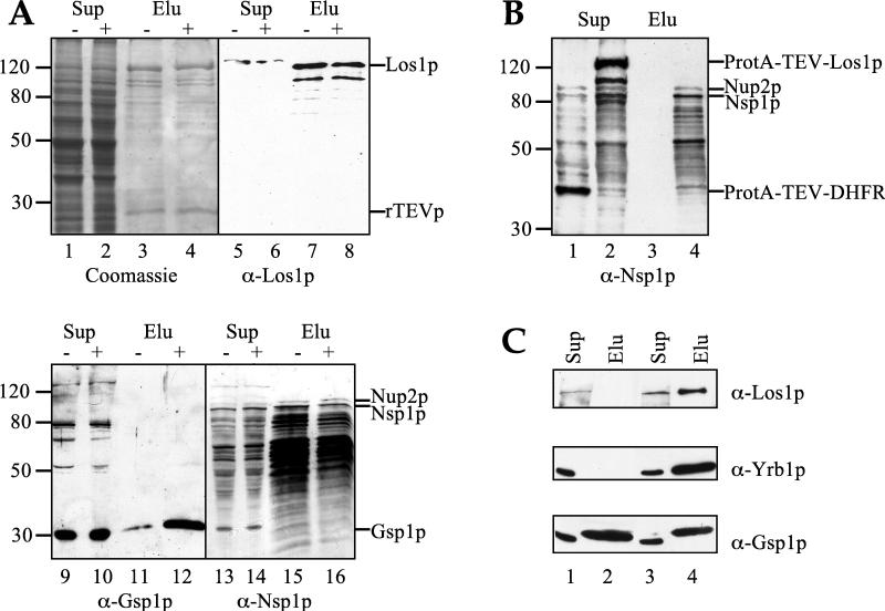
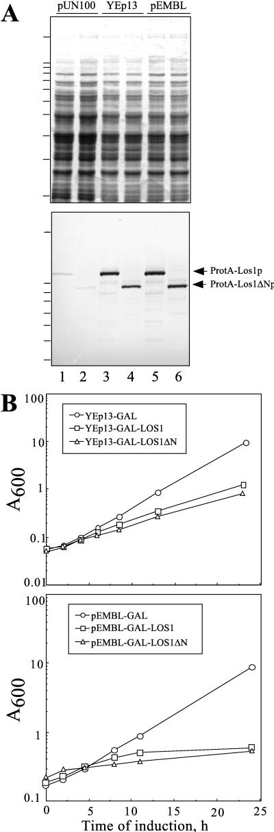
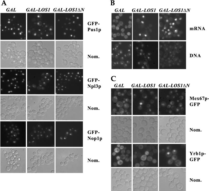
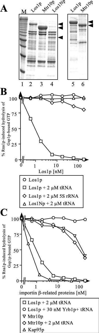
Saccharomyces cerevisiae Los1p, which is genetically linked to the nuclear pore protein Nsp1p and several tRNA biogenesis factors, was recently grouped into the family of importin/karyopherin-beta-like proteins on the basis of its sequence similarity. In a two-hybrid screen, we identified Nup2p as a nucleoporin interacting with Los1p. Subsequent purification of Los1p from yeast demonstrates its physical association not only with Nup2p but also with Nsp1p. By the use of the Gsp1p-G21V mutant, Los1p was shown to preferentially bind to the GTP-bound form of yeast Ran. Furthermore, overexpression of full-length or N-terminally truncated Los1p was shown to have dominant-negative effects on cell growth and different nuclear export pathways. Finally, Los1p could interact with Gsp1p-GTP, but only in the presence of tRNA, as revealed in an indirect in vitro binding assay. These data confirm the homology between Los1p and the recently identified human exportin for tRNA and reinforce the possibility of a role for Los1p in nuclear export of tRNA in yeast.


Figures

FIG. 1
FIG. 1
Two-hybrid interactions of Los1p. Los1p interacts with Gcd11p (A) and Nup2p (B). The yeast screening strain Y190 was transformed with the indicated bait (pAS2) and prey (pACTII) plasmids in various combinations, and in each case two independent transformants were tested for growth (His+) and β-galactosidase activity (LacZ+) on plates of SDC-Trp-Leu-His plus 3AT and X-Gal. Only in the presence of both pAS2-LOS1 as bait and pACTII-GCD11C (codons 368 to 527) or pACTII-NUP2N (codons 42 to 177) as prey were blue-colored yeast cells found growing on selective plates. The bait plasmids pAS1-SNF1 (40) and pAS2-NSP1M (codons 273 to 564 of NSP1) (3a) served as negative controls.
FIG. 2
FIG. 2
Genetic interaction between LOS1 and GCD11. Shuffle strain YKH53 (los1::HIS3 gcd11::HIS3) containing pRS316-GCD11 was transformed with various GCD11 alleles on a LEU2 plasmid and then streaked on 5-FOA-containing medium, on which cells with the URA3 plasmid, which contains the GCD11 wild-type gene, were selected against. The GCD11 point mutations G397A and R510H, combined with a los1::HIS3 disruption, led to synthetic lethality (left), which was overcome by cotransformation with a LOS1 wild-type gene on a TRP1 plasmid (right).
FIG. 3
FIG. 3
Affinity purification of Los1p and Gsp1p. (A) Cells expressing ProtA-TEV-LOS1 and carrying the plasmid YEp352GAL-GSP1-G21V were grown in the absence (−) or presence (+) of galactose. After lysis, the soluble proteins (supernatants [Sup]) were passed over an IgG-Sepharose column, to which ProtA-TEV-Los1p and associated proteins bound. Los1p was then eluted from the column by incubation with recombinant TEV protease, which cleaves and removes the protein A tag. Supernatants and eluates (Elu) were analyzed by SDS-PAGE and Coomassie staining (lanes 1 to 4) or by Western blotting with the indicated antibodies (lanes 5 to 16). The eluates were shown to contain Los1p, Gsp1p, Nsp1p, and Nup2p. The amount of Gsp1p in the eluate was increased when the cells were grown in the presence of galactose and, therefore, expressed the GSP1-G21V mutant form. (B) Soluble proteins (supernatants) derived from cells expressing ProtA-TEV-DHFR (lanes 1 and 3) or ProtA-TEV-LOS1 (lanes 2 and 4) were applied onto an IgG-Sepharose column, and bound proteins were eluted by treatment with TEV protease. The material loaded on the column (Sup) and the eluates (Elu) were analyzed by Western blotting with anti-Nsp1p antibodies. In the two soluble-protein preparations (lanes 1 and 2), the antibody recognized, in addition to Nup2p and Nsp1p, the protein A tag of Los1p and DHFR. Note that Nup2p and Nsp1p specifically copurified with Los1p and were present in the eluate derived from the ProtA-TEV-Los1p-expressing cells (lane 4) but absent from the eluate from Prot-TEV-DHFR-expressing cells (lane 3). (C) Soluble extracts derived from cells expressing ProtA-TEV-GSP1 (lanes 1 and 2) or ProtA-TEV-GSP1-G21V (lanes 3 and 4) were passed through an IgG-Sepharose column, and bound proteins were eluted by TEV-mediated proteolytic cleavage. The SDS-PAGE-separated extracts (Sup) and the eluates (Elu) were probed with anti-Los1p, anti-Yrb1p, and anti-Gsp1p antibodies. Only the relevant parts of the blots are shown. In the extracts (lanes 1 and 3), equal amounts of Los1p, Yrb1p, and chromosomally encoded Gsp1p were evident. The eluate derived from ProtA-TEV-GSP1-expressing cells contained only Gsp1p (lane 2), while the eluate derived from ProtA-TEV-GSP1-G21V-expressing cells contained, besides Gsp1p-G21V (which migrates slower than native Gsp1p, due to the small amino-terminal addition present after TEV cleavage), also Los1p and Yrb1p (lane 4).
FIG. 4
FIG. 4
Inhibition of cell growth by overexpression of LOS1. (A) Total lysates of cells grown for 4 h in the presence of galactose were analyzed by SDS-PAGE and Coomassie staining (upper panel) or Western blotting (lower panel) to determine the level of the protein A-tagged proteins. The cells were carrying the following plasmids: lane 1, pUN100-ProtA-LOS1; lane 2, pUN100-ProtA-LOS1ΔN; lane 3, YEp13-GAL-ProtA-LOS1; lane 4, YEp13-GAL-ProtA-LOS1ΔN; lane 5, pEMBLyex4-ProtA-LOS1; and lane 6, pEMBLyex4-ProtA-LOS1ΔN. The positions of molecular mass markers (10-kDa ladder) are indicated by the bars on the left (highest bar, 200 kDa; next bars, 120, 110, and 100 kDa; etc.) (B) Wild-type cells transformed with YEp13 (upper panel) or pEMBLyex4 (lower panel) containing no inserts (○) or carrying ProtA-LOS1 (□) or the truncation mutant ProtA-LOS1ΔN (▵) under the control of the GAL10-CYC1 inducible promoter were grown in raffinose-containing medium. At time zero, expression was induced by diluting the cultures in medium containing 2% galactose. Growth rates were determined by monitoring the OD600.
FIG. 5
FIG. 5
Inhibition of nuclear transport processes by overexpression of LOS1. (A) Cells containing pEMBlyex4 (GAL), pEMBLyex4-ProtA-LOS1 (GAL-LOS1), or pEMBLyex4-ProtA-LOS1ΔN (GAL-LOS1ΔN) and expressing GFP-PUS1, GFP-NPL3, or GFP-NOP1, as indicated, were grown in the presence of galactose for 7 h. The intracellular location of the GFP-tagged proteins was revealed by fluorescence microscopy. (B) Cells carrying pEMBlyex4 (GAL), pEMBLyex4-ProtA-LOS1 (GAL-LOS1), or pEMBLyex4-ProtA-LOS1ΔN (GAL-LOS1ΔN) were grown in galactose medium for 3 h and then processed for in situ localization of mRNA and stained for DNA with 4′,6-diamidino-2-phenylindole (DAPI). (C) Mex67 cells expressing MEX67-GFP were transformed with pEMBLyex4 (GAL), pEMBLyex4-ProtA-LOS1 (GAL-LOS1), or pEMBLyex4-ProtA-LOS1ΔN (GAL-LOS1ΔN) (upper two rows), and wild-type cells expressing YRB1-GFP were transformed with YEp13-GAL (GAL), YEp13-GAL-ProtA-LOS1 (GAL-LOS1), or YEp13-GAL-ProtA-LOS1ΔN (GAL-LOS1ΔN) (lower two rows). After growth in galactose medium for 7 h, the localization of the GFP-tagged proteins was observed. Nom., Nomarski image.
FIG. 6
FIG. 6
Interaction of recombinant Los1p with Gsp1p-GTP and tRNA. (A) His6-tagged Los1p-, Mtr10p-, and Los1Np-containing E. coli lysates were passed through a Ni-NTA–agarose column, and bound proteins were eluted with 200 mM imidazole, desalted, concentrated, and analyzed by SDS-PAGE and Coomassie staining (lanes 2 to 4). In the case of Los1p and Mtr10p, Ni-NTA column eluates were subsequently passed through an ion-exchange column (Mono Q) and bound proteins were eluted with an NaCl gradient. The fractions containing Los1p or Mtr10p were pooled, desalted, concentrated, and analyzed by SDS-PAGE and Coomassie staining (lanes 5 and 6). Minor bands in these preparations represented mostly degradation products. From top to bottom, arrowheads indicate the positions of Los1p, Mtr10p, and Los1Np, respectively. Lane 1 contains molecular mass standards (M; 10-kDa ladder; the most intense band corresponds to 50 kDa). (B and C) Cooperative binding of Gsp1p-GTP and tRNA to Los1p. Gsp1p-[γ-32P]GTP (50 pM) was preincubated for 15 min either with the importin-β-related proteins alone, with these proteins as mixtures with tRNA or 5S rRNA in the indicated concentrations, or with incubation buffer. Then 20 nM Rna1p was added, and the reaction was allowed to proceed for 2 min. Where indicated, Yrb1p was added immediately before the addition of Rna1p. Hydrolysis of Gsp1p-bound GTP was determined as released [32P]phosphate.

Similar articles

Cited by

References

    1. Aitchison J D, Blobel G, Rout M P. Kap104p: a karyopherin involved in the nuclear transport of messenger RNA binding proteins. Science. 1996;274:624–627. - PubMed
    1. Amberg D C, Goldstein A L, Cole C N. Isolation and characterization of RAT1: an essential gene of Saccharomyces cerevisiae required for the efficient nucleocytoplasmic trafficking of mRNA. Genes Dev. 1992;6:1173–1189. - PubMed
    1. Arts G-J, Fornerod M, Mattaj I W. Identification of a nuclear export receptor for tRNA. Curr Biol. 1998;8:305–314. - PubMed
    1. Bailer, S. Unpublished data.
    1. Baudin A, Ozier-Kaloeropoulos O, Denoul A, Lacroute F, Cullin C. A simple and efficient method for direct gene deletion in Saccharomyces cerevisiae. Nucleic Acids Res. 1993;21:3329–3330. - PMC - PubMed

Publication types

MeSH terms