. 1998 Nov;18(11):6399-407.
doi: 10.1128/MCB.18.11.6399.

The middle subunit of replication protein A contacts growing RNA-DNA primers in replicating simian virus 40 chromosomes

Affiliations

The middle subunit of replication protein A contacts growing RNA-DNA primers in replicating simian virus 40 chromosomes

G Mass et al. Mol Cell Biol. 1998 Nov.

Erratum in

  • Mol Cell Biol 1999 Jan;19(1):957

Abstract

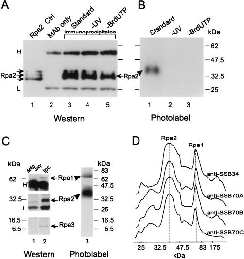
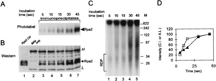
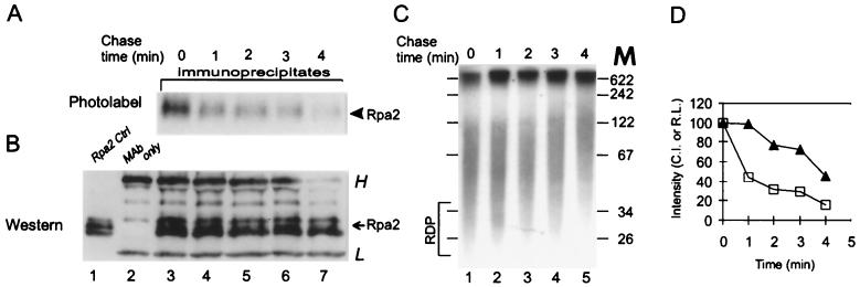
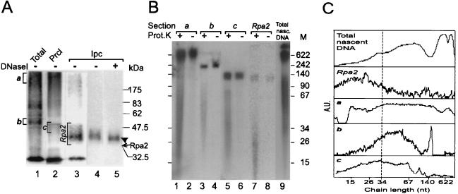
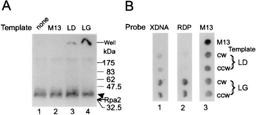
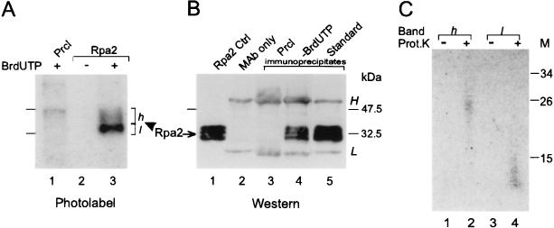
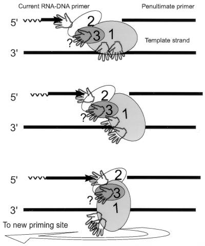
The eukaryotic single-stranded DNA binding protein replication protein A (RPA) participates in major DNA transactions. RPA also interacts through its middle subunit (Rpa2) with regulators of the cell division cycle and of the response to DNA damage. A specific contact between Rpa2 and nascent simian virus 40 DNA was revealed by in situ UV cross-linking. The dynamic attributes of the cross-linked DNA, its size distribution, its RNA primer content, and its replication fork polarity were determined [corrected]. These data suggest that Rpa2 contacts the early DNA chain intermediates synthesized by DNA polymerase alpha-primase (RNA-DNA primers) but not more advanced products. Possible signaling functions of Rpa2 are discussed, and current models of eukaryotic lagging-strand DNA synthesis are evaluated in view of our results.


Figures

FIG. 1
FIG. 1
UV cross-linking of Rpa2 with nascent SV40 DNA. Proteins were photolabeled with nascent DNA within replicating SV40 chromosomes pulse-labeled by BrdUTP and [α-32P]dATP for 90 s. Following DNase I digestion, Rpa2 was immunopurified and detected by immunoblotting and autoradiography, as described in Materials and Methods. (A) Immunoblot probed with anti-SSB34A. Proteins extracted from a nonlabeled control nuclear monolayer (lane 1), antibody alone (lane 2), the immunoprecipitates of cross-linked proteins derived from the standard reaction mixture (lane 3), a nonirradiated control (lane 4), or a control with dTTP instead of BrdUTP (lane 5) are shown. (B) Autoradiogram of proteins immunoprecipitated from the standard mixture (lane 1), a nonirradiated control (lane 2), or the control with dTTP instead of BrdUTP (lane 3). (C) Immunoprecipitation of the photolabeled RPA heterotrimer. RPA was immunoprecipitated under native conditions from the photolabeled protein mixture derived from replicating SV40 chromatin with anti-SSB34. After separation by SDS-PAGE, individual RPA subunits were detected by immunoblotting and photolabeled derivatives were detected by autoradiography. (D) Densitometric tracings of photolabeled RPA precipitated under native conditions by the indicated MAbs and resolved by SDS-PAGE as described for panel C. H and L indicate heavy and light Ig chains, respectively. The arrows and arrowheads point to the ECL signal of Rpa2 and the photolabeled derivative, respectively. Ctrl, control; Ipc, immunoprecipitated cleared Rpa.
FIG. 2
FIG. 2
Behavior of Rpa2’s photolabel in continuous labeling. Protein-DNA conjugates were prepared after the indicated pulse-labeling times. Rpa2 was then immunopurified and detected by autoradiography and Western blot analysis, essentially as described in Materials and Methods and in the legend of Fig. 1. (A) Autoradiogram of photolabeled Rpa2; (B) immunoblot; (C) pattern of nascent-SV40-DNA chains from aliquots of the same replication mixtures; (D) Rpa2 CI (open squares) and absolute labeling (A.L.) of the RDP fraction (filled triangles) versus labeling time. H and L indicate heavy and light Ig chains, respectively. The arrow points to the ECL signal of Rpa2; the arrowhead points to the photolabeled derivative. Ctrl, control. M, DNA size markers (in nucleotides).
FIG. 3
FIG. 3
Pulse-chase kinetics of Rpa2’s photolabel. SV40 DNA pulse-labeled for 90 s with BrdUTP and [α-32P]dATP was chased with dTTP and nonlabeled dATP as indicated. After UV cross-linking, Rpa2 was immunopurified and detected as described in Materials and Methods and in the legend of Fig. 1. (A) Autoradiogram; (B) immunoblot; (C) patterns of nascent SV40 DNA during chase; (D) Rpa2 CI (open squares) and relative labeling (R.L.) of the RDP fraction (filled triangles) versus chase time. The photolabeled protein samples shown in panel A were isolated from the bulk (90%) of the reaction mixture, and the blot showing them was exposed for a week. The total nascent DNA shown in panel C represents 10% of the reaction mixture, and the gel containing it was exposed for 16 h. H and L indicate heavy and light Ig chains, respectively. The arrow points to the ECL signal of Rpa2; the arrowhead points to the photolabeled derivative. M, DNA size markers (in nucleotides).
FIG. 4
FIG. 4
Sizing of nascent-DNA chains cross-linked to Rpa2. (A) Photolabeled Rpa2 not treated with DNase I was isolated from replication mixtures that had been pulse-labeled for 90 s as detailed in Materials and Methods. Crude photolabeled protein mixture (Total; lane 1); photolabeled proteins of the preclearing precipitate (Prcl; lane 2); immunoprecipitated cleared photolabeled Rpa2 (Ipc; lane 3); and nitrocellulose blots of the Rpa2 conjugate separated by SDS-PAGE, before or after DNase I digestion (lanes 4 and 5, respectively), are shown. (B) Portions of the bracketed gel sections a to c and Rpa2 (from panel A, lanes 1 to 3) were incubated with either proteinase K (Prot.K) (lanes 1, 3, 5, and 7) or buffer only (lanes 2, 4, 6, and 8). DNA was phenol extracted from the gel sections and separated by denaturing polyacrylamide-urea gel electrophoresis. Lane 9 contains an aliquot of nascent SV40 DNA (Total nasc. DNA) of the same replication mixture. The arrow marks the position of the ECL signal of Rpa2, and the arrowhead marks the position of the photolabeled derivative M, DNA size markers (in nucleotides). (C) Densitometric tracings of the total and released DNA preparations of panel B. The profile of the total DNA is shown. Net profiles of the DNA populations released from the indicated sections of panel A were obtained by subtracting the profile of an unproteolyzed sample (even-numbered lanes in panel B) from the original profile of the matched proteolyzed sample (odd-numbered lanes). However, residual spikes of large chains remained in the net profiles in sections b and c. These spikes coincide with the free-DNA contaminants and probably result from an excess of proteolyzed over unproteolyzed sample in the paired samples. The size distributions of chains in profiles a to c were estimated by setting their half-maximal heights as lower and upper borders. A.U., arbitrary absorption units.
FIG. 5
FIG. 5
Determination of fork polarity of DNA cross-linked to Rpa2. (A) Rpa2 cross-linked to intact nascent-DNA chains was immunopurified and hybridized to pairs of M13-SV40 ssDNA clones of leading (LD)- or lagging (LG)-strand fork polarity. The mixtures were resolved by SDS-PAGE and autoradiographed as described in Materials and Methods and the legend of Fig. 4. Lanes: 1, not hybridized; 2, vector; 3, mSVBH10 and mSVTB10 (clockwise replication fork [CW] and counterclockwise replication fork [CCW] leading-strand templates, respectively); 4, mSVBH11 and mSVTB11 (CW and CCW lagging-strand templates, respectively). (B) Nascent DNA was released by proteolysis of photolabeled Rpa2 (from Fig. 4B, lane 7). Following extraction from the gel, it was hybridized to dot blots containing the indicated leading- and lagging-strand probes (blot 1), along with a purified RDP fraction from Fig. 2C (blot 2) or vector DNA labeled by random priming (blot 3). XDNA, DNA released from the cross-linked Rpa2-DNA conjugate; RDP, RDP fraction; M13, vector DNA. The arrow indicates the position of the ECL signal of Rpa2, and the arrowhead indicates the photolabeled derivative.
FIG. 6
FIG. 6
Cross-linking of Rpa2 to nascent DNA labeled in the RNA primer moiety. Protein-DNA conjugates radiolabeled from [α-32P]UTP were prepared and detected by autoradiography and Western blotting, and the cross-linked DNAs were sized, as detailed in Materials and Methods. (A) Autoradiogram of a preclearance (Prcl) immunoprecipitate (lane 1), an Rpa2 immunoprecipitate of a control replication mixture containing dTTP instead of BrdUTP (lane 2), or the standard mixture (lane 3). (B) Immunoblot probed with the anti-Rpa2 MAb. Shown are proteins extracted from a nonlabeled control (Cntrl) nuclear monolayer (lane 1), antibody alone (lane 2), a preclearance immunoprecipitate (lane 3), and Rpa2 immunoprecipitates from reaction mixtures without (lane 4) or with (lane 5) BrdUTP. (C) Nascent RNA-DNA released by proteolysis from photolabeled Rpa2 species of bands h and l (from panel A, lane 3) was resolved by gel electrophoresis (lanes 2 [band h] and 4 [band l]) along with respective unproteolyzed controls (lanes 1 and 3). h and l designate slow- and fast-migrating photolabeled Rpa2 species, respectively. H and L indicate heavy and light Ig chains, respectively. Prot.K proteinase K; M, protein size markers (in thousands). The arrow indicates the ECL signal of Rpa2, and the arrowhead indicates at the photolabeled derivative.
FIG. 7
FIG. 7
Proposed contacts of RPA with DNA during the RDP cycle.

Similar articles

Cited by

References

    1. Blackwell L J, Borowiec J A, Mastrangelo I A. Single-stranded-DNA binding alters human replication protein A structure and facilitates interaction with DNA-dependent protein kinase. Mol Cell Biol. 1996;16:4798–4807. - PMC - PubMed
    1. Blackwell L J, Borowiec J A. Human replication protein A binds single-stranded DNA in two distinct complexes. Mol Cell Biol. 1994;14:3993–4001. - PMC - PubMed
    1. Bochkarev A, Pfuetzner R A, Edwards A M, Frappier L. Structure of the single-stranded-DNA-binding domain of replication protein A bound to DNA. Nature. 1997;385:176–181. - PubMed
    1. Boubnov N V, Weaver D T. scid cells are deficient in Ku and replication protein A phosphorylation by the DNA-dependent protein kinase. Mol Cell Biol. 1995;15:5700–5706. - PMC - PubMed
    1. Braun K A, Lao Y, He Z, Ingles C J, Wold M S. Role of protein-protein interactions in the function of replication protein A (RPA): RPA modulates the activity of DNA polymerase alpha by multiple mechanisms. Biochemistry. 1997;36:8443–8454. - PubMed

Publication types

LinkOut - more resources