. 1998 Nov;18(11):6595-604.
doi: 10.1128/MCB.18.11.6595.

Physical and functional interactions between type I transforming growth factor beta receptors and Balpha, a WD-40 repeat subunit of phosphatase 2A

Affiliations

Physical and functional interactions between type I transforming growth factor beta receptors and Balpha, a WD-40 repeat subunit of phosphatase 2A

I Griswold-Prenner et al. Mol Cell Biol. 1998 Nov.

Abstract

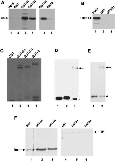
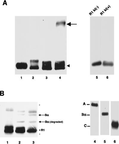
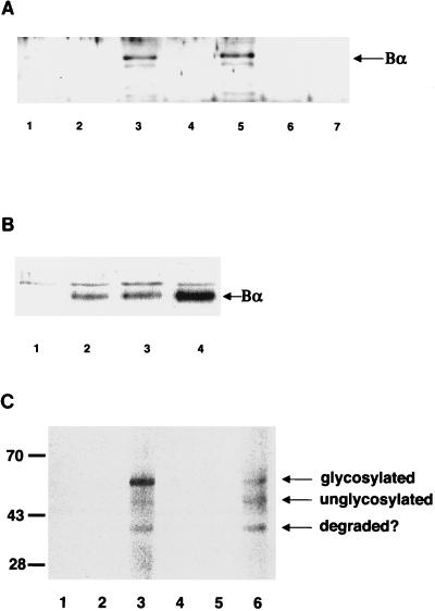
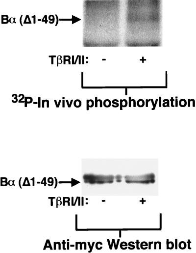
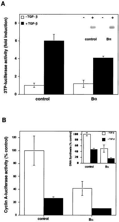
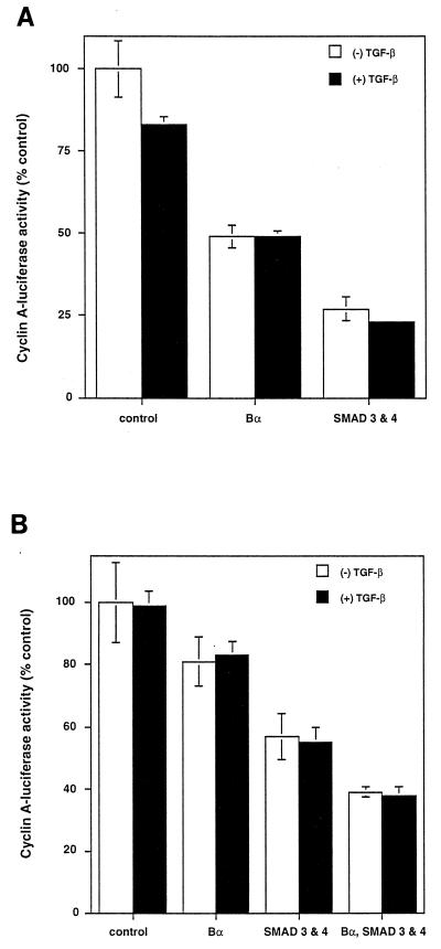
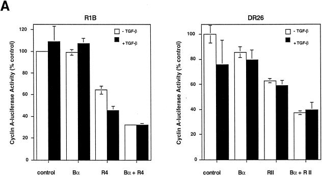
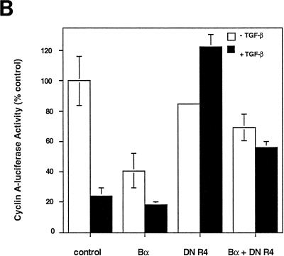
We have previously shown that a WD-40 repeat protein, TRIP-1, associates with the type II transforming growth factor beta (TGF-beta) receptor. In this report, we show that another WD-40 repeat protein, the Balpha subunit of protein phosphatase 2A, associates with the cytoplasmic domain of type I TGF-beta receptors. This association depends on the kinase activity of the type I receptor, is increased by coexpression of the type II receptor, which is known to phosphorylate and activate the type I receptor, and allows the type I receptor to phosphorylate Balpha. Furthermore, Balpha enhances the growth inhibition activity of TGF-beta in a receptor-dependent manner. Because Balpha has been characterized as a regulator of phosphatase 2A activity, our observations suggest possible functional interactions between the TGF-beta receptor complex and the regulation of protein phosphatase 2A.


Figures

FIG. 1
FIG. 1
In vitro association of Bα with type I receptors. (A) Association of in vitro-translated Bα with GST-receptor fusion proteins. In vitro-translated 35S-labeled Bα was incubated with glutathione-Sepharose-coupled GST (lane 2) or GST fused to the cytoplasmic domains of Tsk7L/R1 (lanes 3 and 5), TβRI/R4 (lane 4), or TβRII (lane 6). Bound proteins were eluted and separated by SDS-PAGE. Lane 1 shows 1/50 of the input 35S-labeled Bα used in each of the GST adsorption experiments. (B) Lack of interaction of TRIP-1 with the cytoplasmic domain of a type I receptor fused to GST (GST-R1) or with GST itself. (C) Gel electrophoretic analysis of the purified GST fusion experiments used in panel A and other experiments. Whereas the GST protein itself corresponded to a single band on the gel, the fusion proteins ran as several bands, the largest of which corresponded to the full-size proteins, and the smaller bands are presumably degradation products. (D) Covalent cross-linking of Bα in the ABαC complex with the cytoplasmic domain of Tsk7L/R1. Purified ABαC complex was incubated without cross-linking (lane 1) or was chemically cross-linked in the absence (lane 2) or presence (lane 3) of purified cytoplasmic domain of Tsk7L/R1. Cross-linked proteins were separated by SDS-PAGE and immunoblotted with anti-Bα antibody. The arrow points to the cross-linked complex of Bα with Tsk7L/R1 which is detected as an anti-Bα immunoreactive band. The arrowhead points to noncomplexed Bα. (E) Covalent cross-linking of Bβ in the ABβC complex with the cytoplasmic domain of Tsk7L/R1. Purified ABβC was chemically cross-linked in the absence (lane 1) or presence (lane 2) of the His6-tagged cytoplasmic domain Tsk7L/R1. The arrow points to the cross-linked complex of Bβ with Tsk7L/R1 which is detected as the anti-Bβ immunoreactive band, and the arrowhead points to noncomplexed Bβ. (F) In vitro-translated B′ does not bind GST-receptor fusion proteins. In vitro-translated 35S-labeled Bα (lanes 1 to 3) and B′ (lanes 4 to 6) were tested for their association with GST (lanes 1 and 4) or GST fusion proteins with the cytoplasmic domains of Tsk7L/R1 (lanes 2 and 5) or TβRI/R4 (lanes 3 and 6) as described for panel A.
FIG. 2
FIG. 2
Interaction of Bα with the type I receptor cytoplasmic domain depends on its kinase activity and allows phosphorylation of Bα by the receptor kinase. (A) Bα interacts with kinase-active [ki(+)], not with kinase-inactive [ki(−)], Tsk7L/R1. Bα in the absence of the cytoplasmic domain of the receptor without (lane 1) or after (lane 2) chemical cross-linking is shown. Bα incubated with the kinase-inactive (lane 3) or kinase-active (lane 4) cytoplasmic domain of the receptor and stabilized by chemical cross-linking is also shown. The arrow points to the cross-linked complex of Bα with Tsk7L/R1 which is detected as the anti-Bα immunoreactive band, and the arrowhead points to noncomplexed Bα. Lanes 5 and 6 show that equal amounts of the kinase-negative or -positive cytoplasmic domains of Tsk7L/R1, purified as His6-tagged proteins, were used in lanes 3 and 4. (B) Tsk7L/R1 phosphorylates Bα. The purified cytoplasmic domain of Tsk7L/R1 was incubated alone (lane 1), with Bα (lane 2), or with ABαC (lane 3) in a kinase reaction in the presence of [γ-32P]ATP. Proteins were separated by SDS-PAGE, and 32P-phosphorylated substrates were visualized by autoradiography. Besides Bα, a degradation product of Bα [Bα (degraded)] was also phosphorylated. The phosphorylated band above the Bα band is a protein that copurified with the receptor cytoplasmic domain. The gray arrowheads denote the positions of the A (upper) and C (lower) subunits, which were not phosphorylated. Lanes 4 to 6 show Western blots of purified and electrophoretically separated ABαC (26), which was used in lane 3, with antibodies specific for A, Bα, or C, respectively.
FIG. 3
FIG. 3
In vivo association of Bα with type I receptors. (A) In vivo association of Bα with type I receptors. Bα was expressed alone (lane 1) or coexpressed with His6-tagged kinase-inactive (lanes 2, 4, and 6) or kinase-active (lanes 3, 5, and 7) cytoplasmic domains of the type I receptor Tsk7L/R1 (lanes 2 and 3) or TβRI/R4 (lanes 4 and 5) or the type II receptor TβRII (lanes 6 and 7) in transfected COS-1 cells. His6-tagged cytoplasmic domains and associated proteins were purified on Co2+-Sepharose beads and, following elution, separated by SDS-PAGE, and Bα was detected by immunoblotting with anti-Bα antibody. (B) TβRII coexpression enhances the association of Bα with TβRI. Bα was expressed alone (lane 1) or was coexpressed with the His6-tagged cytoplasmic domain of TβRI/R4 (lanes 2 to 4) in transfected COS-1 cells, in the absence (lane 2) or presence (lanes 3 and 4) of TβRII (1 [lane 3] or 10 [lane 4] μg of transfected plasmid DNA). As in panel A, the proteins associated with TβRI purified on Co2+-Sepharose beads were separated by SDS-PAGE and associated Bα was detected by immunoblotting. (C) Interaction of Bα with full-size type I receptor at the cell surface. The full-size type I receptor Tsk7L/R1 (lanes 2 and 3) or TβRI/R4 (lanes 5 and 6) was expressed in COS-1 cells in the presence (lanes 3 and 6) or absence (lanes 2 and 5) of Bα in transfected cells. The corresponding cytoplasmically truncated Tsk7L/R1 (lane 1) or TβRI/R4 (lane 4) was also coexpressed with Bα. Cell surface proteins were labeled by surface biotinylation of intact cells, and immunoprecipitations were carried out by using anti-Bα antibody, followed by visualization of coprecipitated, biotinylated proteins. The full-size glycosylated and unglycosylated type I receptor and an often-observed degradation product (degraded?) are indicated.
FIG. 4
FIG. 4
N-terminally truncated Bα is phosphorylated upon coexpression of TβRI and TβRII. Myc-epitope-tagged Bα, lacking amino acids 1 to 49, was coexpressed in the presence (+) or absence (−) of TβRI/R4 and TβRII (TβRI/II) in transfected COS cells. The in vivo phosphorylation of this deletion mutant of Bα was assessed by in vivo 32P labeling followed by immunoprecipitation with anti-Myc antibody 9E10 and detection by SDS-PAGE and autoradiography (upper lanes). The lower lanes illustrate equal expression levels of the Myc-tagged Bα mutant as assessed by anti-Myc Western blotting of anti-Myc immunoprecipitated protein. Left lanes, without TβRI/TβRII expression; right lanes, with TβRI/TβRII expression.
FIG. 5
FIG. 5
Increased Bα expression does not alter the overall phosphorylation state of TGF-β receptors. FLAG-tagged type II receptors (TβRII) and type I receptor TβRI/R4 (R4) were coexpressed with Bα in transfected L17 cells. The in vivo phosphorylation of the receptors was assessed by in vivo 32P labeling followed by immunoprecipitation with FLAG antibody and detection by SDS-PAGE and autoradiography. Lane 1, without increased Bα expression; lane 2, with increased Bα expression.
FIG. 6
FIG. 6
Effect of Bα on TGF-β responsiveness. (A) Effect of Bα on luciferase expression from the 3TP promoter. The Bα expression plasmid pRK7-Bα or the control plasmid pRK7 was cotransfected with p3TP-lux in HaCaT cells. Luciferase expression was measured in the absence or presence of TGF-β. The inset shows that PAI-1 protein levels are not altered by Bα in stably transfected cell lines. 35S-labeled PAI-1 protein secreted from HaCaT cells, stably transfected with pRK7 (control) or pRK7-Bα in the absence (−) or presence (+) of 100 pM TGF-β, was used. (B) Bα enhances the inhibition of luciferase activity by TGF-β from the cyclin A promoter. HaCaT cells were cotransfected with the pCal2 luciferase reporter plasmid and the control plasmid pRK7 or the Bα expression plasmid pRK7-Bα. Luciferase expression from the cyclin A promoter, a measure of cell proliferation, was measured in the absence or presence of TGF-β. the inset shows that Bα increases the inhibition of DNA synthesis by TGF-β in stably transfected cells. DNA synthesis, measured by [3H]thymidine incorporation in HaCaT cells, stably transfected with pRK7 (control) or pRK7-Bα in the absence (−) or presence (+) of 100 pM TGF-β, is shown. Data are presented relative to those of untreated, control transfected cells. Standard deviations were based on triplicate measurements.
FIG. 7
FIG. 7
Bα enhances the inhibition of cyclin A-luciferase activity independently from Smads. Bα increases the inhibition of cyclin A-luciferase activity comparably to Smad3 and -4. HaCaT (A) or Smad4-deficient SW480.7 (B) cells were cotransfected with pCal2 and pRK7 (control), pRK7-Bα, or pRK5 expression plasmids for Smad3 and -4. Luciferase expression from the cyclin A promoter was measured in the absence (−) or presence (+) of TGF-β. Bα inhibition of cyclin A-luciferase activity occurs in the absence of Smad4 and is therefore Smad independent (B), whereas Bα and Smad3-Smad4 additively inhibit cyclin A-luciferase activity in these cells.
FIG. 8
FIG. 8
Dependence of the growth inhibition effect of Bα on functional TGF-β receptors. (A) The growth inhibition effect of Bα is TGF-β receptor dependent. R1B cells, an Mv1Lu derivative lacking functional TβRI, or DR26 cells, an Mv1Lu derivative lacking functional TβRII, were cotransfected with the pCal2 cyclin A-luciferase plasmid and pRK7 expression plasmids for Bα, TβRI/R4 (in R1B cells), or TβRII (in DR26 cells). Luciferase expression from the cyclin A promoter was measured in the absence (−) or presence (+) of TGF-β. (B) Dominant-negative inhibition of TβR1/R4 activity blocks growth inhibition by Bα. Mv1Lu cells were cotransfected with the pCal2 cyclin A-luciferase plasmid and pRK7 expression plasmids for Bα or a cytoplasmically truncated form of TβRI (DN R4) or both. Luciferase expression from the cyclin A promoter was measured in the absence or presence of TGF-β. Luciferase values, normalized for transfection efficiency, are presented relative to those of untreated, control transfected cells. Standard deviations were based on triplicate measurements.
FIG. 8
FIG. 8
Dependence of the growth inhibition effect of Bα on functional TGF-β receptors. (A) The growth inhibition effect of Bα is TGF-β receptor dependent. R1B cells, an Mv1Lu derivative lacking functional TβRI, or DR26 cells, an Mv1Lu derivative lacking functional TβRII, were cotransfected with the pCal2 cyclin A-luciferase plasmid and pRK7 expression plasmids for Bα, TβRI/R4 (in R1B cells), or TβRII (in DR26 cells). Luciferase expression from the cyclin A promoter was measured in the absence (−) or presence (+) of TGF-β. (B) Dominant-negative inhibition of TβR1/R4 activity blocks growth inhibition by Bα. Mv1Lu cells were cotransfected with the pCal2 cyclin A-luciferase plasmid and pRK7 expression plasmids for Bα or a cytoplasmically truncated form of TβRI (DN R4) or both. Luciferase expression from the cyclin A promoter was measured in the absence or presence of TGF-β. Luciferase values, normalized for transfection efficiency, are presented relative to those of untreated, control transfected cells. Standard deviations were based on triplicate measurements.

References

    1. Alessi D R, Gomez N, Moorhead G, Lewis T, Keyse S M, Cohen P. Inactivation of p42 MAP kinase by protein phosphatase 2A and a protein tyrosine phosphatase, but not CL100, in various cell lines. Curr Biol. 1995;5:283–295. - PubMed
    1. Andersson S, Davis D L, Dahlback H, Jornvall H, Russell D W. Cloning, structure, and expression of the mitrochondrial cytochrome P-450 sterol 26-hydroxylase, a bile acid biosynthetic enzyme. J Biol Chem. 1989;264:8222–8229. - PubMed
    1. Bassing C H, Yingling J M, Howe D J, Wang T, He W W, Gustafson M L, Shah P, Donahoe P K, Wang X-F. A transforming growth factor β type I receptor that signals to activate gene expression. Science. 1993;263:87–89. - PubMed
    1. Brand T, Schneidert M D. Inactive type II and type I receptors for TGF β are dominant inhibitors of TGF beta-dependent transcription. J Biol Chem. 1995;270:8274–8284. - PubMed
    1. Cairns J, Qin S, Philp R, Tan Y H, Guy G R. Dephosphorylation of the small heat shock protein Hsp27 in vivo by protein phosphatase 2A. J Biol Chem. 1994;269:9176–9183. - PubMed

Publication types

MeSH terms

Substances

LinkOut - more resources