. 1998 Nov;18(11):6653-65.
doi: 10.1128/MCB.18.11.6653.

Direct interaction of SRY-related protein SOX9 and steroidogenic factor 1 regulates transcription of the human anti-Müllerian hormone gene

Affiliations

Direct interaction of SRY-related protein SOX9 and steroidogenic factor 1 regulates transcription of the human anti-Müllerian hormone gene

P De Santa Barbara et al. Mol Cell Biol. 1998 Nov.

Abstract

For proper male sexual differentiation, anti-Müllerian hormone (AMH) must be tightly regulated during embryonic development to promote regression of the Müllerian duct. However, the molecular mechanisms specifying the onset of AMH in male mammals are not yet clearly defined. A DNA-binding element for the steroidogenic factor 1 (SF-1), a member of the orphan nuclear receptor family, located in the AMH proximal promoter has recently been characterized and demonstrated as being essential for AMH gene activation. However, the requirement for a specific promoter environment for SF-1 activation as well as the presence of conserved cis DNA-binding elements in the AMH promoter suggest that SF-1 is a member of a combinatorial protein-protein and protein-DNA complex. In this study, we demonstrate that the canonical SOX-binding site within the human AMH proximal promoter can bind the transcription factor SOX9, a Sertoli cell factor closely associated with Sertoli cell differentiation and AMH expression. Transfection studies with COS-7 cells revealed that SOX9 can cooperate with SF-1 in this activation process. In vitro and in vivo protein-binding studies indicate that SOX9 and SF-1 interact directly via the SOX9 DNA-binding domain and the SF-1 C-terminal region, respectively. We propose that the two transcription factors SOX9 and SF-1 could both be involved in the expression of the AMH gene, in part as a result of their respective binding to the AMH promoter and in part because of their ability to interact with each other. Our work thus identifies SOX9 as an interaction partner of SF-1 that could be involved in the Sertoli cell-specific expression of AMH during embryogenesis.


Figures

FIG. 1
FIG. 1
Deletional analysis of the AMH proximal promoter region. (A) Sequence comparison among human, mouse, rat, and bovine AMH proximal promoter regions. The characteristic TATA box, the SF-1-binding site (SF-1-BS), the SOX-binding site (SOX-BS), and a putative GATA site (GATA) that are conserved among the different sequences are indicated. Numbers below the sequences correspond to the human gene. (B) Intrinsic activity of the human AMH proximal promoter in NT2/D1 cells. Various human AMH promoter-CAT reporter gene constructs were transfected in NT2/D1 and COS-7 cells. CAT reporter activities were quantitated after cotransfection of 1 μg of constructs containing either the bp −154 (p154CAT), the bp −123 human AMH promoter (p123CAT), or the bp −154 AMH promoter mutated on the SOX-binding site (p154MUTSOXCAT) and 0.2 μg of pCMV–β-galactosidase. The values shown represent the means of four transfection experiments. Increases in activation are shown relative to pEMBL empty vector. Standard deviations are indicated by bars.
FIG. 2
FIG. 2
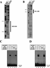
DNase I footprint analysis of the AMH proximal promoter by using SOX9 and SF-1 recombinant proteins. (A) SOX9 DNase I footprint. The upper strand of the 164-bp AMH promoter was 32P labeled; 104 cpm of the probe was incubated in the presence of 1 μg of either free GST or GST-SOX9. In each case, the reaction mix was resolved on a 6% polyacrylamide sequencing gel. The lane labeled Ladder designates a G+A Maxam-Gilbert sequence ladder obtained with the probe. The protected regions are indicated by a box. (B) SF-1 DNase I footprint. The same radiolabeled probe was incubated with 1 μg of either free GST or GST–SF-1. The lane labeled Ladder represents the G+A Maxam-Gilbert sequence ladder. The protected regions are indicated by a box. (C) Affinity binding of GST-SOX9 to the SOX-binding site (SOX-BS) in the AMH proximal promoter probe. The double-stranded SOX-binding-site oligonucleotide was end labeled and incubated with 10 ng of purified GST-SOX9 protein in the absence (lane 2) or presence (lanes 4 and 5) of a 50-fold molar excess of unlabeled competitors. Free GST was used as the control (lane 3). (D) Affinity binding of GST–SF-1 recombinant protein to the SF-1-binding site (SF-1-BS) in the AMH promoter. The double-stranded SF-1-binding-site oligonucleotide was end labeled and incubated with 10 ng of purified GST-SOX9 protein in the absence (lane 2) or presence (lanes 4 and 5) of a 50-fold molar excess of unlabeled competitors. Free GST was used as control (lane 3).
FIG. 3
FIG. 3
Cooperation between SOX9 and SF-1 proteins in the activation of the AMH minimal promoter in COS-7 cells. (A) Cotransfection assay in COS-7 cells of a reporter plasmid with the CAT gene under the control of an AMH minimal promoter (p154CAT), a constant amount of pcDNA3-SOX9 (10 ng), and increasing amounts of the pcDNA3–SF-1 construct. (B) The specificity of SOX9 activity was tested with a deleted version of pcDNA3-SOX9 (10 ng) or with pcDNA3-SRY (10 ng). (C) Comparison of the SOX9–SF-1 activity on the wild-type AMH proximal promoter (p154CAT) with that on the same promoter mutated on the SOX-binding site p154MUTSOX. The CAT activity of the reporter plasmid alone was set as 1. All values represent the means of three separate transfection experiments (± standard errors).
FIG. 4
FIG. 4
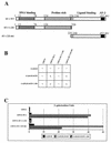
SOX9 interacts with the carboxy-terminal region of the SF-1 protein. The SOX9–SF-1 interaction was scored by a yeast two-hybrid assay (see Materials and Methods). (A) Schematic diagrams of SF-1 wild type, deletion constructs, and functional domains of the human SF-1 protein. (B) Qualitative histidine assays. A positive interaction results in the ability of the Y187-Hf7c diploid expressing the designated constructs to grow (+) or not grow (−) on a medium depleted of tryptophan, leucine, and histidine. Assays were done for three independent diploids. All the SF-1 constructs were tested against the empty pGADGH vector as a negative control, and the SOX9-derived constructs were tested against the empty pGBT11 vector. (C) Quantitative β-galactosidase assays for these interactions were conducted on the same diploids as those used in the histidine assays. Mean values are given in relative β-galactosidase units.
FIG. 5
FIG. 5
SOX9 and SF-1 interact in vitro. (A) The diagram shows the different SOX9 deletion mutants which were used to investigate the physical interaction between SOX9 and SF-1. In each case, the respective percentage of protein bound to the GST–SF-1 phase was determined by phosphorimager analysis. (B) The SOX9 polypeptides depicted in panel A were translated in vitro in the presence of [35S]methionine and analyzed for binding to either GST, GST–SF-1 or GST–c-erbA fusion proteins bound to glutathione-Sepharose. The GST pulldown assay was performed as described in Materials and Methods. The 10% input (left lane) and bound proteins (the other lanes) were separated by SDS-PAGE analysis and then autoradiographed. (C) Binding of the GST–SF-1 fusion protein to the SF-1-binding-site (SF-1-BS) probe (lane 2) is shifted after preincubation with in vitro-translated TNT-SOX9 protein as shown by EMSA (lane 4). The specificity was assessed by the use of either TNT or TNT plus GST alone (lanes 1 and 3). Lane 5 shows the absence of TNT-SOX9 protein binding to the SF-1-binding-site probe in the presence of 2 μg of poly(dI-dC). WT, wild type.
FIG. 5
FIG. 5
SOX9 and SF-1 interact in vitro. (A) The diagram shows the different SOX9 deletion mutants which were used to investigate the physical interaction between SOX9 and SF-1. In each case, the respective percentage of protein bound to the GST–SF-1 phase was determined by phosphorimager analysis. (B) The SOX9 polypeptides depicted in panel A were translated in vitro in the presence of [35S]methionine and analyzed for binding to either GST, GST–SF-1 or GST–c-erbA fusion proteins bound to glutathione-Sepharose. The GST pulldown assay was performed as described in Materials and Methods. The 10% input (left lane) and bound proteins (the other lanes) were separated by SDS-PAGE analysis and then autoradiographed. (C) Binding of the GST–SF-1 fusion protein to the SF-1-binding-site (SF-1-BS) probe (lane 2) is shifted after preincubation with in vitro-translated TNT-SOX9 protein as shown by EMSA (lane 4). The specificity was assessed by the use of either TNT or TNT plus GST alone (lanes 1 and 3). Lane 5 shows the absence of TNT-SOX9 protein binding to the SF-1-binding-site probe in the presence of 2 μg of poly(dI-dC). WT, wild type.
FIG. 6
FIG. 6
SOX9 and SF-1 form a common protein-DNA complex. Nuclear extracts were prepared from NT2/D1 cells. In an EMSA reaction, α-32P-labeled SF-1-binding-site (SF-1-BS) oligonucleotide and 2 μg of nuclear extract were incubated together. The specificity of the retarded bands was controlled by the use of either an excess of cold SF-1-binding-site probe (lane 2) or a mutated form of this oligonucleotide (lane 10). Supershift experiments were performed after preincubation of either 1 μl of SF-1 antibody (SF-1-Ab.) (lane 4) or 1 μl of SOX9 antibody (SOX9-Ab.) (lane 6). The corresponding preimmune serum (SF-1-Pre-imm. and SOX9-Pre-imm.) (lanes 3 and 5) or 1 μl of a specific anti-SRY antibody (SRY-Ab.) (lane 7) was used as the control. The specificity of the observed shifts with the different antibodies was assessed by the use of the corresponding antibody only (lanes 11 and 12). Gel retardation of the TNT–SF-1 protein after binding to the SF-1-binding-site probe was used as a control (lane 8).
FIG. 7
FIG. 7
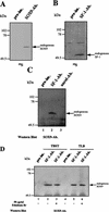
Coimmunoprecipitation of endogenous SOX9 and SF-1 from metabolically 35S-labeled NT2/D1 cells. After labeling, equal amounts of NT2/D1 cell extracts (prepared as described in Materials and Methods) were immunoprecipitated. (A) Immunoprecipitation with SOX9 antibodies (SOX9-Ab.) or preimmune (pre-im.) antibodies. (B) Immunoprecipitation with SF-1 antibodies (SF-1-Ab.) or preimmune (pre-im.) antibodies. (C) Western blot analysis with a SOX9-specific rat antibody of an SF-1 immunoprecipitate (SF-1-Ab.). As a control, an unrelated antibody (unrel-Ab.) was introduced in the precipitation step before revelation with the SOX9 antibody. First-step immunoprecipitation with preimmune (pre-im.) antibodies is also shown. (D) Similar experiments comparing two buffers containing two different detergents, TBST (lanes 1 to 3) or TLB (lanes 4 to 6) (see Materials and Methods), and including or not including ethidium bromide. Lanes 1 and 4 show immunoprecipitation with preimmune (pre-im.) antibodies as controls.
FIG. 8
FIG. 8
Immunolocalization of SOX9 and SF-1 protein in cultured human NT2/D1 cells. Immunostaining of SF-1 in green (A and D) and of SOX9 in red (B and E) or both (panel F) is shown. As a control, AMH expression was checked by using the corresponding rabbit antibody (in red) along with a counterstaining of cell nuclei with Hoechst 33258 in blue (C). Scale bars, 10 μm.

Similar articles

Cited by

References

    1. Ambrosetti D-C, Basilico C, Dailey L. Synergistic activation of the fibroblast growth factor 4 enhancer by Sox2 and Oct-3 depends on protein-protein interactions facilitated by a specific spatial arrangement of factor binding sites. Mol Cell Biol. 1997;17:6321–6329. - PMC - PubMed
    1. Bell D M, Leung K K H, Wheatley S C, Ng L J, Zhou S, Ling K W, Sham M H, Koopman P, Tam P P L, Cheah K S E. SOX9 directly regulates the type-II collagen gene. Nat Genet. 1997;16:174–178. - PubMed
    1. Carré-Eusèbe D, Di Clemente N, Rey R, Pieau C, Vigier B, Josso N, Picard J-Y. Cloning and expression of the chick anti-Müllerian hormone gene. J Biol Chem. 1996;271:4798–4804. - PubMed
    1. Clépet C, Schafer A J, Sinclair A H, Palmer M S, Lovell-Badge R, Goodfellow P N. The human SRY transcript. Hum Mol Genet. 1993;2:2007–2012. - PubMed
    1. Crawford P A, Dorn C, Sadovsky Y, Milbrandt J. Nuclear receptor DAX-1 recruits nuclear receptor corepressor N-CoR to steroidogenic factor 1. Mol Cell Biol. 1998;18:2949–2956. - PMC - PubMed

Publication types

MeSH terms