. 1998 Nov;18(11):6816-25.
doi: 10.1128/MCB.18.11.6816.

Cytoplasmic dynein intermediate-chain isoforms with different targeting properties created by tissue-specific alternative splicing

Affiliations

Cytoplasmic dynein intermediate-chain isoforms with different targeting properties created by tissue-specific alternative splicing

D I Nurminsky et al. Mol Cell Biol. 1998 Nov.

Abstract

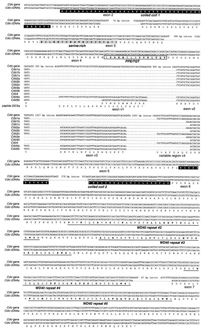
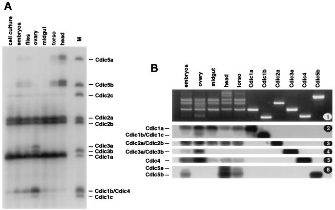
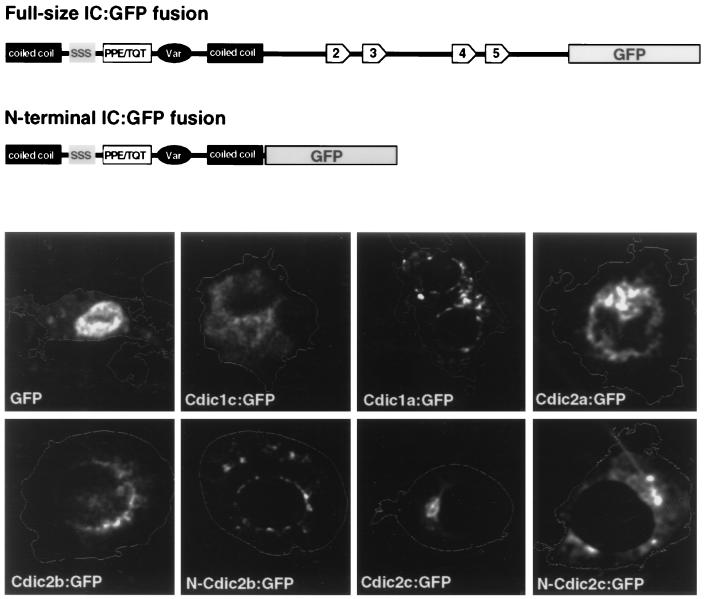
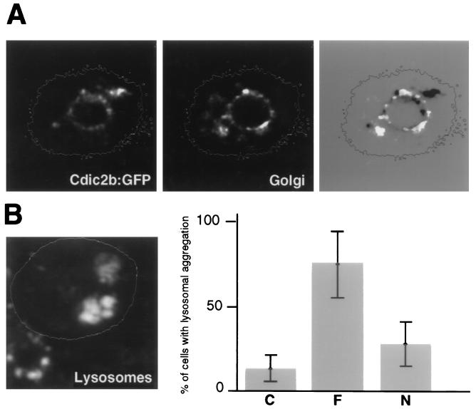
The intermediate chains (ICs) are the subunits of the cytoplasmic dynein that provide binding of the complex to cargo organelles through interaction of their N termini with dynactin. We present evidence that in Drosophila, the IC subunits are represented by at least 10 structural isoforms, created by the alternative splicing of transcripts from a unique Cdic gene. The splicing pattern is tissue specific. A constitutive set of four IC isoforms is expressed in all tissues tested; in addition, tissue-specific isoforms are found in the ovaries and nervous tissue. The structural variations between isoforms are limited to the N terminus of the IC molecule, where the interaction with dynactin takes place. This suggests differences in the dynactin-mediated organelle binding by IC isoforms. Accordingly, when transiently expressed in Drosophila Schneider-3 cells, the IC isoforms differ in their intracellular targeting properties from each other. A mechanism is proposed for the regulation of dynein binding to organelles through the changes in the content of the IC isoform pool.


Figures

FIG. 1
FIG. 1
A. Dynein IC transcripts in Drosophila. Samples (10 μg) of total RNA isolated from various developmental stages of D. melanogaster, as indicated at the top, along with RNA samples from the heads of D. melanogaster adults or D. simulans adults, were separated in a 1% agarose–formaldehyde gel. Hybridization with probe A (Fig. 2) revealed two major bands, corresponding to the Cdic and Sdic transcripts. Only the Cdic transcripts were detected in D. simulans. (B) Control hybridization with the probe for the constitutively expressed gene oxen (1a) shows sufficient RNA loading on all lanes. The numbers on the right indicate the sizes of transcripts in kilobases.
FIG. 2
FIG. 2
Cloning and characterization of Cdic and Sdic cDNAs. λZAP clones are indicated by thin black bars, and RACE products are indicated by shaded bars. In the composite cDNAs, coding regions are black. The positions for the DIC-U and DIC-LL primers, used to amplify cDNAs for Cdic isoforms, and for the DICr-U and DIC-LL primers, used for Sdic, are shown. A and B are the Cdic/Sdic-specific and Cdic-specific probes, respectively.
FIG. 3
FIG. 3
Comparison of the sequence of the N-terminal regions of Cdic and other known cytoplasmic ICs from rats (GenBank accession no. U39046) and Dictyostellium (accession no. U25116). Conserved amino acids are outlined and presented in the consensus line. Coiled-coil domains, shown by solid boxes, were predicted with the PAIRCOILS (3) and COILS (13) algorithms. The serine-rich domain and PPE/TQT conserved block are outlined by boxes. The positions of variable regions in the rat IC (var-1 and var-2) and the beginning of the variable region in Cdic (var) are indicated. Cdic isoform shown is Cdic5b.
FIG. 4
FIG. 4
Sequence comparison shows that Cdic cDNA represents the transcripts from the Cdic gene and that Sdic cDNA corresponds to the transcripts from the annexin-dynein repeat. The entire Cdic gene sequence is presented; for cDNAs and the annexin-dynein repeat, only the differences are shown. Gaps introduced in the sequences are marked with dots.
FIG. 5
FIG. 5
Exon-intron structure of the Cdic gene. The genomic sequence is shown at the top, with exons indicated by boxes. Coding sequences are shown as solid boxes. The promoter is shown as triangle. 1 to 7, constitutive exons present in all Cdic mRNAs. v1 to v3, variable exons. Five classes of Cdic transcripts are shown below the sequence.
FIG. 6
FIG. 6
Sequence alignment of coding regions of the Cdic gene with Cdic cDNAs. The genomic sequence is at the top, with the gaps introduced in place of introns. cDNA sequences are below, shown as a single line in “constitutive” regions where they are identical and shown individually in the variable region. A conceptual protein sequence is shown at bottom; for the isoform Cdic5a, a frameshifted translation of exon v1 is included (peptide 5a). Coiled-coil donains, outlined as black boxes, were predicted with the PAIRCOILS (3) and COILS (13) algorithms. In the N-terminal region, serine-rich domain and the PPE/TQT conserved region are boxed. In the C-terminal region, four of five WD-40 repeats (27) are underlined.
FIG. 7
FIG. 7
Tissue specificity of Cdic isoforms. RT-PCR fragments were amplified across the variable region of Cdic transcripts. (A) PCR fragments were labeled at one DNA strand with 32P and separated in a 5% acrylamide sequencing gel. The source of the RNA is indicated at the top. Lane M contains marker fragments generated from the of cDNA clones representing Cdic isoforms. (B) PCR fragments were separated in a 3% agarose gel (1) and, after Southern transfer, hybridized with oligonucleotides "iso2" (2), "iso2‴ (3), "iso3" (4), "iso4" (5), and "iso5" (6). The source of the RNA is indicated at the top; individual cDNA clones Cdic1a, Cdic1b, Cdic2a, Cdic3a, Cdic4, and Cdic5b were used to generate the marker fragments in the six right-hand lanes.
FIG. 8
FIG. 8
Localization of the Cdic-GFP fusion proteins in cultured Schneider-3 cells. A schematic representation of the full-size fusion and N-terminal fusion proteins is shown at the top. Cells were transfected with plasmids expressing fusion proteins under the control of the cytomegalovirus promoter and stained with propidium iodide. Staining of the cellular content with propidium iodide was detected in the rhodamine channel, and the image was converted to the contour of the cell. GFP fluorescence was detected in the fluorescein isothiocyanate channel and pseudocolored in white. IC isoforms are indicated; for example, Cdic1a:GFP is a full-size fusion of Cdic1a isoform with GFP, and N-Cdic2b and N-Cdic2c are the N-terminal fusions. The localization of the GFP expressed alone is shown for comparison.
FIG. 9
FIG. 9
(A) Tight association of the Cdic2b fusion protein with the nucleus. In this case, Golgi staining mostly reveals the perinuclear elements. On the right, the GFP fluorescence image was converted to the negative and overlaid on the Golgi staining image. Note that the GFP fusion protein is located more proximal than the Golgi elements. (B) Redistribution of the lysosomes in the cells overexpressing the Cdic2c:GFP fusion protein. On the left, two transfected cells are contoured. Staining with Lysotracker DND-99 revealed aggregation of the lysosomes in transfected cells, in contrast to the random and mostly juxtranuclear distribution in nontransfected cell in the left lower corner. On the right, the frequency of the lysosomal aggregation in cells expressing Cdic2c:GFP (F) or N-Cdic2c:GFP (N) versus nontransfected cells (C) is shown. Error bars represent 95% confidence intervals.

References

    1. Aniento F, Emans N, Griffiths G, Gruenberg J. Cytoplasmic dynein-dependent vesicular transport from early to late endosomes. J Cell Biol. 1993;123:1373–1387. - PMC - PubMed
    1. Benevolenskaya, E. Unpublished data.
    1. Benevolenskaya E V, Nurminsky D I, Gvozdev V A. Structure of the Drosophila melanogaster annexin X gene. DNA Cell Biol. 1994;14:349–357. - PubMed
    1. Berger B, Wilson D B, Wolf E, Tonchev T, Milla M, Kim P S. Predicting coiled coils by use of pairwise residue correlations. Proc Natl Acad Sci USA. 1995;92:8259–8263. - PMC - PubMed
    1. Bruno K S, Tinsley J H, Minke P F, Plamann M. Genetic interactions among cytoplasmic dynein, dynactin, and nuclear distribution mutants of Neurospora crassa. Proc Natl Acad Sci USA. 1996;93:4775–4780. - PMC - PubMed

Publication types