. 1998 Oct;153(4):1099-112.
doi: 10.1016/S0002-9440(10)65654-4.

Angiogenesis in mice with chronic airway inflammation: strain-dependent differences

Affiliations

Angiogenesis in mice with chronic airway inflammation: strain-dependent differences

G Thurston et al. Am J Pathol. 1998 Oct.

Abstract

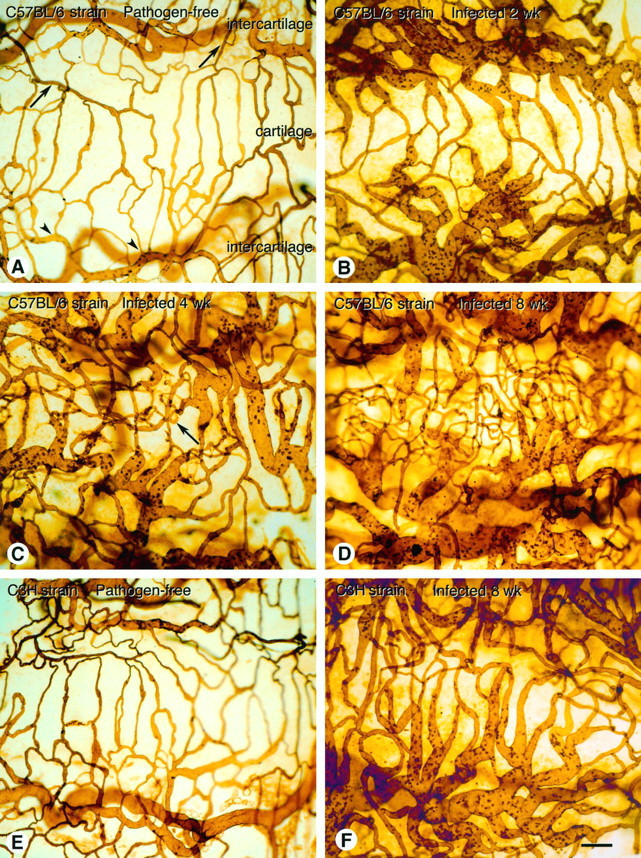
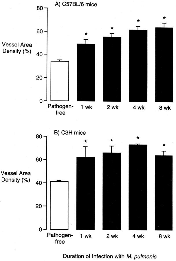
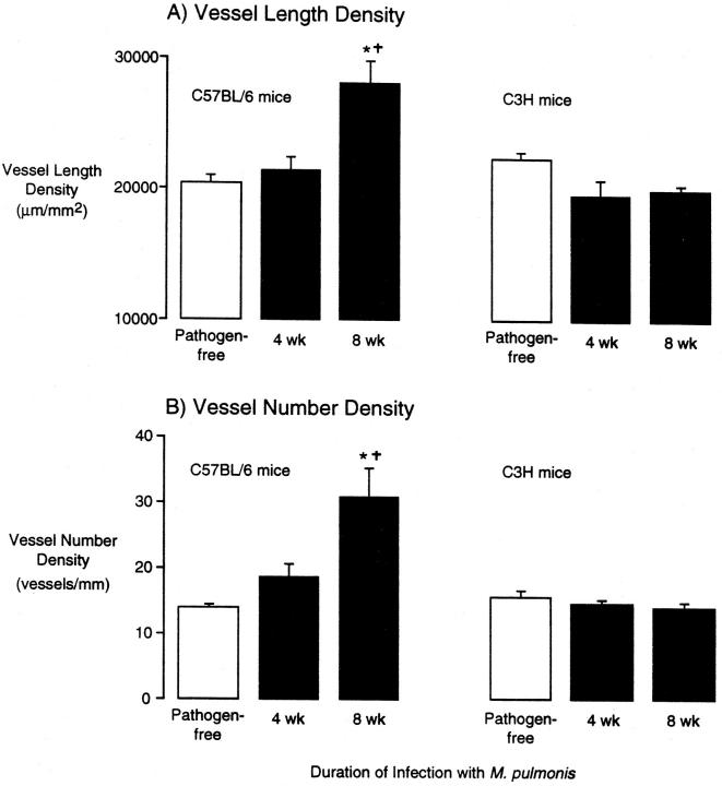
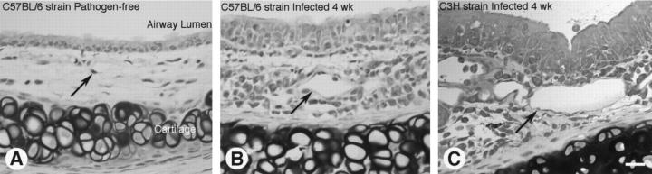
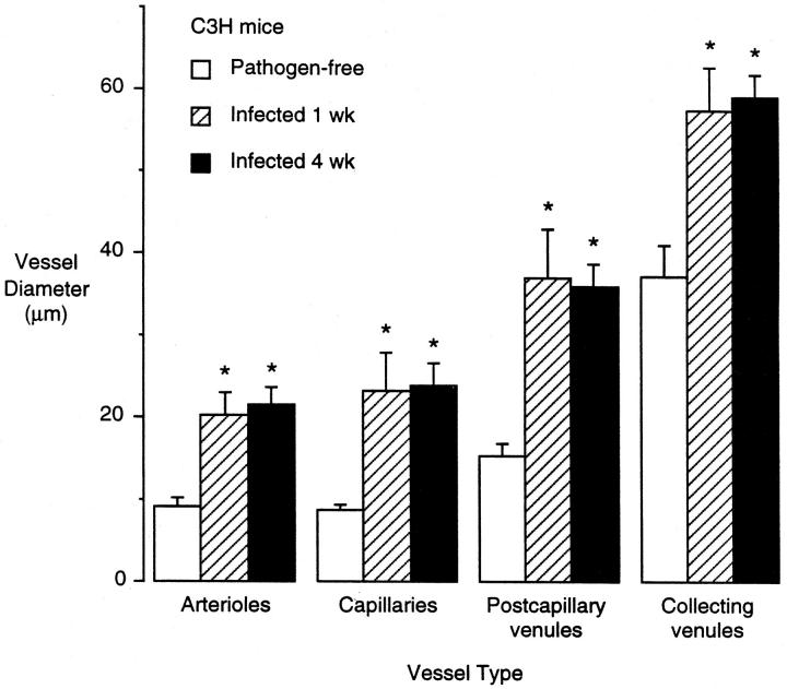
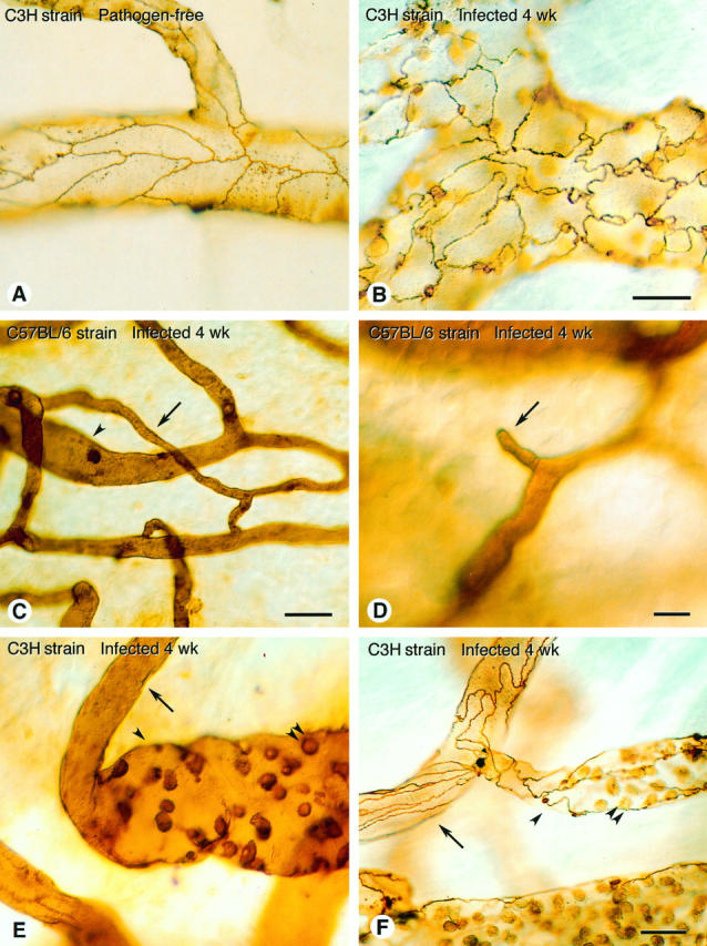
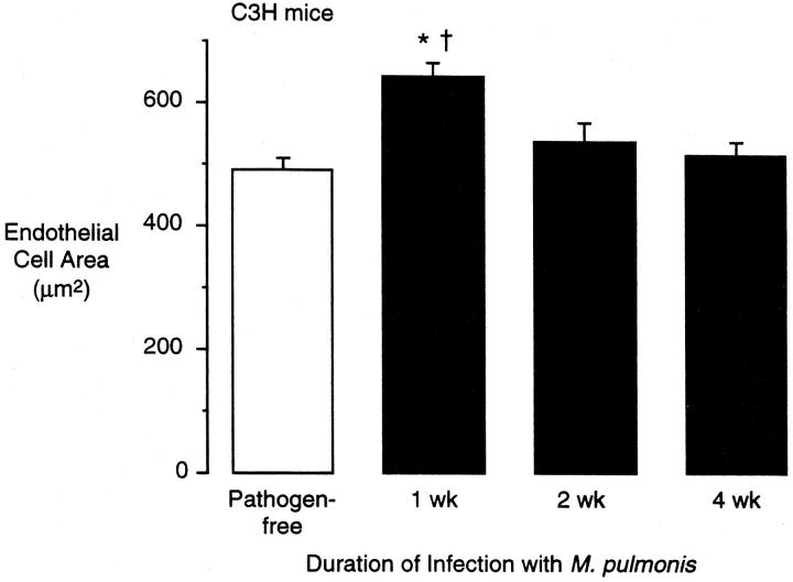
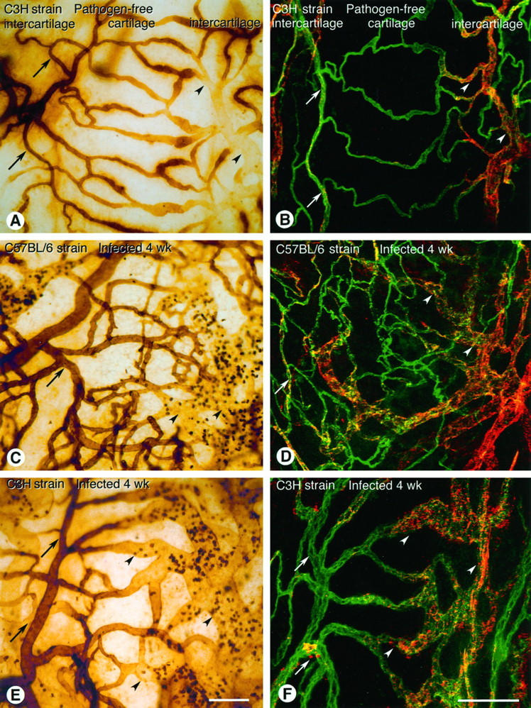
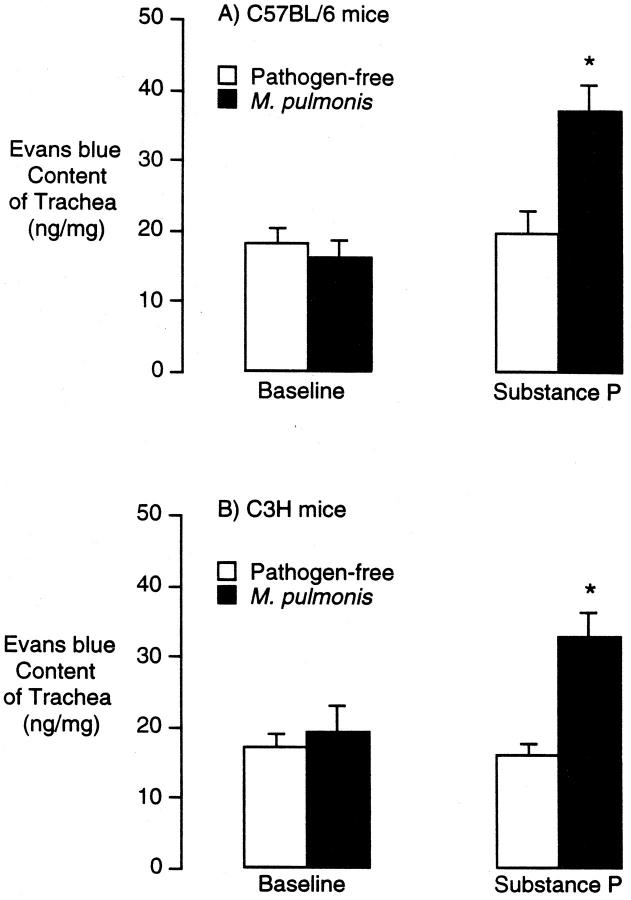
Chronic inflammation is associated with blood vessel proliferation and enlargement and changes in vessel phenotype. We sought to determine whether these changes represent different types of angiogenesis and whether they are stimulus dependent. Chronic airway inflammation, produced by infection with Mycoplasma pulmonis, was compared in strains of mice known to be resistant (C57BL/6) or susceptible (C3H). Tracheal vascularity, assessed in whole mounts after Lycopersicon esculentum lectin staining, increased in both strains at 1, 2, 4, and 8 weeks after infection, but the type of vascular remodeling was different. The number of vessels doubled in tracheas of C57BL/6 mice, with corresponding increases of capillaries and venules. In contrast, neither the number nor the length of vessels changed in C3H mice. Instead, vessel diameter and endothelial cell number doubled, and the proportion of venules doubled with a corresponding decrease of capillaries. Although the infection had no effect on baseline plasma leakage, in both strains it potentiated the leakage produced by substance P. We conclude that the same stimulus can result in blood vessel proliferation or enlargement, depending on the host response. Endothelial cells proliferate in both cases, but in one case new capillaries form whereas in the other capillaries convert to venules.


Figures

Figure 1.
Figure 1.
Tracheal vasculature in pathogen-free and M. pulmonis-infected C57BL/6 and C3H mice. The vasculature was perfusion stained with biotinylated L. esculentum lectin and ABC/DAB peroxidase reaction and visualized by light microscopy in tracheal whole mounts. A: Organized pattern of mucosal vessels in trachea of pathogen-free C57BL/6 mouse, showing capillaries across a cartilaginous ring (cartilage), fed from arterioles (arrows) and drained by venules (arrowheads) in intercartilaginous regions (intercartilage). B: Enlarged vessels in trachea of C57BL/6 mouse at 2 weeks after infection C: Enlarged vessels and regions with increased numbers of capillary-sized vessels (arrow) in trachea of C57BL/6 mouse at 4 weeks after infection. D: Numerous capillary-sized vessels and enlarged vessels in trachea of C57BL/6 mouse at 8 weeks after infection. Many capillary-sized vessels are out of focus as vessels are no longer confined to plane of epithelium. E: Trachea of pathogen-free C3H mouse, showing organized pattern of mucosal vessels. F: Enlarged vessels in trachea of C3H mouse at 8 weeks after infection. All segments of the vasculature appear enlarged. Similar enlarged vessels were seen at 1, 2, and 4 weeks after inoculation of C3H mice. Scale bar (A–F), 100 μm.
Figure 2.
Figure 2.
Vessel area density in tracheal mucosa of pathogen-free and M. pulmonis-infected C57BL/6 (A) and C3H (B) mice. Vessel area density was measured by morphometric point counting in L. esculentum lectin-stained tracheal whole mounts. Values represent mean ± SE of 6 to 10 microscope fields per trachea and four tracheas per group (n = 4). *Significantly different from corresponding pathogen-free value.
Figure 3.
Figure 3.
Vessel length density (A) and number of vessels (B) in tracheal mucosa of pathogen-free and M. pulmonis-infected C57BL/6 and C3H mice. Vessel length density and number of vessels were measured morphometrically in L. esculentum lectin-stained tracheal whole mounts. Values represent mean ± SE of 6 to 10 microscope fields per trachea and four tracheas per group (n = 4). *Significantly different from corresponding pathogen-free value; significantly different from corresponding value in C3H mice.
Figure 4.
Figure 4.
Histological sections of tracheas from pathogen-free and M. pulmonis-infected mice. Tracheas were fixed by perfusion, embedded in glycol methacrylate, sectioned, and stained with toluidine blue. A–C: Trachea from pathogen-free C57BL/6 mouse (A), 4-week-infected C57BL/6 mouse (B), and 4-week-infected C3H mouse (C). Arrows mark vessels, which are enlarged in the infected mice (B and C). Scale bar (A–C) 20 μm.
Figure 5.
Figure 5.
Vessel luminal diameter in tracheal mucosa of pathogen-free and M. pulmonis-infected C3H mice. Vessel diameter was measured morphometrically in L. esculentum lectin-stained tracheal whole mounts. Values represent mean ± SE of 10 vessels of each type per trachea and four tracheas per group (n = 4). *Significantly different from corresponding pathogen-free value.
Figure 6.
Figure 6.
Tracheal vasculature in pathogen-free and M. pulmonis-infected C3H and C57BL/6 mice. A: Smooth endothelial cell borders in tracheal venules of pathogen-free C3H mouse, stained by perfusion of silver nitrate. B: Irregular endothelial cell borders in silver-stained tracheal venules of C3H mouse infected for 4 weeks. Although borders are very irregular compared with pathogen-free mice, average luminal area of endothelial cells is similar. C: High-magnification view of trachea of C57BL/6 mouse infected for 8 weeks showing small capillary-like vessel (arrow) connected to venule (arrowhead). D: Example of blind vascular sprout (arrow) emanating from small vessel in trachea of C57BL/6 mouse infected for 4 weeks. E and F: Abrupt transitions from arterioles (arrows) to venule-like vessels (arrowheads) with little or no intervening capillary in tracheal vasculature of C3H mice infected for 4 weeks and stained with L. esculentum lectin (E) or silver nitrate (F). Adherent leukocytes in venules (double arrowheads ). Scale bars, 20 μm (A and B), 20 μm (C), 10 μm (D), and 20 μm (E and F).
Figure 7.
Figure 7.
Luminal endothelial cell surface area in tracheal mucosa of pathogen-free and M. pulmonis-infected C3H mice. Endothelial cell area was measured in silver-nitrate-stained vessels in tracheal whole mounts by tracing cell borders on video microscopic projections with a digitizing tablet. Values represent mean ± SE of 10 venular endothelial cells per trachea and four tracheas per group (n = 4). *Significantly different from pathogen-free value; significantly different from 4-week-infected value.
Figure 8.
Figure 8.
WGA-stained and vWF-immunoreactive vessels in tracheal whole mounts of pathogen-free and M. pulmonis-infected C57BL/6 and C3H mice. Biotinylated WGA was perfused and stained with ABC/DAB peroxidase reaction (A, C, and E). vWF was stained immunofluorescently using polyclonal primary antibody and Cy3-labeled secondary antibody (red/orange) on tracheas after the vessels were labeled by perfusion of fluorescein L. esculentum lectin (green; B, D, and F). vWF was imaged by confocal microscopy. A: Pathogen-free C3H mouse. WGA lectin binds strongly to arterioles (arrows) and capillaries across cartilaginous ring but weakly or not at all to venules (arrowheads). B: Pathogen-free C3H mouse, showing weak vWF immunoreactivity (red/orange) of capillaries across cartilaginous ring, strong immunoreactivity of venules (arrowheads), and moderate immunoreactivity of arterioles (arrows). C: C57BL/6 mouse infected for 4 weeks. WGA binds to arterioles (arrow) and the numerous small vessels across the cartilaginous ring, with little or no binding in enlarged vessels (arrowheads). Adherent leukocytes in venules show strong staining. D: C57BL/6 mouse infected for 8 weeks, showing moderate vWF immunoreactivity in arterioles (arrow), weak vWF immunoreactivity in the numerous small vessels across the cartilaginous ring, and strong immunoreactivity in enlarged venules (arrowhead). E: C3H mouse infected for 4 weeks. WGA binds strongly to endothelium in arterioles (arrows) but binds weakly or not at all to enlarged venules across the cartilaginous ring and in intercartilaginous regions (arrowheads). Adherent leukocytes in venules bind strongly. F: C3H mouse infected for 4 weeks, showing strong vWF immunoreactivity of the enlarged venules across the cartilaginous ring and in intercartilaginous regions (arrowheads) and much less in arterioles (arrows). Scale bars, 100 μm.
Figure 9.
Figure 9.
Evans blue leakage from tracheal vessels in pathogen-free and M. pulmonis-infected C57BL/6 and C3H mice. Evans blue was injected 5 minutes before injection of substance P (5 μg/kg) or vehicle, and the animals were perfused 5 minutes later. Evans blue was extracted from trachea and measured spectrophotometrically. Values represent the mean ± SE of six tracheas per group (n = 6). *Significantly different from corresponding pathogen-free values.

Comment in

References

    1. Risau W: Mechanisms of angiogenesis. Nature 1997, 386:671-674 - PubMed
    1. Folkman J, Brem H: Angiogenesis and inflammation. ed 2 Gallin JI Goldstein IM Snyderman R eds. Inflammation: Basic Principles and Clinical Correlates, 1992, :pp 821-839 Raven Press, New York
    1. Colville-Nash PR, Scott DL: Angiogenesis and rheumatoid arthritis: pathogenic and therapeutic implications. Ann Rheum Dis 1992, 51:919-925 - PMC - PubMed
    1. Jackson JR, Seed MP, Kircher CH, Willoughby DA, Winkler JD: The co-dependence of angiogenesis and chronic inflammation. FASEB J 1997, 11:457-465 - PubMed
    1. Dunnill MS: The pathology of asthma, with special references to changes in the bronchial mucosa. J Clin Pathol 1960, 13:27-33 - PMC - PubMed

Publication types

MeSH terms

LinkOut - more resources