

Advanced glycation end products in Alzheimer's disease and other neurodegenerative diseases
- PMID: 9777946
- PMCID: PMC1853056
- DOI: 10.1016/S0002-9440(10)65659-3
Advanced glycation end products in Alzheimer's disease and other neurodegenerative diseases
Abstract
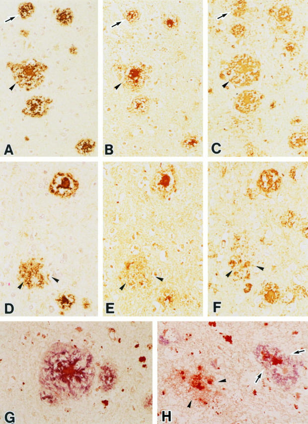
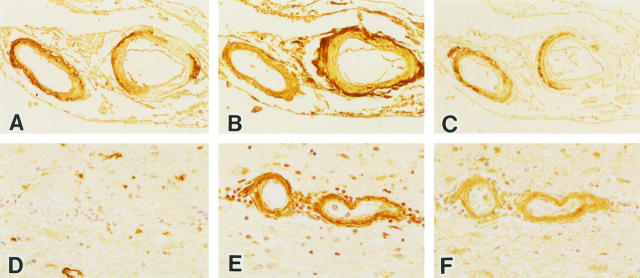
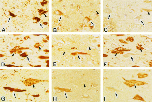
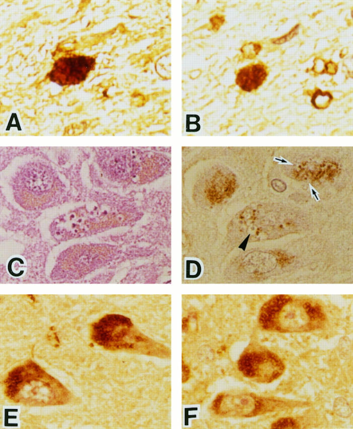
Advanced glycation end products (AGEs) have been implicated in the chronic complications of diabetes mellitus and have been reported to play an important role in the pathogenesis of Alzheimer's disease. In this study, we examined the immunohistochemical localization of AGEs, amyloid beta protein (A beta), apolipoprotein E (ApoE), and tau protein in senile plaques, neurofibrillary tangles (NFTs), and cerebral amyloid angiopathy (CAA) in Alzheimer's disease and other neurodegenerative diseases (progressive supranuclear palsy, Pick's disease, and Guamanian amyotrophic lateral sclerosis/Parkinsonism-dementia complex). In most senile plaques (including diffuse plaques) and CAA from Alzheimer's brains, AGE and ApoE were observed together. However, approximately 5% of plaques were AGE positive but A beta negative, and the vessels without CAA often showed AGE immunoreactivity. In Alzheimer's disease, AGEs were mainly present in intracellular NFTs, whereas ApoE was mainly present in extracellular NFTs. Pick's bodies in Pick's disease and granulovacuolar degeneration in various neurodegenerative diseases were also AGE positive. In non-Alzheimer neurodegenerative diseases, senile plaques and NFTs showed similar findings to those in Alzheimer's disease. These results suggest that AGE may contribute to eventual neuronal dysfunction and death as an important factor in the progression of various neurodegenerative diseases, including Alzheimer's disease.
Figures
References
-
- Monnier VM, Cerami A: Nonenzymatic browning in vivo: possible process for aging of long-lived proteins. Science 1981, 211:491-493 - PubMed
-
- Brownlee M, Vlassara H, Cerami A: Nonenzymatic glycosylation and the pathogenesis of diabetic complications. Ann Intern Med 1984, 101:527-537 - PubMed
-
- Ledl F, Schleicher E: New aspects of the Maillard reaction in foods and in the human body. Angew Chem Int Ed Engl 1990, 6:565-706
-
- Vlassara H, Bucala R, Striker L: Pathogenic effects of advanced glycosylation: biochemical, biologic, and clinical implications for diabetes and aging. Lab Invest 1994, 70:138-151 - PubMed
-
- Selkoe DJ: Normal and abnormal biology of the β-amyloid precursor protein. Annu Rev Neurosci 1994, 17:489-517 - PubMed
Publication types
MeSH terms
Substances
LinkOut - more resources
Full Text Sources
Other Literature Sources
Medical
Miscellaneous
