

Role of vascular endothelial growth factor in ovarian cancer: inhibition of ascites formation by immunoneutralization
- PMID: 9777956
- PMCID: PMC1853065
- DOI: 10.1016/S0002-9440(10)65669-6
Role of vascular endothelial growth factor in ovarian cancer: inhibition of ascites formation by immunoneutralization
Abstract
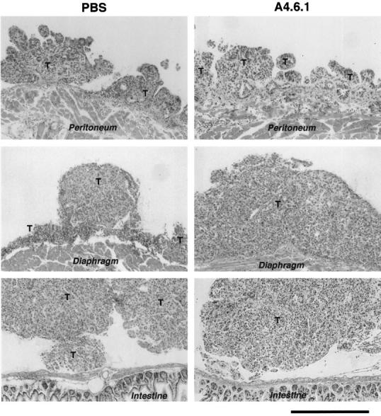
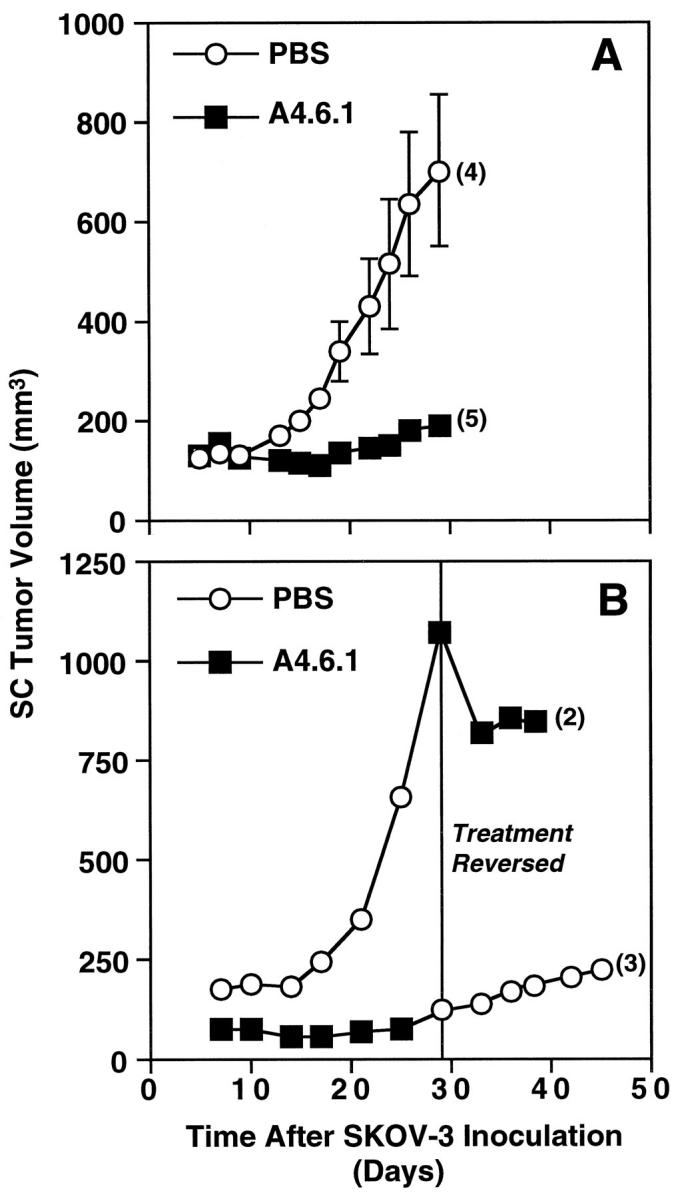
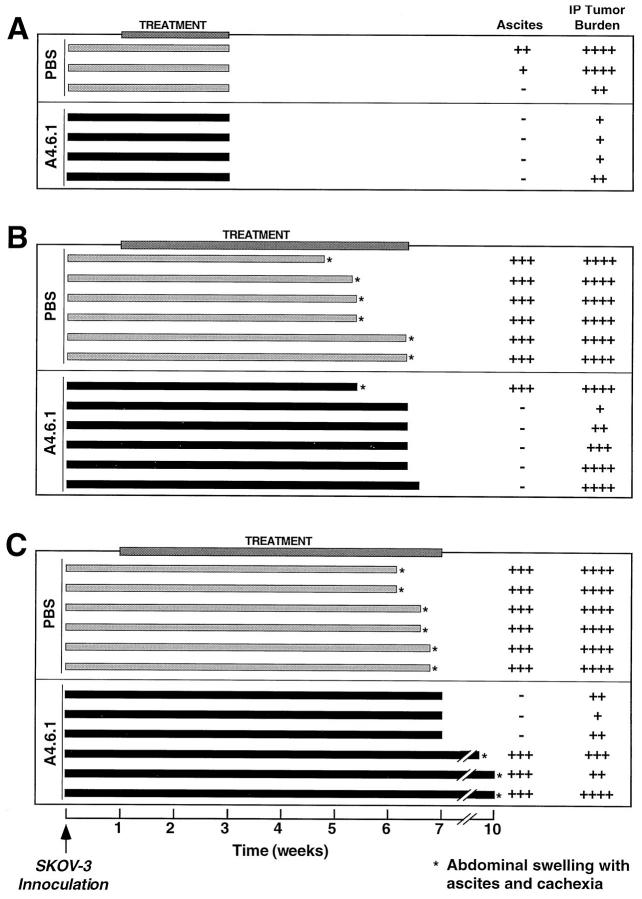
Ovarian cancer is characterized by the rapid growth of solid intraperitoneal tumors and large volumes of ascitic fluid. Vascular endothelial growth factor (VEGF) augments tumor growth by inducing neovascularization and may stimulate ascites formation by increasing vascular permeability. We examined the role of VEGF in ovarian carcinoma using in vivo models in which intraperitoneal or subcutaneous tumors were induced in immunodeficient mice using the human ovarian carcinoma cell line SKOV-3. After tumor engraftment (7 to 10 days), some mice were treated with a function-blocking VEGF antibody (A4.6.1) specific for human VEGF. A4.6.1 significantly (P < 0.05) inhibited subcutaneous SKOV-3 tumor growth compared with controls. However, tumor growth resumed when A4.6.1 treatment was discontinued. In mice bearing intraperitoneal tumors (IP mice), ascites production and intraperitoneal carcinomatosis were detected 3 to 7 weeks after SKOV-3 inoculation. Importantly, A4.6.1 completely inhibited ascites production in IP mice, although it only partially inhibited intraperitoneal tumor growth. Tumor burden was variable in A4.6.1-treated IP mice; some had minimal tumor, whereas in others tumor burden was similar to that of controls. When A4.6.1 treatment was stopped, IP mice rapidly (within 2 weeks) developed ascites and became cachectic. These data suggest that in ovarian cancer, tumor-derived VEGF is obligatory for ascites formation but not for intraperitoneal tumor growth. Neutralization of VEGF activity may have clinical application in inhibiting malignant ascites formation in ovarian cancer.
Figures
Similar articles
-
Vascular endothelial growth factor immunoneutralization plus Paclitaxel markedly reduces tumor burden and ascites in athymic mouse model of ovarian cancer.Am J Pathol. 2002 Nov;161(5):1917-24. doi: 10.1016/S0002-9440(10)64467-7. Am J Pathol. 2002. PMID: 12414537 Free PMC article.
-
Differential inhibition of fluid accumulation and tumor growth in two mouse ascites tumors by an antivascular endothelial growth factor/permeability factor neutralizing antibody.Cancer Res. 1998 Jun 15;58(12):2594-600. Cancer Res. 1998. PMID: 9635584
-
Involvement of VEGF and its receptors in ascites tumor formation.Cancer Chemother Pharmacol. 1999;43 Suppl:S72-7. doi: 10.1007/s002800051102. Cancer Chemother Pharmacol. 1999. PMID: 10357563
-
The role of vascular endothelial growth factor in angiogenesis.Acta Haematol. 2001;106(4):148-56. doi: 10.1159/000046610. Acta Haematol. 2001. PMID: 11815711 Review.
-
Targeting vascular endothelial growth factor (VEGF) for anti-tumor therapy, by anti-VEGF neutralizing monoclonal antibodies or by VEGF receptor tyrosine-kinase inhibitors.Cancer Metastasis Rev. 1999;18(4):473-81. doi: 10.1023/a:1006358220123. Cancer Metastasis Rev. 1999. PMID: 10855790 Review.
Cited by
-
Vascular endothelial growth factor and other signaling pathways in developmental and pathologic angiogenesis.Int J Hematol. 2004 Jul;80(1):7-20. doi: 10.1532/ijh97.04065. Int J Hematol. 2004. PMID: 15293563 Review.
-
Targeted therapies in epithelial ovarian cancer.Cancers (Basel). 2010 Feb 23;2(1):88-113. doi: 10.3390/cancers2010088. Cancers (Basel). 2010. PMID: 24281034 Free PMC article.
-
The VEGF pathway and the AKT/mTOR/p70S6K1 signalling pathway in human epithelial ovarian cancer.Br J Cancer. 2009 Mar 24;100(6):971-8. doi: 10.1038/sj.bjc.6604921. Epub 2009 Feb 24. Br J Cancer. 2009. PMID: 19240722 Free PMC article.
-
Anti-tumour activity of tivozanib, a pan-inhibitor of VEGF receptors, in therapy-resistant ovarian carcinoma cells.Sci Rep. 2017 Apr 6;7:45954. doi: 10.1038/srep45954. Sci Rep. 2017. PMID: 28383032 Free PMC article.
-
Real-time monitoring biomarker expression of carcinoma cells by surface plasmon resonance biosensors.Chem Commun (Camb). 2012 Oct 28;48(84):10389-91. doi: 10.1039/c2cc34853e. Epub 2012 Sep 6. Chem Commun (Camb). 2012. PMID: 22957340 Free PMC article.
References
-
- Folkman J, Watson K, Ingber D, Hanahan D: Induction of angiogenesis during the transition from hyperplasia to neoplasia. Nature 1989, 339:58-61 - PubMed
-
- Liotta LA, Kleinerman J, Saidel GM: Quantitative relationships of intravascular tumor cells, tumor vessels, and pulmonary metastases following tumor implantation. Cancer Res 1974, 4:997-1004 - PubMed
-
- Weidner N, Semple JP, Welch WR, Folkman J: Tumor angiogenesis and metastasis: correlation in invasive breast carcinoma. N Engl J Med 1991, 324:1-8 - PubMed
-
- Folkman J, Klagsbrun M: Vascular physiology: a family of angiogenic peptides. Nature 1987, 329:671-672 - PubMed
-
- Folkman J: What is the evidence that tumors are angiogenesis dependent? J Natl Cancer Inst 1990, 82:4-6 - PubMed
Publication types
MeSH terms
Substances
Grants and funding
LinkOut - more resources
Full Text Sources
Other Literature Sources
Medical
