. 1998 Oct;153(4):1257-66.
doi: 10.1016/S0002-9440(10)65670-2.

Modulation of endothelial cell function by normal polyspecific human intravenous immunoglobulins: a possible mechanism of action in vascular diseases

Affiliations

Modulation of endothelial cell function by normal polyspecific human intravenous immunoglobulins: a possible mechanism of action in vascular diseases

C Xu et al. Am J Pathol. 1998 Oct.

Abstract

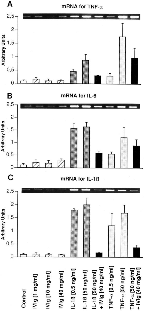
Intravenous immunoglobulin (IVIg) is increasingly used in the treatment of autoimmune and inflammatory diseases, including vasculitides and Kawasaki disease. However, the outcome of IVIg interaction with endothelial cells of the vascular bed is not clear as yet. We have investigated the effect of IVIg on the in vitro activation of human endothelial cells, as assessed by cell proliferation and reverse transcription-polymerase chain reaction-detected expression of mRNA coding for adhesion molecules (intercellular adhesion molecule-1 and vascular cellular adhesion molecule-1), chemokines (monocyte chemoattractant protein-1, macrophage colony-stimulating factor, and granulocyte-macrophage colony-stimulating factor), and proinflammatory cytokines (tumor necrosis factor-alpha, interleukin-1beta, and interleukin-6). IVIg inhibited proliferation of endothelial cells in a time-dependent manner. This effect was dependent on both Fc and F(ab')2 fragments of the immunoglobulin molecule and was fully reversible. Tumor necrosis factor-alpha and interleukin-1beta also inhibited thymidine incorporation, but to a lesser degree. IVIg had no effect on basal levels of mRNA coding for the adhesion molecules, chemokines, and proinflammatory cytokines. IVIg fully down-regulated the expression induced by tumor necrosis factor-alpha or interleukin-1beta of mRNA coding for these molecules. Thus, blockade of cellular proliferation and of cytokine-induced expression of adhesion molecules, chemokines, and cytokines may explain the therapeutic effect of IVIg in vascular and inflammatory disorders.


Figures

Figure 1.
Figure 1.
Kinetics of the inhibitory effect of IVIg on EC proliferation. IVIg added to the culture medium for various periods of time inhibited [3H]thymidine incorporation in a dose- and time-dependent manner. Maximal inhibition was attained at 6 hours when low concentration of IVIg was used, or after 30-hour incubation for higher concentrations, and partly disappeared afterwards, except for 40 mg/ml IVIg. Time effect, P = 0.001; group effect, P = 0.001 (n = 6 cords).
Figure 2.
Figure 2.
Comparative effect of different sources of IVIg on EC proliferation. Sandoglobulin, Gammagard, and Endobulin added to the culture medium for 24 hours inhibited [3H]thymidine incorporation in a dose-dependent manner. Gammagard and Endobulin were more potent inhibitors than Sandoglobulin. Group effect: P = 0.034 (n = 4 cords).
Figure 3.
Figure 3.
Inhibitory effect of IVIg on EC proliferation. As assessed by trypan blue exclusion, the number of live cells was significantly reduced by IVIg in a dose-dependent manner. Nevertheless, 20 or 30 mg/ml of IVIg had less effect when added for 48 hours than for 24 hours. The number of dead cells slightly increased with IVIg concentration but did not exceed 8% at the end of the incubation period. Live cells: time effect, P = 0.001; concentration effect, P = 0.001; dead cells: time effect, P = 0.004; concentration effect, P = 0.001 (n = 3 cords).
Figure 4.
Figure 4.
FACScan analysis of cell viability after IVIg treatment. The number of dead cells as determined by propidium iodide uptake did not exceed 6%, whatever the time of incubation and/or the concentration of agonists. Each curve represents a typical experiment. For each condition, the ratio of dead cells to live cells (mean ± standard error of the mean) is given above the graph. Duration of treatment effect, P = 0.4527; group effect, P = 0.3841 (n = 3 cords).
Figure 5.
Figure 5.
Effect of IVIg washout on EC proliferation inhibition. After a 24-hour (A) or 48-hour (B) incubation period of cells with IVIg, [3H]thymidine incorporation was monitored at 0, 24, or 48 hours after IVIg was withdrawn. When cells were incubated for 24 hours (A) with 20 and 30 mg/ml IVIg, but not with 40 mg/ml, the uptake of thymidine was fully restored 24 hours after IVIg had been withdrawn. When IVIg was used for 48 hours (B), the uptake of thymidine was fully recovered 24 hours after IVIg removal. After a 24- or 48-hour IVIg incubation: time effect, P = 0.001; group effect, P = 0.001 (n = 3 cords).
Figure 6.
Figure 6.
TNF-α- and IL-1β-induced inhibition of EC proliferation. Both cytokines inhibited [3H]thymidine incorporation in a dose-dependent manner. The maximum inhibitory effect was twofold less pronounced than the IVIg effect. When HUVECs were coincubated with a mixture of TNF-α or IL-1β and IVIg prepared just before addition onto the cells (A columns) or were pretreated for 15 minutes with TNF-α or IL-1β alone before the addition of IVIg into the culture medium (B columns), IVIg plus cytokines led to a more efficient inhibition of cell proliferation than IVIg alone (group effect, P = 0.0012; n = 3), but no difference was seen depending on the way in which IVIg was added to the cytokines (P = 0.082; n = 3).
Figure 7.
Figure 7.
Expression of mRNA encoding VCAM-1 and ICAM-1 adhesion molecules. IL-1β and TNF-α induced an overexpression of mRNA coding for VCAM-1 (B) and ICAM-1 (C) in a dose-dependent manner. Although IVIg alone had no significant effect on this synthesis, it down-regulated the mRNA expression induced by IL-1β or TNF-α. The intensities of the cDNA bands for each protein were normalized to the GAPDH band intensities (A). Experiments were run five times for each cell extract, with cell lines coming from three to five different umbilical cords.
Figure 8.
Figure 8.
Expression of mRNA encoding MCP-1, M-CSF, and GM-CSF chemokines. IL-1β and TNF-α induced an overexpression of mRNA coding for MCP-1 (A), M-CSF (B), and GM-CSF (C) in a dose-dependent manner. Although IVIg alone had no significant effect on this synthesis, it down-regulated the mRNA expression induced by IL-1β or TNF-α. The intensities of the cDNA bands for each protein were normalized to the GAPDH band intensities depicted in Figure 7A ▶ .
Figure 9.
Figure 9.
Expression of mRNA encoding proinflammatory cytokines TNF-α, IL-6, and IL-1β. IL-1β and TNF-α induced an overexpression of mRNA coding for TNF-α (A), IL-6 (B), and IL-1β (C) in a dose-dependent manner. Although IVIg alone had no significant effect on this synthesis, it down-regulated the mRNA expression induced by IL-1β or TNF-α. The intensities of the cDNA bands for each protein were normalized to the GAPDH band intensities depicted in Figure 7A ▶ .

References

    1. Carlos TM, Harlan JM: Leukocyte-endothelial adhesion molecules. Blood 1994, 84:2068-2101 - PubMed
    1. Demuth K, Myara I, Moatti N: Biologie de la cellule endothéliale et athérogenèse. Ann Biol Clin 1995, 53:171-189 - PubMed
    1. Matsushima KV, Oppenheim JJ: Interleukin-8 and MCAF: novel inflammatory cytokines induced by TNF and Il-1. Cytokines 1989, 1:2-10 - PubMed
    1. Brady HR: Leukocyte adhesion molecules and kidney diseases. Kidney Int 1994, 45:1285-1300 - PubMed
    1. Munro JM, Pober JS, Cotran RS: Tumor necrosis factor and interferon-γ induce distinct patterns of endothelial activation and associated leukocyte accumulation in skin of Papio anubis. Am J Pathol 1989, 135:121-133 - PMC - PubMed

Publication types

MeSH terms