. 1998 Oct;153(4):1267-76.
doi: 10.1016/S0002-9440(10)65671-4.

Bikunin present in human peritoneal fluid is in part derived from the interaction of serum with peritoneal mesothelial cells

Affiliations

Bikunin present in human peritoneal fluid is in part derived from the interaction of serum with peritoneal mesothelial cells

G J Thomas et al. Am J Pathol. 1998 Oct.

Abstract

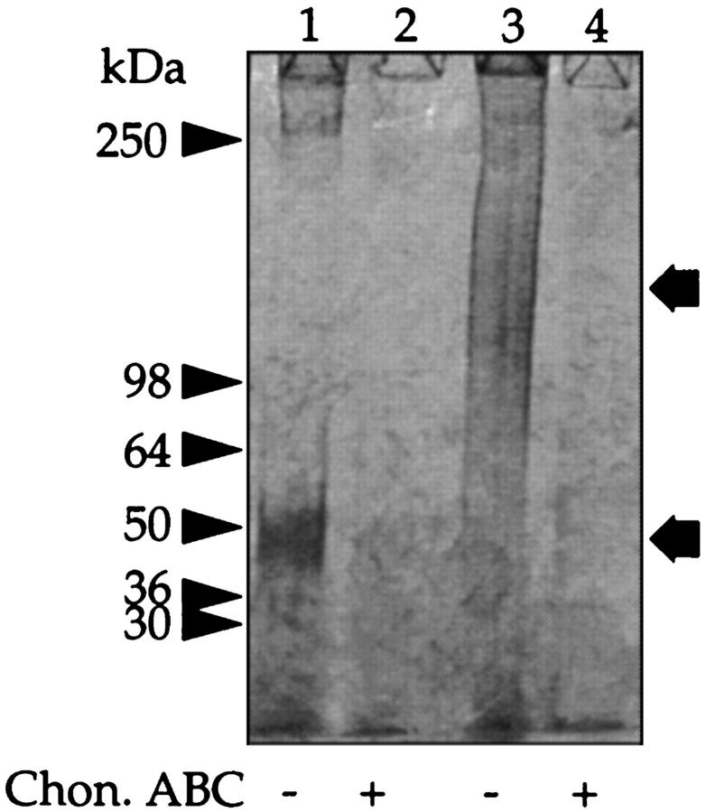
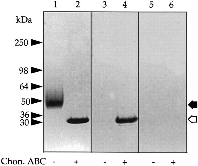
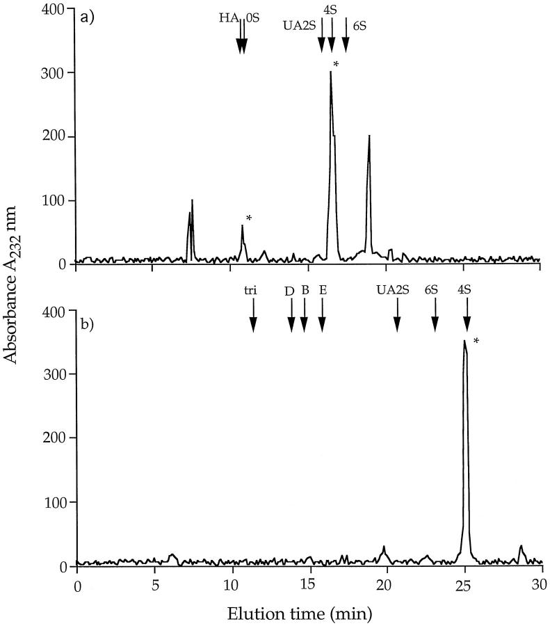
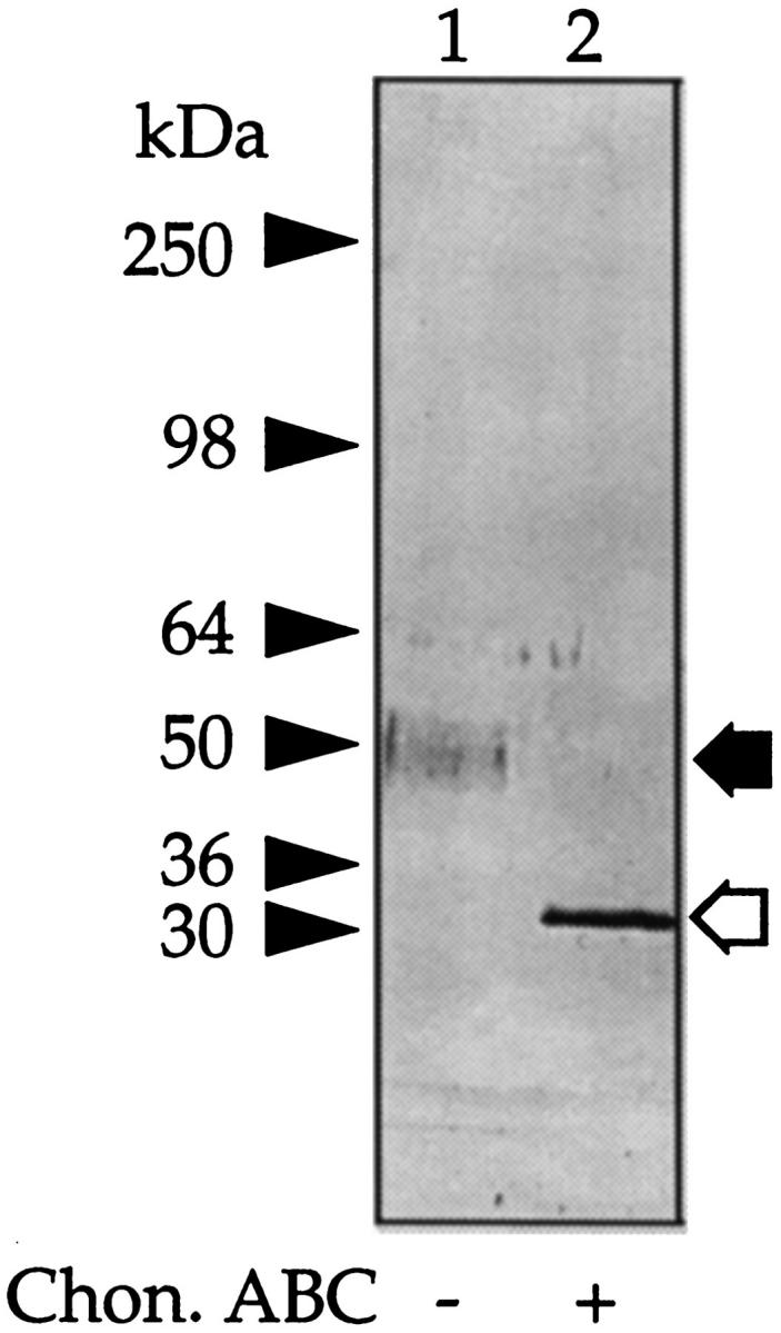
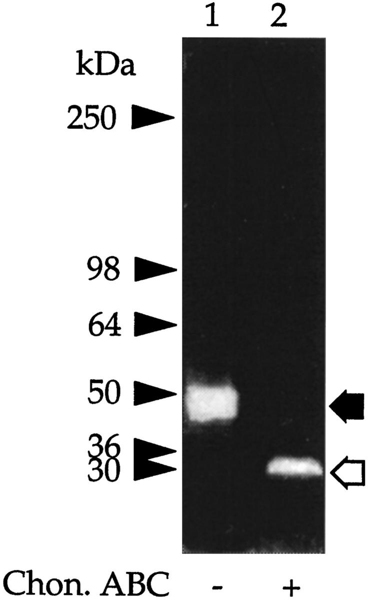
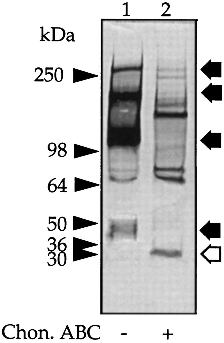
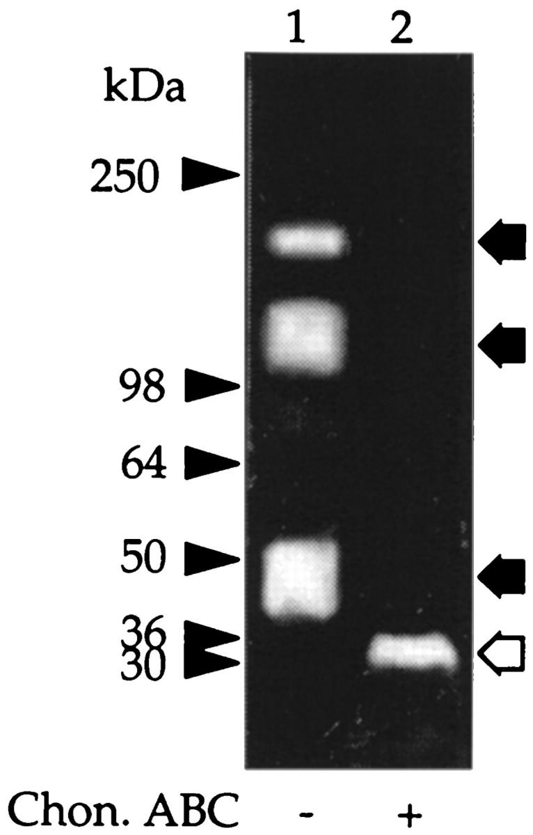
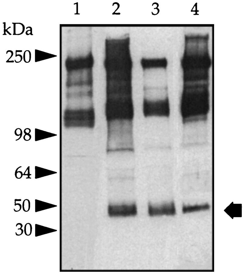
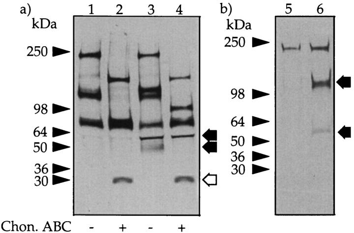
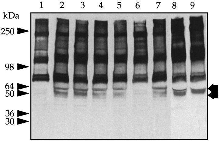
We recently reported that peritoneal fluid mainly contains two proteoglycans; one is the interstitial proteoglycan referred to as decorin, and the other an uncharacterized small chondroitin sulfate proteoglycan. In the present study, we have used a two-step process to isolate the small chondroitin sulfate proteoglycan free of decorin. The purified molecule ran as a single band on sodium dodecyl sulfate-polyacrylamide gel electrophoresis with apparent molecular mass 50 kd made up of a chondroitin-4-sulfate glycosaminoglycan chain and a 30-kd core protein. NH2-terminal analysis of the core protein showed significant sequence homology with bikunin, a component of the human inter-alpha-trypsin inhibitor (IalphaI) family. A Western blot analysis using anti-human inter-alpha-trypsin inhibitor confirmed the identity of the small chondroitin sulfate proteoglycan as bikunin, and a trypsin inhibitor counterstain assay confirmed its anti-trypsin activity. Examination of serum from patients receiving continuous peritoneal dialysis suggests that free bikunin in peritoneal fluid may be the result of leakage of serum proteins into the peritoneum. Our findings further show that the interaction of serum with peritoneal mesothelial cells offers a new and novel explanation for the presence of bikunin in peritoneal fluid.


Figures

Figure 1.
Figure 1.
SDS-PAGE of peritoneal fluid PGs. Peritoneal fluid was passed over an EconoPac Q column, and the nonbound material and GAGs were eluted with increasing concentrations of NaCl (see Materials and Methods). The materials that eluted with 0.4 mol/L NaCl (F1) and 0.6 mol/L NaCl (F2) were separately pooled. Aliquots of F1 (lanes 1 and 2) and F2 (lanes 3 and 4) were incubated with buffer alone (lanes 1 and 3) or chondroitin ABC lyase (lanes 2 and 4) and subjected to SDS-PAGE under reducing conditions on a 3 to 12% gradient gel and stained for GAG with Alcian blue. The prestained molecular mass markers are indicated with arrowheads, and the resolved PGs are indicated with arrows.
Figure 2.
Figure 2.
Western blot analysis of the 50-kd CSPG. Peritoneal fluid was concentrated by precipitation with CTAB and F1 obtained as outlined in Figure 1 ▶ . Aliquots were either incubated with buffer alone (lanes 1, 3, and 5) or chondroitin ABC lyase (lanes 2, 4, and 6) and run on a 3 to 12% gradient gel. The gel was cut as indicated and stained for either protein with Coomassie brilliant blue (lanes 1 and 2) or blotted and probed with mAb 2B6 (lanes 3 and 4) or mAb 3B3 (lanes 5 and 6). The prestained molecular mass markers are indicated with arrowheads and the resolved PGs with arrows. The open arrows indicate the core protein released after incubation with chondroitin ABC lyase.
Figure 3.
Figure 3.
Capillary electrophoresis. An aliquot (1 mg/ml) of the 50-kd CSPG was incubated with chondroitin ABC lyase, and the released disaccharides were electrophoresed under normal polarity (15 kV) (a) or reverse polarity (−15 kV) (b) at 40°C for 30 minutes. The peaks were monitored at 232 nm and compared with standard disaccharides: HA, hyalurono-ΔDi-0S; 0S, chondro-ΔDi-0S; 4S, chondro-ΔDi-4S; 6S, chondro-ΔDi-6S; 2S, chondro-ΔDi-UA2S; B, dermato-ΔDi-di-4S, UA2S; D, chondro-ΔDi-di6S, UA2S; E, chondro-ΔDi-di-4,6S; and tri, chondro-ΔDi-tri-4,6S, UA2S.
Figure 4.
Figure 4.
Identification of the 50-kd CSPG as bikunin. Aliquots of the 50-kd CSPG were incubated with buffer alone (lane 1) or chondroitin ABC lyase (lane 2), electrophoresed on 3 to 12% gels, blotted onto nitrocellulose, and probed with an antiserum raised to human serum IαI. The prestained molecular mass markers are indicated with arrowheads and the resolved PGs with arrows. The open arrows indicate the core protein released after incubation with chondroitin ABC lyase.
Figure 5.
Figure 5.
Trypsin inhibitory activity of the 50-kd CSPG. Aliquots of the CSPG were incubated with buffer alone (lane 1) or chondroitin ABC lyase (lane 2) and electrophoresed under nonreducing conditions on 3 to 12% gels, and the trypsin inhibitory activity was determined using the TIC assay. The clear areas on the gel represent trypsin inhibitory activity. The prestained molecular mass markers are indicated with arrowheads and the resolved PGs with arrows. The open arrows indicate the core protein released after incubation with chondroitin ABC lyase.
Figure 6.
Figure 6.
Western blot of peritoneal fluid. Peritoneal fluid (5 μl) was incubated with buffer alone (lane 1) or with chondroitin ABC lyase (lane 2), and a Western blot was generated with anti-human IαI antibody. The prestained molecular mass markers are indicated with arrowheads and the resolved PGs with arrows. The open arrows indicate the core protein released after incubation with chondroitin ABC lyase.
Figure 7.
Figure 7.
Trypsin inhibitory activity in peritoneal fluid. Fresh peritoneal fluid was concentrated as outlined in Figure 1 ▶ . Aliquots were incubated with buffer alone (lane 1) or with chondroitin ABC lyase (lane 2), electrophoresed under nonreducing conditions on 3 to 12% gels, and the trypsin inhibitory activity was determined using the TIC assay. The prestained molecular mass markers are indicated with arrowheads and the resolved PGs with arrows. The open arrows indicate the core protein released after incubation with chondroitin ABC lyase.
Figure 8.
Figure 8.
IαI of normal and uremic serum. Aliquots of normal serum (0.5 μl) (lane 1), serum from a patient receiving peritoneal dialysis (0.5 μl) (lane 2), peritoneal fluid from the same patient (5 μl) (lane 3), and serum from a patient with advanced renal failure not receiving CAPD (0.5 μl) (lane 4) were run on 3 to 12% gels, and a Western blot was generated with anti-human IαI antibody. Arrow: Free bikunin; arrowheads: prestained molecular mass markers.
Figure 9.
Figure 9.
Mesothelial cells generate bikunin from serum IαI. Heat-treated human serum (10%) was incubated in Medium 199 in the presence and absence of confluent human peritoneal mesothelial cells at 37°C for 24 hours. The supernatants were collected, and the cell layer was extracted with 1% SDS. Samples of the supernatant before and after digestion with chondroitin ABC lyase were electrophoresed on a 5 to 15% gel, and a Western blot was generated with anti-human IαI antibody. a: The supernatants obtained in the absence (lanes 1 and 2) or presence (lanes 3 and 4) of cells. Prestained molecular mass markers are indicated with arrowheads and released H chains (60 kd) and free bikunin (50 kd) with arrows. The open arrows indicate the core protein released after incubation with chondroitin ABC lyase. b: Control incubation (lane 5) and cell extract of mesothelial cells incubated with serum (lane 6). Prestained molecular mass markers are indicated with arrowheads and bound proteins with arrows.
Figure 10.
Figure 10.
Mesothelial cell-conditioned medium releases bikunin from serum IαI. Conditioned medium was obtained from mesothelial cells as described in Materials and Methods and incubated in Medium 199 containing 10% heat-inactivated normal human serum in the absence (lane 2) or presence (lanes 3 to 7 of the following proteinase inhibitors at 37°C for 20 hours: 1 mmol/L phenylmethylsulfonyl fluoride (lane 3), 1 mmol/L benzamidine (lane 4), 0.01 mmol/L leupeptin (lane 5), 10 mmol/L ethylenediaminetetraacetic acid (lane 6), 0.36 mmol/L pepstatin (lane 7), 250 ng/ml TIMP-1 (lane 8), and 250 ng/ml TIMP-2 (lane 9). Lane 1: Serum incubated with Medium 199 alone. Aliquots were electrophoresed on 5 to 15% gels, and a Western blot was generated with anti-human IαI antibody. Prestained molecular mass markers are indicated with arrowheads and released H chains (60 kd) and free bikunin (50 kd) with arrows.

Similar articles

Cited by

References

    1. Dobbie JW: New concepts in molecular biology and ultrastructural pathology of the peritoneum: their significance for peritoneal dialysis. Am J Kidney Dis 1990, 15:97-109 - PubMed
    1. Dobbie JW, Pavlina T, Lloyd J, Johnson RC: Phosphatidylcholine synthesis by peritoneal mesothelium: its implications for peritoneal dialysis. Am J Kidney Dis 1988, 12:31-36 - PubMed
    1. Beavis J, Harwood J, Coles G, Williams J: Synthesis of phospholipids by human peritoneal mesothelial cells. Perit Dial Int 1994, 14:348-355 - PubMed
    1. Mizusawa Y, Thomas K, Hills B, Burke J, Mizushima W, Rigby R, Crawford C, Freeman J: Proteolipid in peritoneal effluent of CAPD patients. Perit Dial Int 1998, 18:225-228 - PubMed
    1. Douvdevani A, Rapoport J, Konforty A, Argov S, Ovnat A, Chaimovitz C: Human peritoneal mesothelial cells synthesize IL-1α and IL-1β. Kidney Int 1994, 46:993-1001 - PubMed

Publication types

MeSH terms