. 1998 Oct;153(4):1311-8.
doi: 10.1016/S0002-9440(10)65676-3.

Spontaneous autoimmune gastritis in C3H/He mice: a new mouse model for gastric autoimmunity

Affiliations

Spontaneous autoimmune gastritis in C3H/He mice: a new mouse model for gastric autoimmunity

F Alderuccio et al. Am J Pathol. 1998 Oct.

Abstract

Autoimmune gastritis is the underlying pathological lesion of pernicious anemia in humans. The lesion is characterized by a chronic inflammatory infiltrate in the gastric mucosa with loss of parietal and zymogenic cells. It is associated with circulating autoantibodies to the gastric H/K-ATPase, the enzyme responsible for acidification of gastric juice. Experimental models of autoimmune gastritis have previously been produced in mice after a variety of manipulations, including thymectomy. Here we report for the first time a spontaneous mouse model of autoimmune gastritis in C3H/He mice. The spontaneous gastritis is also accompanied by circulating autoantibodies to the gastric H/K-ATPase. The spontaneous mouse model should be useful for studies directed toward the immunopathogenesis and treatment of autoimmune gastritis.


Figures
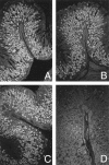
Figure 1.
Figure 1.
Indirect immunofluorescence staining on paraffin embedded-normal mouse stomach sections. Parietal cell reactivity in sera from a C3H/He mouse (A), a neonatally thymectomized BALB/cCrSlc mouse (B), a patient with pernicious anemia (C), and a C3H/He mouse without parietal cell reactivity (D). Magnification, objective ×20.
Figure 2.
Figure 2.
H&E-stained sections of paraffin-embedded mouse stomachs showing gastritis in C3H/He (A) and neonatally thymectomized BALB/c mice (B). Areas of mononuclear cell infiltration are indicated by arrows. (C) Stained section of a nongastritic C3H/He mouse stomach. Magnification, objective ×25.
Figure 3.
Figure 3.
Modified Maxwell’s staining of paraffin-embedded mouse stomachs from nongastritic (A) and gastritic (B) C3H/He mice. Parietal cells stain blue-green, zymogenic (chief) cells stain red-purple, and mucus-secreting cells stain yellow. Note the change in cellular composition in C3H/He mice with gastritis. Magnification, objective ×25.
Figure 4.
Figure 4.
Breeding program used to generate C3H/He mouse line with higher incidence of autoimmune gastritis. Mice with parietal cell autoantibodies from each generation were selected for breeding. Sequential generations of mice are labeled A through E. ○, females without parietal cell reactivity; •, females with parietal cell reactivity; □, males without parietal cell reactivity; ▪, males with parietal cell reactivity.
Figure 5.
Figure 5.
Incidence of gastritis in C3H/He mice with parietal cell- and H/K-ATPase-reactive autoantibodies. Anti-H/K-ATPase reactivity was detected by ELISA (filled bars). Parietal cell autoantibodies were detected by indirect immunofluorescence (IIF) on paraffin-embedded stomach sections. Anti-parietal cell reactivity was scored as follows: −, negative; +, weakly positive; ++, positive; and +++, strongly positive. Gastritis was assessed by histology and indicated by a filled square. Mice without gastritis are indicated by an open square.
Figure 6.
Figure 6.
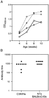
A: Development of anti-H/K-ATPase antibodies in C3H/He mice. Sera were collected from four C3H/He mice at 4, 6, 8, and 10 weeks of age and assayed for gastric H/K ATPase reactivity by ELISA. Each symbol represents the antibody reactivity with gastric H/K ATPase for an individual mouse over the course of the study. B: Serum titer of antibodies to the gastric H/K-ATPase in C3H/He mice and neonatally thymectomized (NTx) BALB/cCrSlc mice. Serum antibody titers were determined by ELISA on gastric H/K-ATPase-coated plates and taken as the highest dilution that produced an optical density reading above baseline.

Similar articles

Cited by

References

    1. Gleeson PA, Toh BH: Molecular targets in pernicious anaemia. Immunol Today 1991, 12:233-238 - PubMed
    1. Toh BH, van Driel IR, Gleeson PA: Pernicious anemia. N Engl J Med 1997, 337:1441-1448 - PubMed
    1. Strickland R, Mackay IR: A reappraisal of the nature and significance of chronic atrophic gastritis. Am J Digest Dis 1973, 18:426-440 - PubMed
    1. Toh BH, Gleeson PA, Simpson RJ, Moritz RL, Callaghan JM, Goldkorn I, Jones CM, Martinelli TM, Mu F-T, Humphris DC, Pettitt JM, Mori Y, Masuda T, Sobieszczuk P, Weinstock J, Mantamadiotis T, Baldwin GS: The 60- to 90-kd parietal cell autoantigen associated with autoimmune gastritis is a β subunit of the gastric H+/K+-ATPase (proton pump). Proc Natl Acad Sci USA 1990, 87:6418-6422 - PMC - PubMed
    1. Callaghan JM, Khan MA, Alderuccio F, van Driel IR, Gleeson PA, Toh BH: α and β subunits of the gastric H+/K+-ATPase are concordantly targeted by parietal cell autoantibodies associated with autoimmune gastritis. Autoimmunity 1993, 16:289-295 - PubMed

Publication types