. 1998 Oct 19;188(8):1413-9.
doi: 10.1084/jem.188.8.1413.

Fractalkine and CX3CR1 mediate a novel mechanism of leukocyte capture, firm adhesion, and activation under physiologic flow

Affiliations

Fractalkine and CX3CR1 mediate a novel mechanism of leukocyte capture, firm adhesion, and activation under physiologic flow

A M Fong et al. J Exp Med. .

Abstract

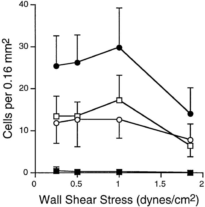
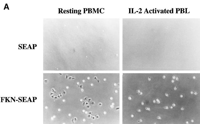
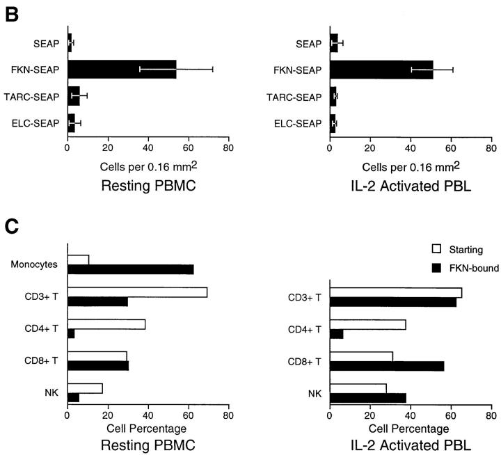
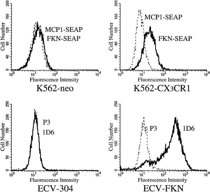
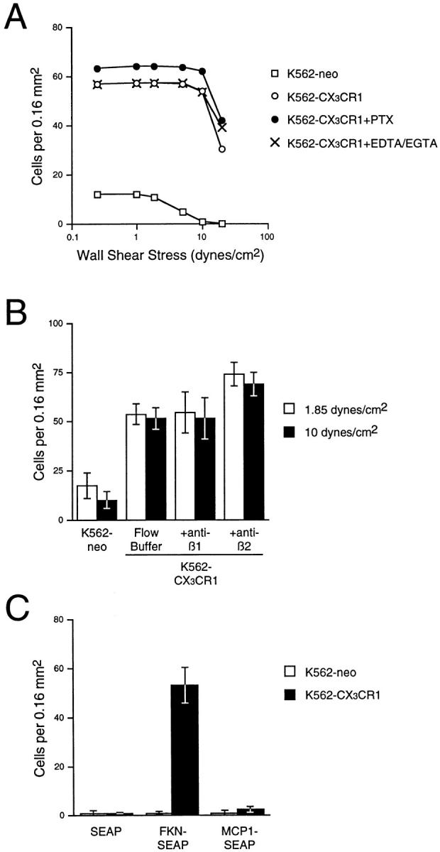
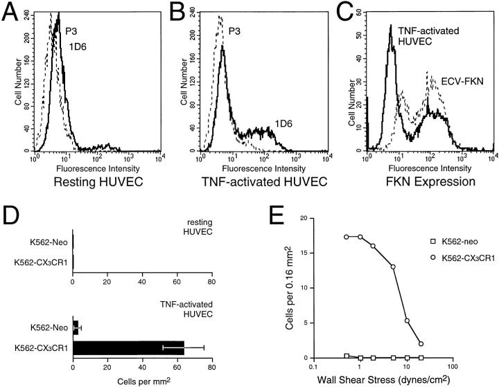
Leukocyte migration into sites of inflammation involves multiple molecular interactions between leukocytes and vascular endothelial cells, mediating sequential leukocyte capture, rolling, and firm adhesion. In this study, we tested the role of molecular interactions between fractalkine (FKN), a transmembrane mucin-chemokine hybrid molecule expressed on activated endothelium, and its receptor (CX3CR1) in leukocyte capture, firm adhesion, and activation under physiologic flow conditions. Immobilized FKN fusion proteins captured resting peripheral blood mononuclear cells at physiologic wall shear stresses and induced firm adhesion of resting monocytes, resting and interleukin (IL)-2-activated CD8(+) T lymphocytes and IL-2-activated NK cells. FKN also induced cell shape change in firmly adherent monocytes and IL-2-activated lymphocytes. CX3CR1-transfected K562 cells, but not control K562 cells, firmly adhered to FKN-expressing ECV-304 cells (ECV-FKN) and tumor necrosis factor alpha-activated human umbilical vein endothelial cells. This firm adhesion was not inhibited by pertussis toxin, EDTA/EGTA, or antiintegrin antibodies, indicating that the firm adhesion was integrin independent. In summary, FKN mediated the rapid capture, integrin-independent firm adhesion, and activation of circulating leukocytes under flow. Thus, FKN and CX3CR1 mediate a novel pathway for leukocyte trafficking.


Figures

Figure 1
Figure 1
PBMCs are captured by immobilized FKN-SEAP under physiologic wall shear stresses. Freshly isolated PBMCs were perfused over immobilized FKN-SEAP and SEAP fusion proteins at wall shear stresses of 0.25, 0.5, 1, or 1.85 dynes/cm2 for 5 min. After perfusing the chamber with flow buffer at 10 dynes/cm2 for 3 min, the number of adherent cells was determined. Also shown are the numbers of PBMCs captured by FKN-SEAP (•), monocytes captured by FKN-SEAP (○), lymphocytes captured by FKN-SEAP (□) and PBMCs captured by control SEAP fusion protein (▪). The error bars represent the mean ± SD of the number of cells bound. Data are representative of three independent experiments.
Figure 2
Figure 2
Resting monocytes, resting and IL-2–activated CD8+ T cells, and IL-2–activated NK cells firmly adhere to FKN under flow. Immobilized FKN was tested for its ability to capture and induce the firm adhesion of freshly isolated PBMCs in the parallel plate flow chamber. Cells were perfused over immobilized proteins at 0.25 dynes/cm2 for 5 min and washed at 10 dynes/cm2. Resting PBMCs and IL-2–activated PBLs were characterized by three-color flow cytometry and firmly adherent cells were characterized by two-color IF microscopy. (A) Photomicrographs of PBMCs and IL-2–activated PBLs bound to immobilized SEAP and FKN-SEAP. (B) Numbers of PBMCs and IL-2–activated PBLs remaining bound to immobilized SEAP, FKN-SEAP, TARC-SEAP, and ELC-SEAP at 10 dynes/cm2. (C) Percentages of leukocyte cell types binding to FKN-SEAP under flow. The cell types measured and quantified were: CD14+ monocytes, CD3+CD16/56 T cells, CD3+CD4+ T cells, CD3+CD8+ T cells, and CD16/56+ NK cells. The percentage of leukocyte subsets in the starting material as measured by multicolor flow cytometry is depicted by white bars, and the percentage of cell subsets in the FKN-bound fraction as measured by two-color IF microscopy is depicted by black bars. Leukocyte subsets from both resting PBMCs and IL-2 activated PBLs bound firmly and specifically to FKN-SEAP under flow. FKN preferentially bound resting monocytes, resting and IL-2–activated CD8+ T cells, and IL-2–activated NK cells. The majority of FKN-bound, IL-2–activated PBLs formed pseudopods. Data are representative of three experiments performed. The error bars represent the mean ± SD of the number of cells bound.
Figure 2
Figure 2
Resting monocytes, resting and IL-2–activated CD8+ T cells, and IL-2–activated NK cells firmly adhere to FKN under flow. Immobilized FKN was tested for its ability to capture and induce the firm adhesion of freshly isolated PBMCs in the parallel plate flow chamber. Cells were perfused over immobilized proteins at 0.25 dynes/cm2 for 5 min and washed at 10 dynes/cm2. Resting PBMCs and IL-2–activated PBLs were characterized by three-color flow cytometry and firmly adherent cells were characterized by two-color IF microscopy. (A) Photomicrographs of PBMCs and IL-2–activated PBLs bound to immobilized SEAP and FKN-SEAP. (B) Numbers of PBMCs and IL-2–activated PBLs remaining bound to immobilized SEAP, FKN-SEAP, TARC-SEAP, and ELC-SEAP at 10 dynes/cm2. (C) Percentages of leukocyte cell types binding to FKN-SEAP under flow. The cell types measured and quantified were: CD14+ monocytes, CD3+CD16/56 T cells, CD3+CD4+ T cells, CD3+CD8+ T cells, and CD16/56+ NK cells. The percentage of leukocyte subsets in the starting material as measured by multicolor flow cytometry is depicted by white bars, and the percentage of cell subsets in the FKN-bound fraction as measured by two-color IF microscopy is depicted by black bars. Leukocyte subsets from both resting PBMCs and IL-2 activated PBLs bound firmly and specifically to FKN-SEAP under flow. FKN preferentially bound resting monocytes, resting and IL-2–activated CD8+ T cells, and IL-2–activated NK cells. The majority of FKN-bound, IL-2–activated PBLs formed pseudopods. Data are representative of three experiments performed. The error bars represent the mean ± SD of the number of cells bound.
Figure 3
Figure 3
Expression of FKN in ECV-304 cells and CX3CR1 in K562 cells. The top shows histograms depicting the binding of anti-FKN mAb 1D6 (solid lines) and control mAb P3 (dashed lines) to untransfected ECV-304 cells and to transfected ECV-FKN cells. The bottom shows histograms depicting the binding of FKN-SEAP (solid lines) and MCP-1– SEAP (dashed lines) to control K562-neo cells and transfected K562-CX3CR1 cells.
Figure 4
Figure 4
Integrin-independent firm adhesion of K562-CX3CR1 cells to ECV-FKN cells and immobilized FKN-SEAP under physiologic flow conditions. (A) K562-CX3CR1 cells remain firmly adherent to ECV-FKN cells under physiologic wall shear stresses, and this FKN-mediated firm adhesion is PTX insensitive and integrin independent. K562-neo cells and K562-CX3CR1 cells (± EDTA/EGTA and ± PTX treatment) were perfused over ECV-FKN monolayers for 10 min at 0.25 dynes/cm2 and subjected to increasing wall shear stresses up to 20 dynes/cm2. Shown are the numbers of firmly adherent cells at various shear stresses. (B) Anti-β1 and β2 integrin mAbs have no effect on the firm adhesion of K562-CX3CR1 cells to ECV-FKN cells. The numbers of adherent K562-neo and K562-CX3CR1 cells at 1.85 dynes/ cm2 and 10 dynes/cm2 in the absence and presence of anti-β1 and β2 integrin mAbs are shown. (C) K562-CX3CR1 cells bind to immobilized FKN under flow. FKN-SEAP, MCP-1–SEAP, and SEAP fusion proteins were immobilized by binding to glass coverslips coated with anti–alkaline phosphatase mAbs and were tested for their ability to support firm adhesion of K562-CX3CR1 cells under flow. Cells were perfused over immobilized SEAP fusion proteins for 10 min at 0.25 dynes/ cm2, and exposed to a wall shear stress of 10 dynes/cm2. Shown are the numbers of K562 and K562-CX3CR1 cells remaining bound to immobilized chemokine-SEAP fusion proteins at 10 dynes/cm2. Error bars represent the mean ± SD. Data are representative of three experiments performed.
Figure 5
Figure 5
Expression of FKN by TNF-activated HUVECs and their ability to support arrest of K562-CX3CR1 cells under flow conditions. FKN expression on HUVECs, either resting (A) or stimulated with 100 ng/ml TNF-α for 12 h (B), was measured by IF staining with mAb 1D6. Shown are histograms of the reactivity of anti-FKN (1D6) and control (P3) mAbs. Cells were also counterstained with anti-CD31-PE to ensure they were endothelial cells. FKN was expressed on a subset of CD31+ TNF-activated HUVECs. (C) Comparison of the level of FKN expression by TNF-activated HUVECs and ECV-FKN cells. Shown are histograms of the reactivity of mAb 1D6 with 12-h TNF-activated HUVECs and ECV-FKN cells. (D) K562-CX3CR1 cells bind to TNF- activated HUVECs but not to resting HUVECs. K562-neo cells and K562-CX3CR1 cells were perfused over HUVECs and TNF-activated HUVEC monolayers for 5 min at 0.5 dynes/ cm2, and subjected to a wall shear stress of 1.85 dynes/cm2. Shown are the numbers of firmly adherent cells to HUVECs and TNF-activated HUVECs at 1.85 dynes/cm2. Error bars represent the mean ± SD. (E) K562-CX3CR1 cells bind firmly to TNF-activated HUVECs at high wall shear stresses. K562-neo cells and K562-CX3CR1 cells were perfused over TNF-activated HUVECs monolayers for 5 min at 0.5 dynes/cm2 and were subjected to increasing wall shear stresses up to 20 dynes/cm2. Shown are the numbers of firmly adherent cells to TNF-activated HUVECs at various shear stresses from a representative experiment. All data are representative of at least three independent experiments.

References

    1. Springer TA. Traffic signals for lymphocyte recirculation and leukocyte emigration, the multistep paradigm. Cell. 1994;76:301–314. - PubMed
    1. Butcher EC, Picker LJ. Lymphocyte homing and homeostasis. Science. 1996;272:60–66. - PubMed
    1. Ley K, Tedder TF. Leukocyte interactions with vascular endothelium: new insights into selectin mediated attachment and rolling. J Immunol. 1995;155:525–528. - PubMed
    1. Tedder TF, Steeber DA, Chen A, Engel P. The selectins: vascular adhesion molecules. FASEB (Fed Am Soc Exp Biol) J. 1995;9:866–873. - PubMed
    1. Bargatze RF, Butcher EC. Rapid G protein– regulated activation event involved in lymphocyte binding to high endothelial venules. J Exp Med. 1993;178:367–372. - PMC - PubMed

Publication types