. 1998 Oct 19;188(8):1521-8.
doi: 10.1084/jem.188.8.1521.

CD1d-mediated recognition of an alpha-galactosylceramide by natural killer T cells is highly conserved through mammalian evolution

Affiliations

CD1d-mediated recognition of an alpha-galactosylceramide by natural killer T cells is highly conserved through mammalian evolution

L Brossay et al. J Exp Med. .

Abstract

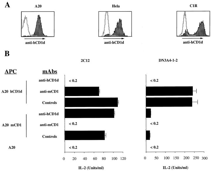
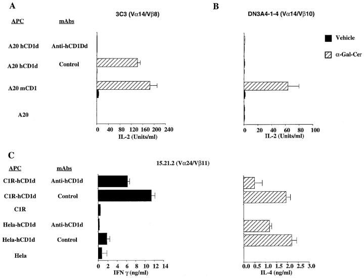
Natural killer (NK) T cells are a lymphocyte subset with a distinct surface phenotype, an invariant T cell receptor (TCR), and reactivity to CD1. Here we show that mouse NK T cells can recognize human CD1d as well as mouse CD1, and human NK T cells also recognize both CD1 homologues. The unprecedented degree of conservation of this T cell recognition system suggests that it is fundamentally important. Mouse or human CD1 molecules can present the glycolipid alpha-galactosylceramide (alpha-GalCer) to NK T cells from either species. Human T cells, preselected for invariant Valpha24 TCR expression, uniformly recognize alpha-GalCer presented by either human CD1d or mouse CD1. In addition, culture of human peripheral blood cells with alpha-GalCer led to the dramatic expansion of NK T cells with an invariant (Valpha24(+)) TCR and the release of large amounts of cytokines. Because invariant Valpha14(+) and Valpha24(+) NK T cells have been implicated both in the control of autoimmune disease and the response to tumors, our data suggest that alpha-GalCer could be a useful agent for modulating human immune responses by activation of the highly conserved NK T cell subset.


Figures

Figure 1
Figure 1
hCD1d stimulates mouse NK T cell hybridomas. (A) Flow cytometry profiles of hCD1d-transfected A20, Hela, and C1R cell lines. Stainings were carried out with biotinylated 42.2 anti-hCD1d mAb, followed by PE-conjugated streptavidin. Before each staining, cells were incubated with 1% human serum. Each panel is an overlay of the isotype control (open histogram) and the anti-hCD1d mAb (filled histogram). (B) Interspecies cross-reactivity of mouse Vα14/Vβ8 T cell hybridomas. The 2C12 and DN3A4-1.2 mCD1-autoreactive hybridomas were cultured at 5 × 10⁴ cells/well with 105 APCs. Antibody-mediated inhibition was determined using an anti-hCD1d mAb 51.1, anti-mCD1 mAb 1B1, or isotype-matched control mAbs, all at 10 μg/ml. After 16 h of culture, IL-2 release was determined by ELISA. The experiments shown here are representative of nine different experiments.
Figure 2
Figure 2
hCD1d presents α-GalCer to mouse and human NK T cells. (A and B) Response of a Vα14/Vβ8 hybridoma. hCD1d-transfected cells (A20 hCD1d), mCD1.1-transfected cells (A20 mCD1), or untransfected A20 cells were pulsed for 2 h either with α-GalCer or the vehicle control. These APCs were washed twice and plated at 105 cells/well. Antibodies at 10 μg/ml were added immediately before the addition of either the 3C3 (Vα14/Vβ8) or the DN3A4-1-4 (Vα14/Vβ10) T cell hybridomas at 5 × 10⁴ cells/well. After 16 h of culture, IL-2 release was determined by ELISA. The experiments shown here are representative of four different experiments. (C) Response of a human Vα24/Vβ11 clone. hCD1d CIR transfectants (C1R hCD1d), hCD1d-transfected Hela cells (Hela hCD1d), or the untransfected parental control cells were antigen pulsed as described above. Cells were then incubated for 48 h with T cells from clone 15.21.2 at 5 × 10⁴ cells/well. Isotype-matched control or anti-hCD1d mAb 51.1 at 20 μg/ml were added immediately before the addition of the T cells. IFN-γ and IL-4 release were determined by ELISA. The experiments shown here have been performed without the addition of PMA, and they are representative of three different experiments.
Figure 3
Figure 3
NK T cells from human PBMCs expand in response to α-GalCer. Crude PBMCs were cultivated in the presence of 50 U/ml of IL-2 and 100 ng/ml of α-GalCer. At day 10 harvested cells from XC cell line (top) or PB cell line (bottom) were cultured for 5 d in the presence of IL-2 only. Proliferating T cells were then cultured for another 5 d with α-GalCer pulsed C1R-hCD1d transfectants in the presence of 50 U of IL-2. Expansion of the Vα24+ cells was determined upon staining with anti-CD4 and anti-Vα24 mAbs at days 0, 9, and 21.

References

    1. Hughes AL. Evolutionary origin and diversification of the mammalian CD1 antigen genes. Mol Biol Evol. 1991;8:185–201. - PubMed
    1. Bilsland CA, Milstein C. The identification of the β2-microglobulin binding antigen encoded by the human CD1D gene. Eur J Immunol. 1991;21:71–78. - PubMed
    1. Calabi F, Jarvis JM, Martin L, Milstein C. Two classes of CD1 genes. Eur J Immunol. 1989;19:285–292. - PubMed
    1. Bradbury A, Belt KT, Neri TM, Milstein C, Calabi F. Mouse CD1 is distinct from and co-exists with TL in the same thymus. EMBO (Eur Mol Biol Organ) J. 1988;7:3081–3086. - PMC - PubMed
    1. Porcelli SA. The CD1 family: a third lineage of antigen-presenting molecules. Adv Immunol. 1995;59:1–98. - PubMed

Publication types