. 1998 Oct 19;188(8):1529-34.
doi: 10.1084/jem.188.8.1529.

CD1d-restricted recognition of synthetic glycolipid antigens by human natural killer T cells

Affiliations

CD1d-restricted recognition of synthetic glycolipid antigens by human natural killer T cells

F M Spada et al. J Exp Med. .

Abstract

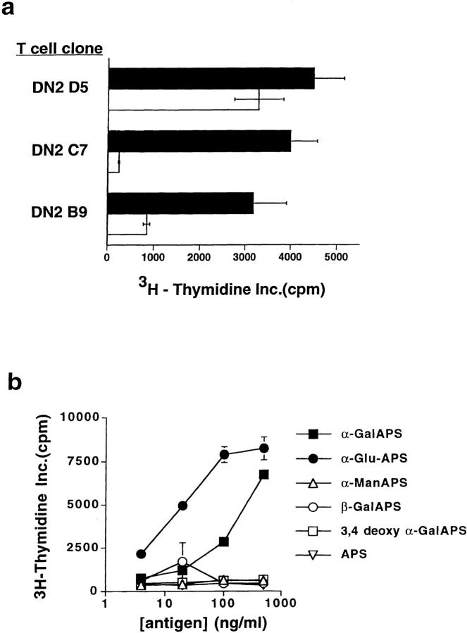
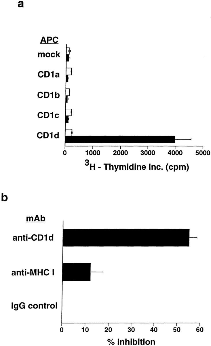
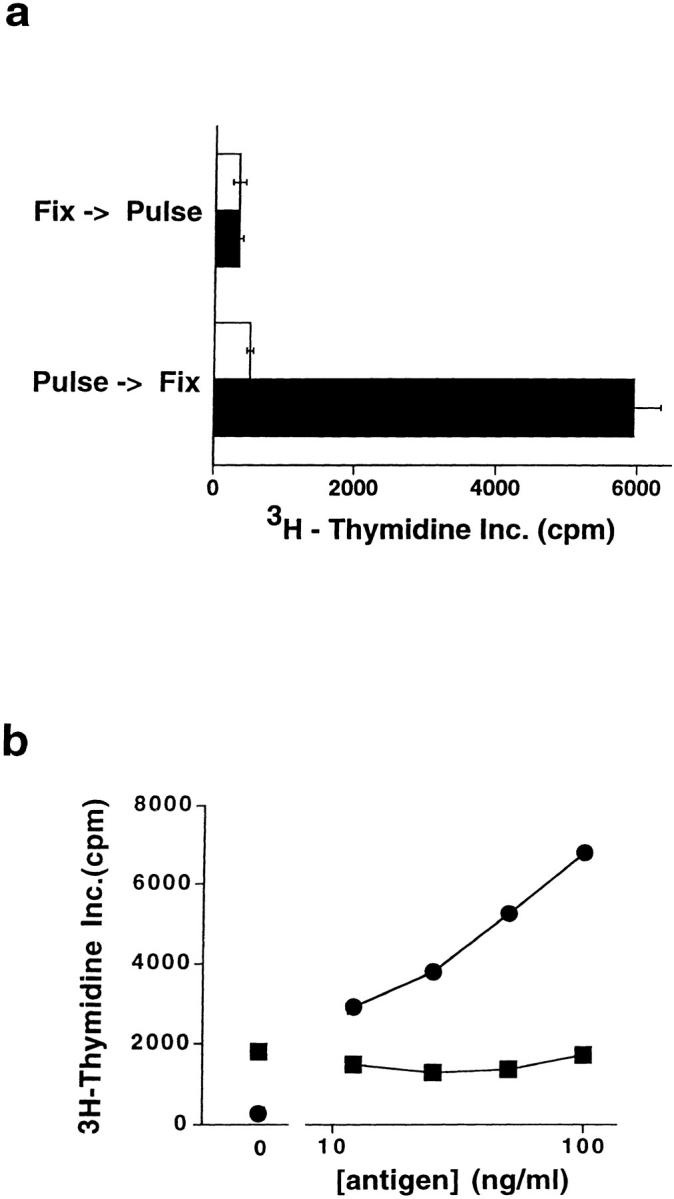
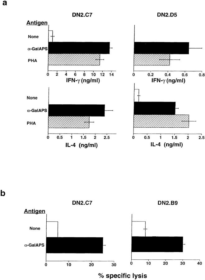
A conserved subset of mature circulating T cells in humans expresses an invariant Valpha24-JalphaQ T cell receptor (TCR)-alpha chain rearrangement and several natural killer (NK) locus-encoded C-type lectins. These human T cells appear to be precise homologues of the subset of NK1.1(+) TCR-alpha/beta+ T cells, often referred to as NK T cells, which was initially identified in mice. Here we show that human NK T cell clones are strongly and specifically activated by the same synthetic glycolipid antigens as have been shown recently to stimulate murine NK T cells. Responses of human NK T cells to these synthetic glycolipids, consisting of certain alpha-anomeric sugars conjugated to an acylated phytosphingosine base, required presentation by antigen-presenting cells expressing the major histocompatibility complex class I-like CD1d protein. Presentation of synthetic glycolipid antigens to human NK T cells required internalization of the glycolipids by the antigen-presenting cell and normal endosomal targeting of CD1d. Recognition of these compounds by human NK T cells triggered proliferation, cytokine release, and cytotoxic activity. These results demonstrate a striking parallel in the specificity of NK T cells in humans and mice, thus providing further insight into the potential mechanisms of immune recognition by NK T cells and the immunological function of this unique T cell subset.


Figures

Figure 1
Figure 1
Human NK T cell clones specifically respond to synthetic glycolipid antigens. (a) Proliferative responses of three different human NK T cell clones to CD1d-transfected HeLa cell APCs were significantly augmented by addition of the synthetic glycolipid αGalAPS. White bars show proliferation in response to CD1d+ HeLa cells plus vehicle (DMSO) alone; black bars show proliferation in response to CD1d+ HeLa cells plus 100 ng/ml αGalAPS. Results are shown as the mean and one standard deviation of triplicate values in this and all subsequent figures. (b) Proliferative responses of human NK T cell clone DN2.C7 to a panel of structurally related synthetic glycolipid antigens. APS is the nonglycosylated acyl-phytosphingosine lipid. αGalAPS, αGluAPS, and αManAPS indicate compounds in which α-anomeric galactose, glucose, and mannose, respectively are conjugated at position 1 of the hexose ring to the APS moiety. 3,4-deoxy αGalAPS is identical to αGalAPS except for the absence of the hydroxyl groups on carbons 3 and 4 of the phytosphingosine base. For complete structures of the synthetic glycolipids, see Kawano et al. (18). Thymidine incorporation in the absence of synthetic glycolipid antigen was 500 cpm in the experiment shown.
Figure 2
Figure 2
CD1d dependence of synthetic glycolipid antigen recognition by human NK T cells. (a) Response of human NK T cell clone DN2.C7 to αGalAPS (100 ng/ml) in the presence of HeLa cells not expressing CD1 (transfected with vector alone; mock) or expressing each of the known protein isoforms of human CD1. T cell proliferation in the absence of αGalAPS is shown by the white bars, and in the presence of αGalAPS by the black bars. Identical results were obtained with two other human NK T cell clones, DN2.B9 and DN2.D5 (data not shown). (b) Inhibition of αGalAPS-stimulated proliferation of human NK T cell clone DN2.B9 by anti-CD1d mAb. Results are shown as the percentage of inhibition of the αGalAPS-dependent proliferation in the presence of CD1d+ HeLa cells. With no antibody present, proliferation in the presence of the αGalAPS was 3,900 cpm, and in the absence of αGalAPS was <200 cpm in the experiment shown. Marginal blocking was observed with anti–MHC class I mAb W6/32 in some experiments. This was also seen in parallel experiments using MHC class II–restricted T cell clones (data not shown), and thus most likely represents a nonspecific effect of this antibody.
Figure 3
Figure 3
Requirement for antigen uptake and endosomal delivery of CD1d in recognition of synthetic glycolipids by human NK T cells. (a) Proliferative responses of clone DN2.C7 to ECDI-fixed APCs. CD1d+ HeLa cells either were pulsed for 12 h with αGalAPS (100 ng/ml) and subsequently fixed by ECDI treatment, or were fixed first followed by αGalAPS pulsing. White bars represent APCs pulsed with vehicle (DMSO) alone, and black bars represent APCs pulsed with αGalAPS. (b) Proliferation of clone DN2.C7 to αGalAPS presented by HeLa cell transfectants expressing wild-type CD1d (circles) versus HeLa transfectants expressing the CD1d/a chimeric protein that lacks an endosomal targeting signal (squares). Both transfectants expressed comparable levels of immunoreactive CD1d on the cell surface. Proliferation in the absence of APCs was <200 cpm in this experiment. Note that DN2.C7 showed a weak but significant response to the CD1d/a transfectant in the absence of added antigen, but no augmentation of this response at any concentration of αGalAPS tested.
Figure 4
Figure 4
Activation of effector functions of human NK T cells by synthetic glycolipid antigens. (a) Stimulation of IL-4 and IFN-γ secretion by αGalAPS. Human NK T cell clones were cultured with CD1d-expressing HeLa cells in the presence (white bars) or absence (black bars) of αGalAPS (100 ng/ml) or with PHA (hatched bars), and supernatants were harvested after 48 h and assayed for cytokines by ELISA. No cytokine production was detected in cultures containing T cells only or CD1d-transfected HeLa cells only (data not shown). (b) Human NK T cell clones specifically lysed CD1d-transfected HeLa cells pulsed for 12 h with αGalAPS (200 ng/ml). No lysis was observed using mock-transfected HeLa cells as targets (data not shown).

References

    1. Bendelac A, Rivera MN, Park SH, Roark JH. Mouse CD1-specific NK1 T cells: development, specificity, and function. Annu Rev Immunol. 1997;15:535–562. - PubMed
    1. Lantz O, Bendelac A. An invariant T cell receptor α chain is used by a unique subset of major histocompatibility complex class I–specific CD4+ and CD4−8−T cells in mice and humans. J Exp Med. 1994;180:1097–1106. - PMC - PubMed
    1. Koseki H, Asano H, Inaba T, Miyashita N, Moriwaki K, Lindahl KF, Mizutani Y, Imai K, Taniguchi M. Dominant expression of a distinctive V14+T-cell antigen receptor α chain in mice. Proc Natl Acad Sci USA. 1991;88:7518–7522. - PMC - PubMed
    1. Exley M, Garcia J, Balk SP, Porcelli S. Requirements for CD1d recognition by human invariant Vα24+ CD4−CD8−T cells. J Exp Med. 1997;186:109–120. - PMC - PubMed
    1. Prussin C, Foster B. TCR Vα24 and Vβ11 coexpression defines a human NK1 T cell analog containing a unique Th0 subpopulation. J Immunol. 1997;159:5862–5870. - PubMed

Publication types