. 1998 Oct 15;12(20):3195-205.
doi: 10.1101/gad.12.20.3195.

SXR, a novel steroid and xenobiotic-sensing nuclear receptor

Affiliations

SXR, a novel steroid and xenobiotic-sensing nuclear receptor

B Blumberg et al. Genes Dev. .

Abstract

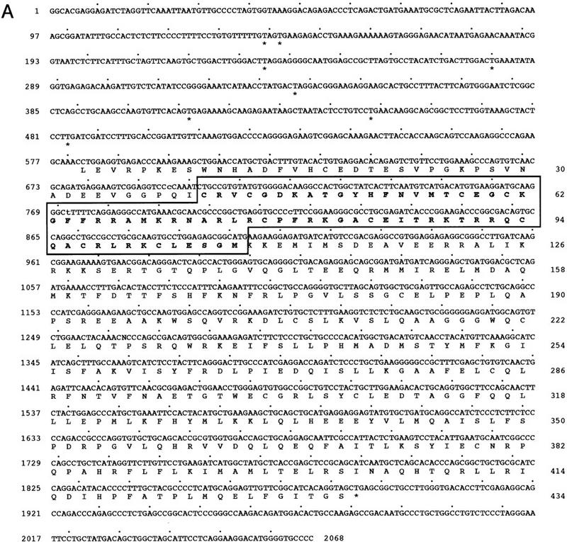
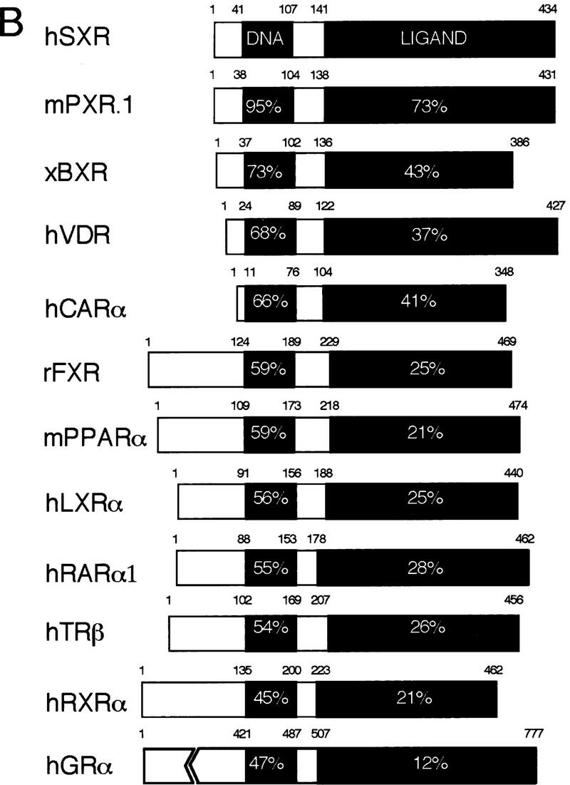
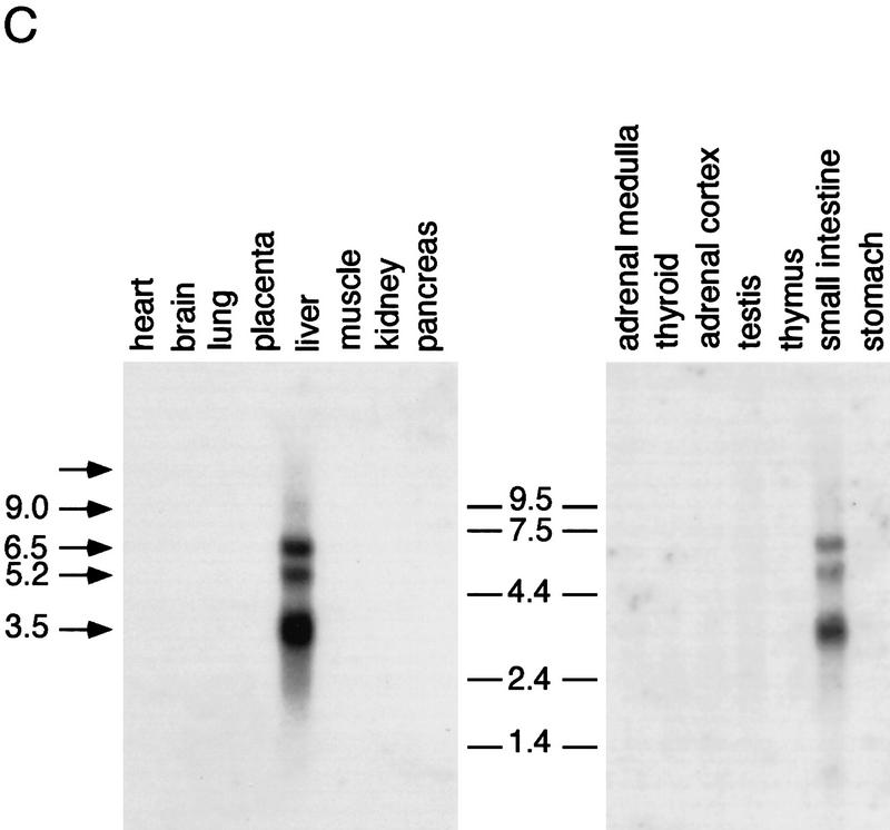
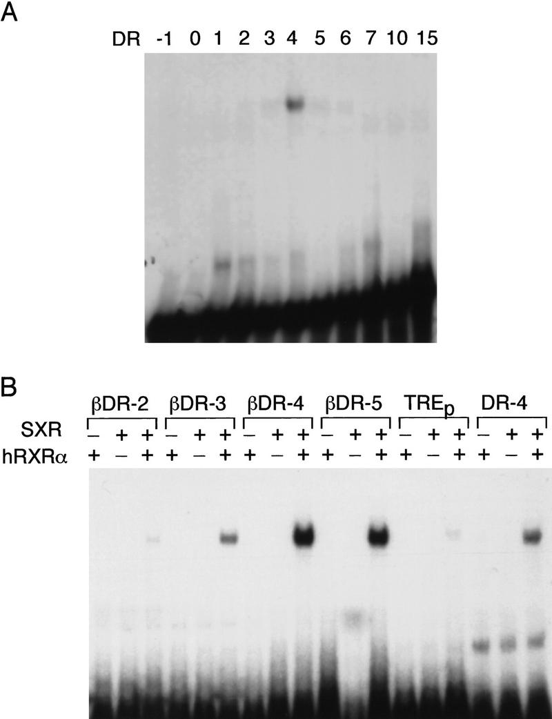
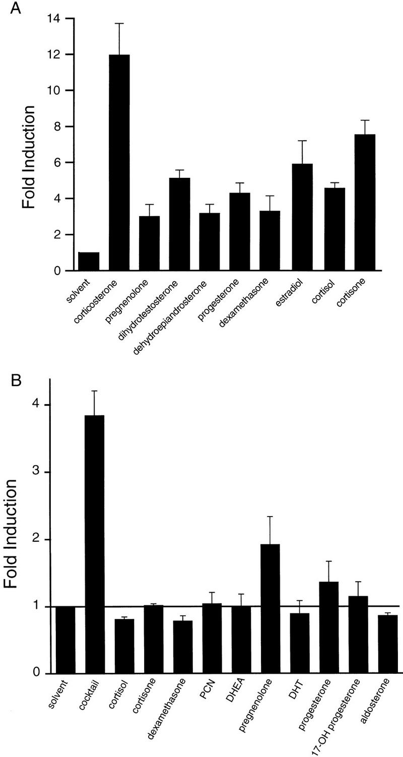
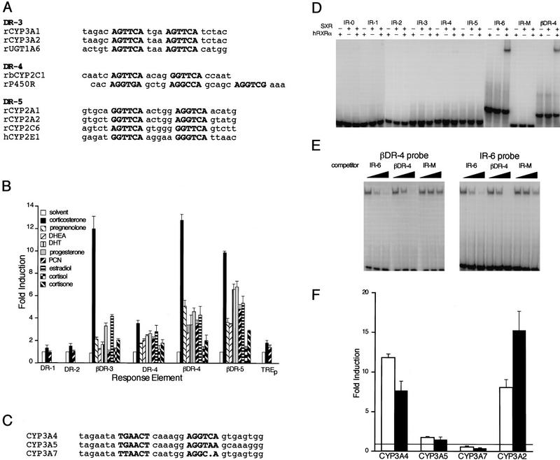
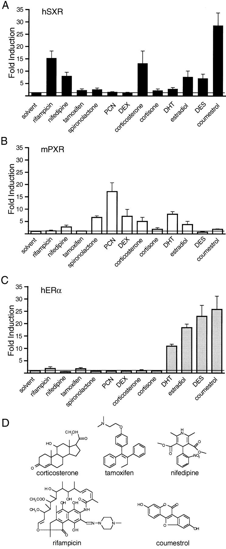
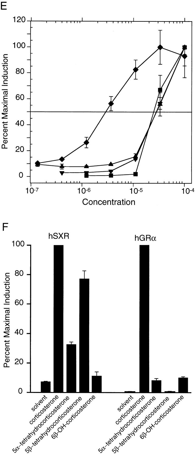
An important requirement for physiologic homeostasis is the detoxification and removal of endogenous hormones and xenobiotic compounds with biological activity. Much of the detoxification is performed by cytochrome P-450 enzymes, many of which have broad substrate specificity and are inducible by hundreds of different compounds, including steroids. The ingestion of dietary steroids and lipids induces the same enzymes; therefore, they would appear to be integrated into a coordinated metabolic pathway. Instead of possessing hundreds of receptors, one for each inducing compound, we propose the existence of a few broad specificity, low-affinity sensing receptors that would monitor aggregate levels of inducers to trigger production of metabolizing enzymes. In support of this model, we have isolated a novel nuclear receptor, termed the steroid and xenobiotic receptor (SXR), which activates transcription in response to a diversity of natural and synthetic compounds. SXR forms a heterodimer with RXR that can bind to and induce transcription from response elements present in steroid-inducible cytochrome P-450 genes and is expressed in tissues in which these catabolic enzymes are expressed. These results strongly support the steroid sensor hypothesis and suggest that broad specificity sensing receptors may represent a novel branch of the nuclear receptor superfamily.


Figures

Figure 1
Figure 1
SXR is a novel orphan nuclear receptor. (A) Sequence of the longest SXR cDNA clone. The DBD is shown in boldface type; upstream termination codons in-frame with the putative initiator leucine are indicated by asterisks. Leu can function as an initiator, as demonstrated by SDS-PAGE analysis of labeled proteins produced from in vitro-transcribed, translated cDNAs. The unmodified cDNAs yielded a translation product indistinguishable in size from that produced when the leucine was changed to methionine, albeit not nearly as efficiently (data not shown). (B) Schematic comparisons among SXR and other RXR partners. Amino acid sequences were aligned using the program GAP (Devereaux et al. 1984). The similarity between SXR and other receptors is expressed as percent amino acid identity. LBD boundaries follow those for the canonical nuclear receptor LBD (Wurtz et al. 1996). (C) SXR mRNA expression. Full-length SXR cDNA was used to probe multiple-tissue Northern blots (Clontech). SXR mRNA is expressed abundantly in liver and strongly, but much less abundantly, in intestine. (Left) Exposed for 4 hr, (right) exposed for 24 hr. Longer exposures did not reveal hybridizing bands in any other tissues on these blots. The sizes of four of five mRNAs are shown; the fifth could not be sized accurately as it is much larger than the largest size marker.
Figure 1
Figure 1
SXR is a novel orphan nuclear receptor. (A) Sequence of the longest SXR cDNA clone. The DBD is shown in boldface type; upstream termination codons in-frame with the putative initiator leucine are indicated by asterisks. Leu can function as an initiator, as demonstrated by SDS-PAGE analysis of labeled proteins produced from in vitro-transcribed, translated cDNAs. The unmodified cDNAs yielded a translation product indistinguishable in size from that produced when the leucine was changed to methionine, albeit not nearly as efficiently (data not shown). (B) Schematic comparisons among SXR and other RXR partners. Amino acid sequences were aligned using the program GAP (Devereaux et al. 1984). The similarity between SXR and other receptors is expressed as percent amino acid identity. LBD boundaries follow those for the canonical nuclear receptor LBD (Wurtz et al. 1996). (C) SXR mRNA expression. Full-length SXR cDNA was used to probe multiple-tissue Northern blots (Clontech). SXR mRNA is expressed abundantly in liver and strongly, but much less abundantly, in intestine. (Left) Exposed for 4 hr, (right) exposed for 24 hr. Longer exposures did not reveal hybridizing bands in any other tissues on these blots. The sizes of four of five mRNAs are shown; the fifth could not be sized accurately as it is much larger than the largest size marker.
Figure 2
Figure 2
SXR DNA-binding specificity. (A) SXR:hRXRα heterodimers prefer DR-4 among a panel of AGGTCA-containing HREs. In vitro-transcribed and -translated SXR was incubated with 32P-labeled oligonucleotides and electrophoresed in native polyacrylamide gels. (B) AGTTCA is preferred to AGGTCA. SXR:hRXRα heterodimers were tested for their ability to bind half-sites of the sequence AGTTCA derived from the RARβ RA–responsive element (Sucov et al. 1990). We found that in addition to a spacing motif of 4 (βDR-4) they bind nearly as well to βDR-5 spacing and significantly to a βDR-3 motif. DR-4 and TREp are shown for reference.
Figure 3
Figure 3
SXR is activated by many steroids. (A) Chimeric receptors composed of the GAL4 DNA-binding domain and the SXR-ligand binding domain were cotransfected into CV-1 cells with the reporter gene tk(MH100)4–luc (Forman et al. 1995). DHEA and pregnenolone activated this chimeric receptor; therefore, other steroids were tested for activation. Results are shown as fold induction over solvent (DMSO) control for 50 μm steroid and represent the averages and standard error from triplicate assays. Reporter alone or reporter plus GAL4–DBD was not activated by any of these compounds (data not shown). Similar results were obtained using full-length receptors and appropriate reporters (see below). (B) The ability of steroidal activators to act additively was tested using full-length SXR and the reporter tk(LXRE)3–luc (Willy et al. 1995). The cocktail contained 10 μm of each steroid for an overall concentration of 100 μm total steroid. The cocktail and its individual components were tested at 100, 10, and 1 μm; results are shown for 100 μm cocktail and 10 μm component steroids. Similar results were obtained using GAL–SXR (not shown).
Figure 4
Figure 4
SXR can activate responsive elements found in steroid and xenobiotic-inducible P-450 enzymes. (A) Putative DR-series response elements are found in inducible cytochrome P-450 enzymes. A database search for repeats of the sequence RGKTCA was performed, and some of the hits for enzymes involved in hepatic steroid hydroxylation are indicated. The standard nomenclature for P-450 enzymes has been used. P-450R is the single P-450 oxidoreductase required for hydroxylation of steroids. UGT1A6 is a rat UDP–glucuronosyltransferase that conjugates glucuronic acid to hydroxylated steroids. (B) SXR has a broad specificity for both response elements and steroidal activators. Full-length SXR was tested in cotransfection experiments for its ability to activate elements similar to those in A in response to a panel of steroids at 50 μm. DR-1, DR-2, and TREp were only very slightly activated; hence, results are shown only for corticosterone and PCN. The actual response elements and the number of copies are as follows, the base vector is tk–luc in all cases (Hollenberg et al. 1985): DR-1, tk(ApoAI)4 (Ladias and Karathanasis 1991); DR-2, tk(Hox-B1-RARE)2 (Ogura and Evans 1995); βDR-3, tk(CYP3A2)3 (Kliewer et al. 1998); DR-4, tk(MLV-TRE)2 (Umesono et al. 1991); βDR-4, tk(LXRE)3 (Willy et al. 1995); βDR-5, tk(βRARE)3 (Sucov et al. 1990); TREp, tk(TREp)2 (Umesono et al. 1991). The data shown are expressed as mean fold induction over solvent control ± s.e. from triplicate assays. (C) Conserved glucocorticoid-responsive elements found in human CYP3 genes. The region of human CYP3A4 shown to be necessary and sufficient for glucocorticoid and rifampicin induction of the full-length promoter is shown along with the corresponding regions of CYP3A5 and CYP3A7 (Barwick et al. 1996). (D) SXR:RXR heterodimers bind to IR-6 elements. The ability of SXR to bind to a panel of IR elements with spacers from 0 to 6 were tested along with βDR-4. SXR binds only as a homodimer with RXR to CYP3A4 IR-6 and βDR-4 elements. (E) SXR:RXR heterodimers bind to βDR-4 and IR-6 elements with similar affinity. Competition binding experiments were performed to estimate the relative affinity of SXR:RXR binding to CYP3A4 IR-6 element or βDR-4. The IR-M competitor has half-site mutations that prevent SXR:RXR binding (D). (F) SXR can activate through inducible, but not uninducible CYP3 promoter elements. The ability of SXR to activate tk–CYP3–luc response elements in response to various inducers was tested. (Open bars) Rifamipicin; (solid bars) corticosterone. Results are shown for 50 μm compound and represent the mean of triplicate determinations.
Figure 5
Figure 5
Pharmacology of SXR activation. (A–C) The ability of a panel of compounds to activate SXR, PXR.1, or ER was tested. Results are shown for 50 μm of compound with the following exceptions: 5 μm tamoxifen was used, DES concentration is 50 μm in A and B and 5 μm in C. (D) Chemical structures of some selected SXR activators from A–C. (E) Efficacy of SXR activation by selected compounds. The ability of a dilution series of compounds to activate full-length SXR was tested using several response elements as in Fig. 4. Results are shown for tk(LXRE)3–luc and represent the mean of triplicate determinations. Similar results were obtained for other response elements that SXR can activate. (♦) Rifamipicin; (▴) corticosterone; (▾) estradiol; (█) coumestrol. (F) Reduction of the 4–5 double bond does not inactivate corticosterone. 6β-hydroxylated, nonreduced, 5α and 5β reduced forms of corticosterone were tested for their ability to activate GAL–SXR on tk(MH100)4–luc and hGRα on MTV–luc at 50 μm. Similar results were obtained using full-length SXR.
Figure 5
Figure 5
Pharmacology of SXR activation. (A–C) The ability of a panel of compounds to activate SXR, PXR.1, or ER was tested. Results are shown for 50 μm of compound with the following exceptions: 5 μm tamoxifen was used, DES concentration is 50 μm in A and B and 5 μm in C. (D) Chemical structures of some selected SXR activators from A–C. (E) Efficacy of SXR activation by selected compounds. The ability of a dilution series of compounds to activate full-length SXR was tested using several response elements as in Fig. 4. Results are shown for tk(LXRE)3–luc and represent the mean of triplicate determinations. Similar results were obtained for other response elements that SXR can activate. (♦) Rifamipicin; (▴) corticosterone; (▾) estradiol; (█) coumestrol. (F) Reduction of the 4–5 double bond does not inactivate corticosterone. 6β-hydroxylated, nonreduced, 5α and 5β reduced forms of corticosterone were tested for their ability to activate GAL–SXR on tk(MH100)4–luc and hGRα on MTV–luc at 50 μm. Similar results were obtained using full-length SXR.

Similar articles

Cited by

References

    1. Altschul SF, Gish W, Miller W, Myers EW, Lipman DJ. Basic local alignment search tool. J Mol Biol. 1990;215:403–410. - PubMed
    1. Baes M, Gulick T, Choi HS, Martinoli MG, Simha D, Moore DD. A new orphan member of the nuclear hormone receptor superfamily that interacts with a subset of retinoic acid response elements. Mol Cell Biol. 1994;14:1544–1551. - PMC - PubMed
    1. Baker ME. Endocrine activity of plant-derived compounds: An evolutionary perspective. Proc Soc Exp Biol Med. 1995;208:131–138. - PubMed
    1. Bammel A, van der Mee K, Ohnhaus EE, Kirch W. Divergent effects of different enzyme-inducing agents on endogenous and exogenous testosterone. Eur J Clin Pharmacol. 1992;42:641–644. - PubMed
    1. Barbarash RA, Bauman JL, Fischer JH, Kondos GT, Batenhorst RL. Near-total reduction in verapamil bioavailability by rifampin. Electrocardiographic correlates. Chest. 1988;94:954–959. - PubMed

Publication types

MeSH terms