. 1998 Oct 15;12(20):3236-51.
doi: 10.1101/gad.12.20.3236.

HSP101 functions as a specific translational regulatory protein whose activity is regulated by nutrient status

Affiliations

HSP101 functions as a specific translational regulatory protein whose activity is regulated by nutrient status

D R Wells et al. Genes Dev. .

Abstract

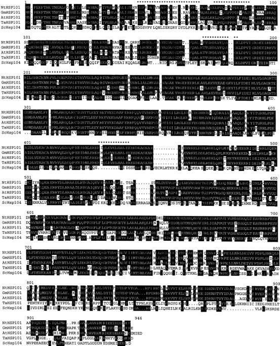
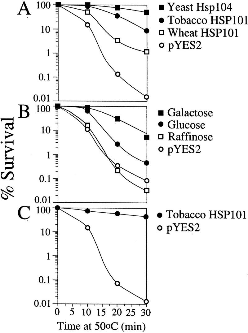
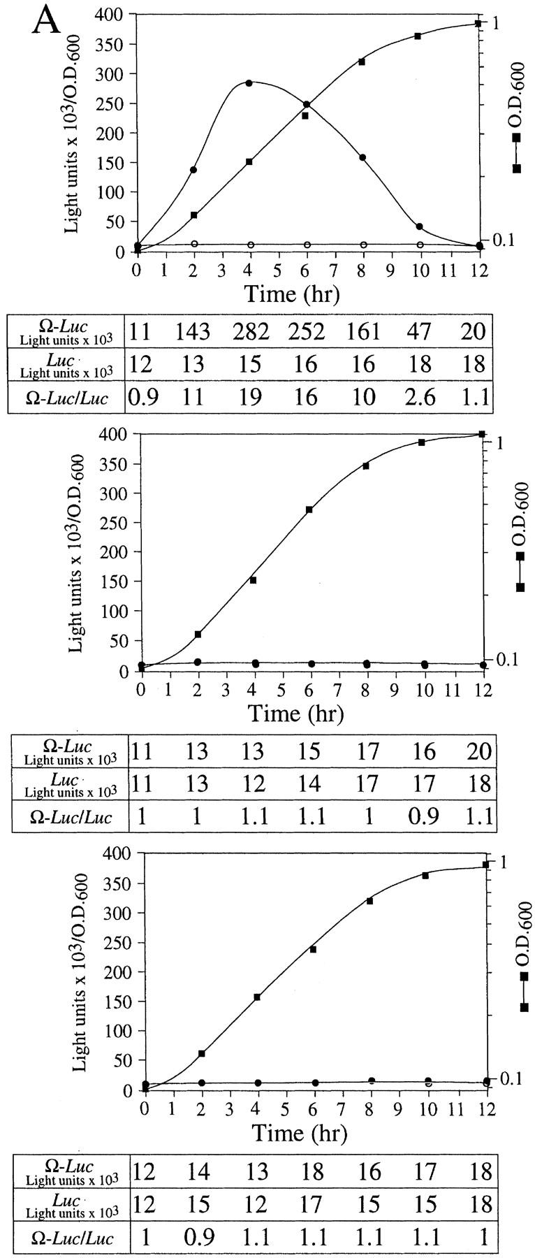
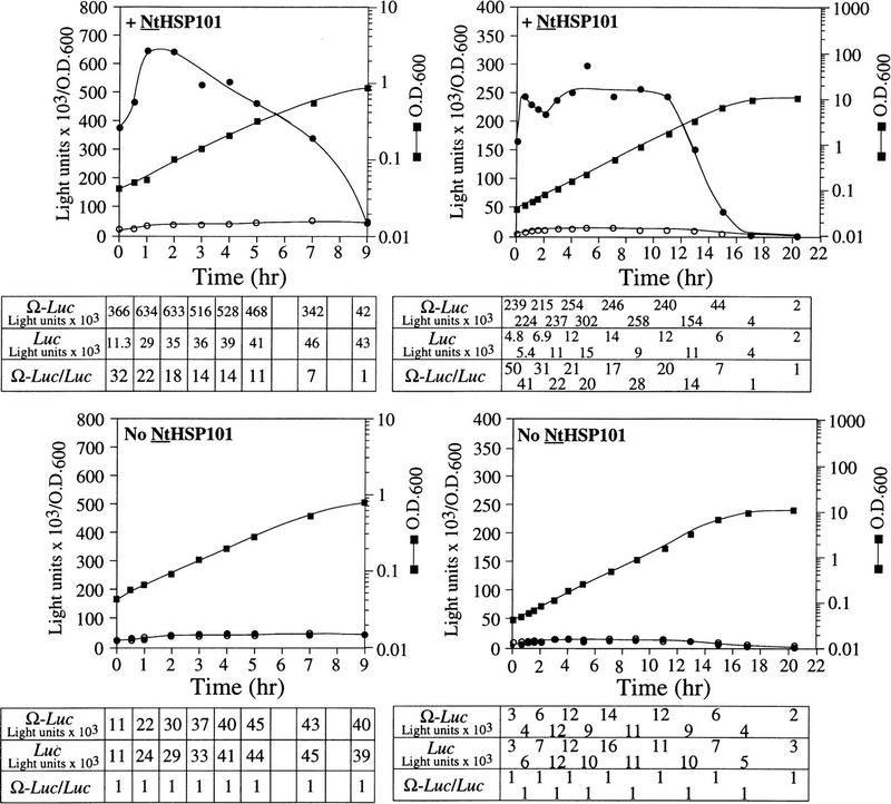
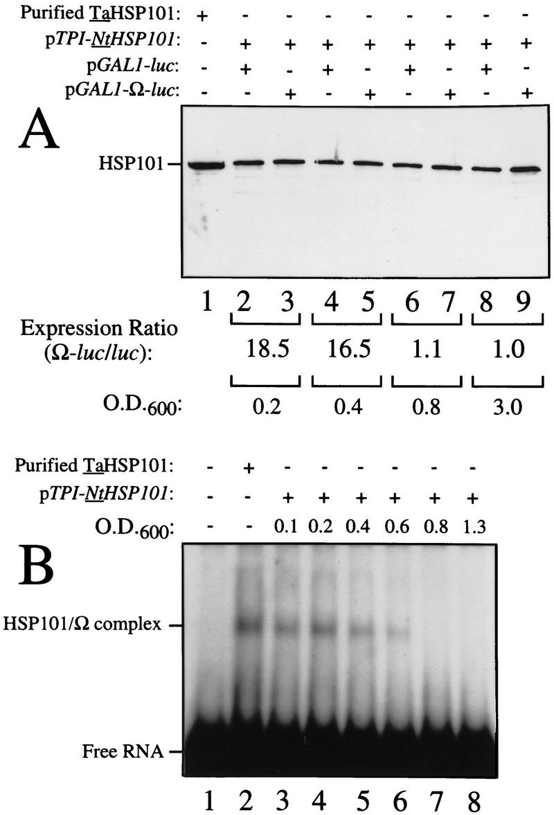
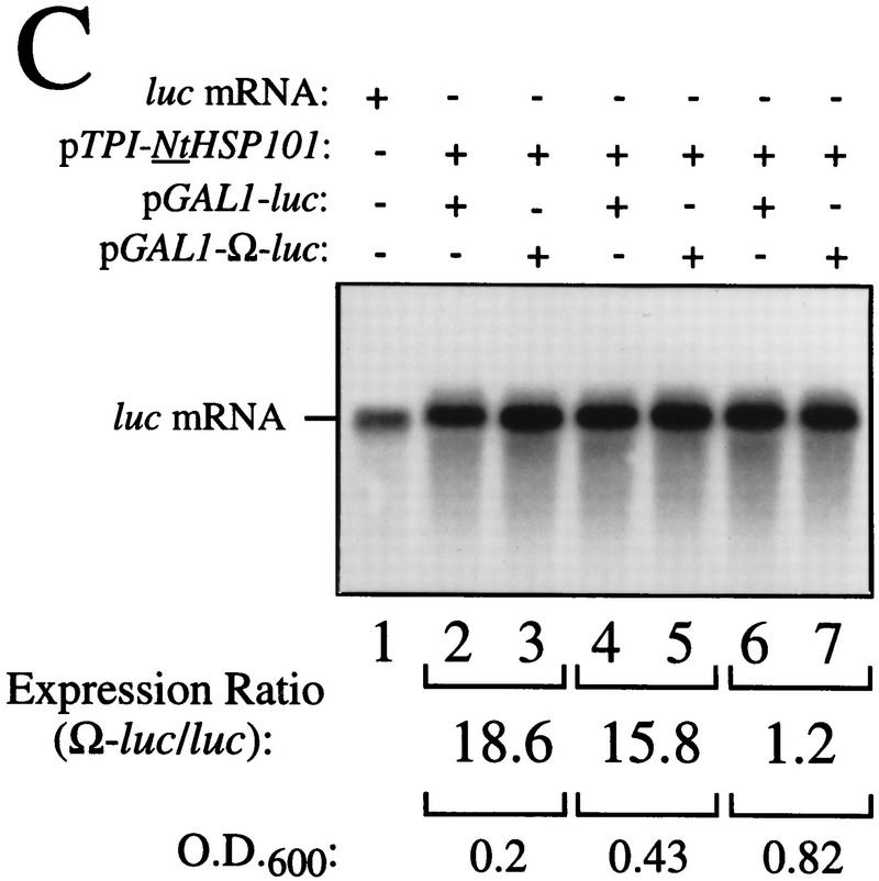
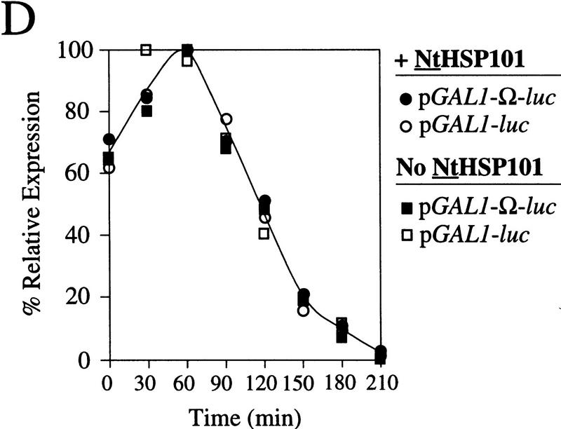
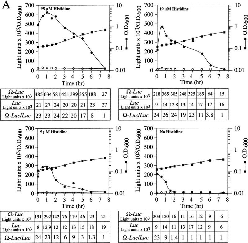
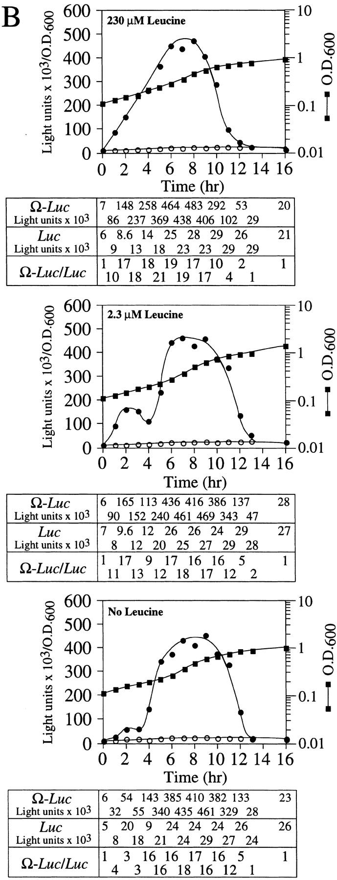
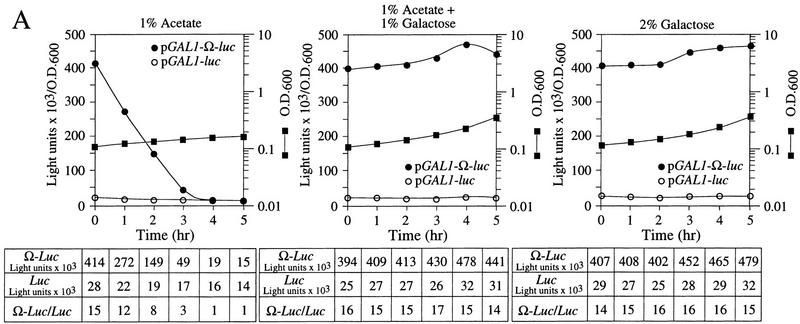
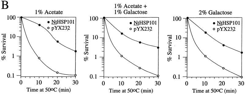
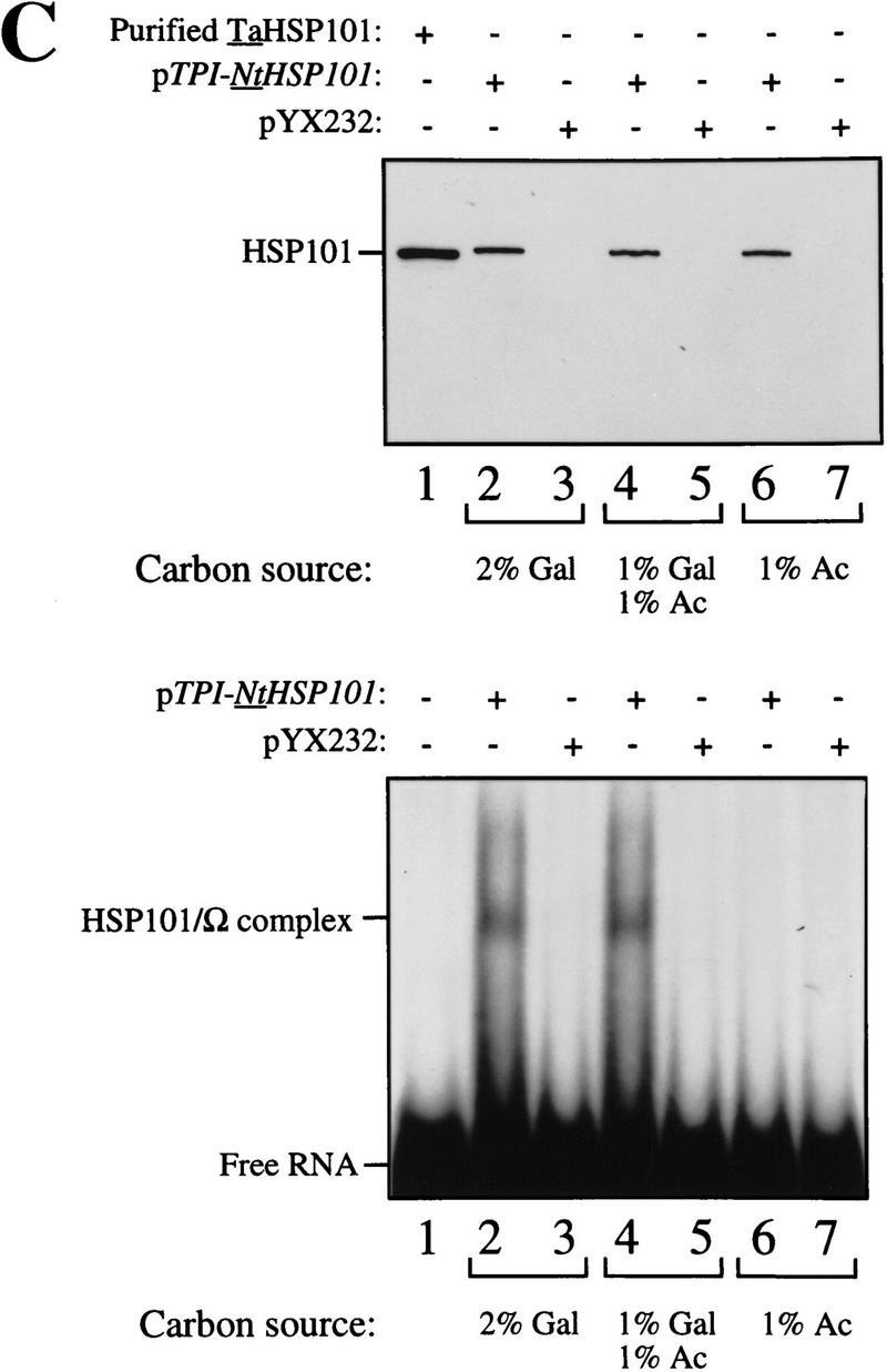
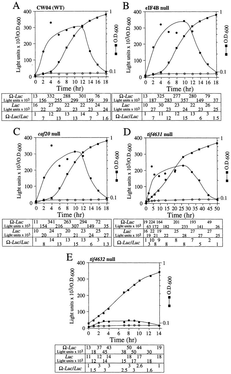
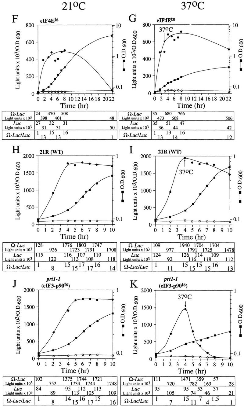
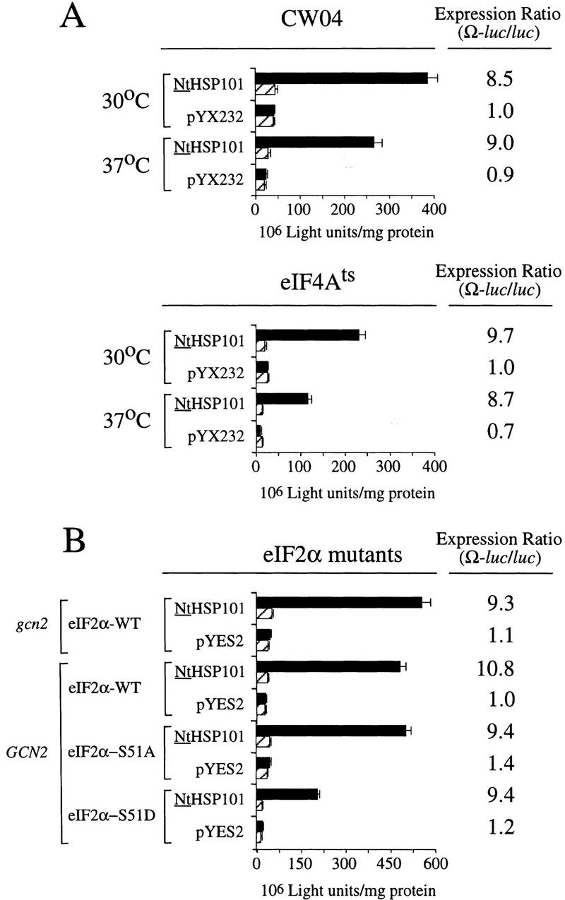
The 5' leader (Omega) of tobacco mosaic viral RNA functions as a translational enhancer. Sequence analysis of a 102-kD protein, identified previously as a specific Omega RNA-binding protein, revealed homology to the HSP101/HSP104/ClpB family of heat shock proteins and its expression in yeast complemented a thermotolerance defect caused by a deletion of the HSP104 gene. Up to a 50-fold increase in the translation of Omega-luc, but not luc mRNA was observed in yeast expressing the tobacco HSP101 whereas Omega failed to enhance translation in the absence of HSP101. Therefore, HSP101 and Omega comprise a two-component translational regulatory mechanism that can be recapitulated in yeast. Analysis of HSP101 function in yeast translation mutants suggested that the initiation factor (eIF) 3 and specifically one (TIF4632) of the two eIF4G proteins were required for the HSP101-mediated enhancement. The RNA-binding and translational regulatory activities of HSP101 were inactive in respiring cells or in cells subject to nutrient limitation, but its thermotolerance function remained unaffected. This is the first identification of a protein required for specific translational enhancement of capped mRNAs, the first report of a translational regulatory function for any heat-shock protein, and the first functional distinction between the two eIF4G proteins present in eukaryotes.


Figures

Figure 1
Figure 1
The predicted amino acid sequence of the tobacco and wheat cDNAs encoding HSP101 and their homology with Arabidopsis (Schirmer et al. 1994), soybean (Lee et al. 1994), and yeast (Parsell et al. 1991) homologs. Regions of homology between the proteins are shaded in black when conserved in at least three of the five proteins. The peptide sequences obtained from amino acid sequencing of the purified wheat and tobacco p102 proteins are indicated by asterisks. The tobacco and wheat cDNA sequences were deposited into GenBank as accession nos. AF083343 and AF083344, respectively.
Figure 2
Figure 2
p102 from tobacco and wheat is a functional HSP101 that complements a thermotolerance defect in yeast. (A) SL304A, the hsp104 mutant containing pGAL1–NtHSP101 or pGAL1–TaHSP101, was grown to an early exponential stage in SGM (an OD of 0.06 or 1.8 × 106 cells/ml) prior to assaying for thermotolerance. The expression vector, pYES2, was used as a negative control and pYS104, containing HSP104, was used as a positive control. The percentage of survival at 50°C was plotted as a function of the length of treatment. (B) SL304A(pGAL1–NtHSP101) was grown in galactose, glucose, or raffinose prior to assaying for thermotolerance. SL304A(pYES2) grown in galactose was included as a negative control. (C) Thermotolerance conferred by tobacco HSP101 in late-exponential yeast (an OD of 1.2 or 3.6 × 107 cells/ml) was examined.
Figure 3
Figure 3
NtHSP101 specifically enhancesexpression from an mRNA when Ω is present as the 5′ leader. (A) Expression from pTPI–Ω-luc (•) or pTPI–luc (○) was followed in SL304A transformed with pGAL1–NtHSP101 (top and middle) or pYES2 (bottom) to examine the regulatory role of NtHSP101 during translation. Transformants were first grown to late-exponential stage (whereupon expression from pTPI–Ω-luc and pTPI–luc was equivalent) and then inoculated into synthetic galactose medium (top and bottom) or synthetic dextrose medium (middle) at the zero time point. Luciferase expression was measured at time points during the growth cycle, normalized to the OD (right) during growth. The expression from pGAL1–Ω-luc, pGAL1–luc, and their expression ratio (i.e., NtHSP101 activity as indicated by the ratio, Ω-luc/luc) are shown below each panel. (B) Tobacco and wheat HSP101 retain RNA-binding activity when expressed in yeast. (Top) SL304A containing pGAL1–NtHSP101 or pGAL1–TaHSP101 were grown in galactose or glucose and crude extracts used in RNA gel shift binding assays with radiolabeled Ω RNA. SL304A(pYES2) was used as a negative control and purified HSP101 was included as a positive control. (Middle) Western analysis of crude extracts from SL304A (containing either pGAL1–NtHSP101, pGAL1–TaHSP101, or pYES2) by use of anti-p102 (i.e., anti-HSP101) antibodies. The lanes correspond to the extracts as indicated at top. The lower molecular weight bands in lane 1 are degradation products of HSP101 that are observed when a high level of the purified protein is analyzed. (Bottom) Titration of HSP101 protein in the Ω RNA gel shift binding assay. The concentration of purified HSP101 used in each binding reaction was decreased twofold in each succeeding lane, whereas the concentration of Ω RNA was held constant.
Figure 3
Figure 3
NtHSP101 specifically enhancesexpression from an mRNA when Ω is present as the 5′ leader. (A) Expression from pTPI–Ω-luc (•) or pTPI–luc (○) was followed in SL304A transformed with pGAL1–NtHSP101 (top and middle) or pYES2 (bottom) to examine the regulatory role of NtHSP101 during translation. Transformants were first grown to late-exponential stage (whereupon expression from pTPI–Ω-luc and pTPI–luc was equivalent) and then inoculated into synthetic galactose medium (top and bottom) or synthetic dextrose medium (middle) at the zero time point. Luciferase expression was measured at time points during the growth cycle, normalized to the OD (right) during growth. The expression from pGAL1–Ω-luc, pGAL1–luc, and their expression ratio (i.e., NtHSP101 activity as indicated by the ratio, Ω-luc/luc) are shown below each panel. (B) Tobacco and wheat HSP101 retain RNA-binding activity when expressed in yeast. (Top) SL304A containing pGAL1–NtHSP101 or pGAL1–TaHSP101 were grown in galactose or glucose and crude extracts used in RNA gel shift binding assays with radiolabeled Ω RNA. SL304A(pYES2) was used as a negative control and purified HSP101 was included as a positive control. (Middle) Western analysis of crude extracts from SL304A (containing either pGAL1–NtHSP101, pGAL1–TaHSP101, or pYES2) by use of anti-p102 (i.e., anti-HSP101) antibodies. The lanes correspond to the extracts as indicated at top. The lower molecular weight bands in lane 1 are degradation products of HSP101 that are observed when a high level of the purified protein is analyzed. (Bottom) Titration of HSP101 protein in the Ω RNA gel shift binding assay. The concentration of purified HSP101 used in each binding reaction was decreased twofold in each succeeding lane, whereas the concentration of Ω RNA was held constant.
Figure 4
Figure 4
Growth in rich medium supports HSP101 activity to higher cell density. (Top) SL304A(pTPI–NtHSP101) and (bottom) SL304A(pYX232) containing either pGAL1–Ω-luc (•) or the GAL1 5′ leader construct, pGAL1–luc (○), were grown in SGM (left) or YPG medium (right). The OD of the cultures is indicated at right. Luciferase expression was normalized to the OD and plotted as a function of the growth phase. The expression from pGAL1–Ω-luc, pGAL1–luc, and their expression ratio (i.e., NtHSP101 activity as indicated by the ratio, Ω-luc/luc) are shown below each panel.
Figure 5
Figure 5
HSP101 RNA-binding activity is regulated during yeast growth without corresponding changes in luciferase mRNA or protein stability. (A) Western analysis of extracts from early, mid, or late-exponential or stationary cells of SL304A(pTPI–NtHSP101) containing either pGAL1–luc or pGAL1–Ω-luc grown in SGM. The expression ratio of Ω-luc/luc at each OD indicated is shown below each pair of lanes. Purified HSP101 from wheat germ (TaHSP101) was loaded in lane 1 as a positive control. (B) NtHSP101 RNA binding to radiolabeled Ω was assayed by RNA gel shift analysis by use of crude extracts of early, mid-, or late exponential SL304A(pTPI–NtHSP101) cells grown in SGM to the OD indicated. Purified TaHSP101, whose RNA-binding properties were characterized previously (Tanguay and Gallie 1996), was included in lane 2 as a positive control. (C) Northern analysis of luc mRNA levels from either pGAL1–Ω-luc or pGAL1–luc in early, mid-, or late-exponential SL304A(pTPI–NtHSP101) cells grown in SGM. Total RNA was used for the analysis and in vitro-synthesized luc mRNA (lane 1) was included to indicate the size of the mRNA. The OD of the cells and expression ratio (i.e., translation from Ω-luc relative to luc mRNA) is indicated below each pair of lanes representing each growth stage examined. (D) The effect of NtHSP101 on luciferase protein stability was determined by switching early exponential SL304A(pTPI–NtHSP101, ○,•) or SL304A(pYX232, □,█) containing either pGAL1–Ω-luc or pGAL1–luc from SGM to SDM and the level of luciferase measured at time points following the transcriptional repression of the GAL1 promoter. Equal volumes of cell culture were used for the measurements to account for the dilution of luciferase protein during cell growth and division. The amount of luciferase activity is shown relative to the level (defined as 100%) present in the cells immediately following introduction into SDM.
Figure 5
Figure 5
HSP101 RNA-binding activity is regulated during yeast growth without corresponding changes in luciferase mRNA or protein stability. (A) Western analysis of extracts from early, mid, or late-exponential or stationary cells of SL304A(pTPI–NtHSP101) containing either pGAL1–luc or pGAL1–Ω-luc grown in SGM. The expression ratio of Ω-luc/luc at each OD indicated is shown below each pair of lanes. Purified HSP101 from wheat germ (TaHSP101) was loaded in lane 1 as a positive control. (B) NtHSP101 RNA binding to radiolabeled Ω was assayed by RNA gel shift analysis by use of crude extracts of early, mid-, or late exponential SL304A(pTPI–NtHSP101) cells grown in SGM to the OD indicated. Purified TaHSP101, whose RNA-binding properties were characterized previously (Tanguay and Gallie 1996), was included in lane 2 as a positive control. (C) Northern analysis of luc mRNA levels from either pGAL1–Ω-luc or pGAL1–luc in early, mid-, or late-exponential SL304A(pTPI–NtHSP101) cells grown in SGM. Total RNA was used for the analysis and in vitro-synthesized luc mRNA (lane 1) was included to indicate the size of the mRNA. The OD of the cells and expression ratio (i.e., translation from Ω-luc relative to luc mRNA) is indicated below each pair of lanes representing each growth stage examined. (D) The effect of NtHSP101 on luciferase protein stability was determined by switching early exponential SL304A(pTPI–NtHSP101, ○,•) or SL304A(pYX232, □,█) containing either pGAL1–Ω-luc or pGAL1–luc from SGM to SDM and the level of luciferase measured at time points following the transcriptional repression of the GAL1 promoter. Equal volumes of cell culture were used for the measurements to account for the dilution of luciferase protein during cell growth and division. The amount of luciferase activity is shown relative to the level (defined as 100%) present in the cells immediately following introduction into SDM.
Figure 6
Figure 6
HSP101 enhancement is specific to Ω-containing mRNAs and is not dependent on a poly(A) tail. (A) In vitro-synthesized luc mRNA constructs terminating with or without a poly(A) tail or with the TMV 3′UTR (indicated in each panel) were electroporated into SL304A (containing either pGAL1–NtHSP101 or pYES2 as indicated at left) and expression measured following the completion of translation. The expression ratio is shown at right. (B) Ω-luc-A50 and luc-A50 mRNAs were introduced into SGM-grown, SL304A(pGAL1–NtHSP101), SL304A(pGAL1–TaHSP101), or SL304A(pYES2) by electroporation and expression measured following the completion of translation. The expression ratio is shown at right. (C) NtHSP101 increases the rate of translation from an Ω-containing mRNA. Ω-luc-A50 and luc-A50 mRNAs were introduced into SGM-grown, SL304A(pGAL1–NtHSP101) cells by electroporation, luciferase expression measured following RNA delivery, and the data plotted as a function of the time of translation. (D) NtHSP101 does not regulate translation from TEV 5′ leader-containing mRNA. TEV-luc-A50, Ω-luc-A50, or luc-A50 mRNAs were electroporated into SL304A containing either pGAL1–NtHSP101 or pYES2. Expression is shown relative to the level obtained from each construct in SL304A(pYES2).
Figure 7
Figure 7
NtHSP101 activity is repressed by amino starvation. SL304A(pTPI–NtHSP101) containing either pGAL1–Ω-luc (•) or pGAL1–luc (○) was grown to an early exponential stage in SGM before introduction into SGM containing (A) 95 μm, 19 μm, 5 μm, or no histidine or (B) 230 μm, 2.3 μm, or no leucine. The expression from pGAL1–Ω-luc, pGAL1–luc, and their expression ratio (i.e., NtHSP101 activity as indicated by the ratio, Ω-luc/luc) are shown below each panel. See A for symbol designation.
Figure 7
Figure 7
NtHSP101 activity is repressed by amino starvation. SL304A(pTPI–NtHSP101) containing either pGAL1–Ω-luc (•) or pGAL1–luc (○) was grown to an early exponential stage in SGM before introduction into SGM containing (A) 95 μm, 19 μm, 5 μm, or no histidine or (B) 230 μm, 2.3 μm, or no leucine. The expression from pGAL1–Ω-luc, pGAL1–luc, and their expression ratio (i.e., NtHSP101 activity as indicated by the ratio, Ω-luc/luc) are shown below each panel. See A for symbol designation.
Figure 8
Figure 8
HSP101 translational regulatory and RNA-binding activity are repressed in respiring cells but retain thermotolerance activity. SL304A(pTPI–NtHSP101) containing either pGAL1–Ω-luc or pGAL1–luc was grown to an early exponential stage in SGM, transferred to synthetic medium with the indicated carbon source supplements and tested for (A) translational regulatory activity by measuring luciferase expression as described in Fig. 4; (B) thermotolerance as described in Fig. 2; or (C) RNA-binding activity as described in Fig. 3B. In C, Western analysis of NtHSP101 expression (top) and NtHSP101 RNA-binding activity (bottom) as revealed by RNA gel shift analysis.
Figure 8
Figure 8
HSP101 translational regulatory and RNA-binding activity are repressed in respiring cells but retain thermotolerance activity. SL304A(pTPI–NtHSP101) containing either pGAL1–Ω-luc or pGAL1–luc was grown to an early exponential stage in SGM, transferred to synthetic medium with the indicated carbon source supplements and tested for (A) translational regulatory activity by measuring luciferase expression as described in Fig. 4; (B) thermotolerance as described in Fig. 2; or (C) RNA-binding activity as described in Fig. 3B. In C, Western analysis of NtHSP101 expression (top) and NtHSP101 RNA-binding activity (bottom) as revealed by RNA gel shift analysis.
Figure 9
Figure 9
Analysis of NtHSP101 translational regulatory activity in yeast mutants affecting individual translation initiation factors. pTPI–NtHSP101 and either pGAL1–Ω-luc (•) or pGAL1–luc (○) were introduced into null and temperature-sensitive mutants affecting translation initiation factors. Transformants first grown to late-exponential stage (whereupon expression from pGAL1–Ω-luc and pGAL1–luc was equivalent) were inoculated into fresh SGM at the zero time point. The expression from pGAL1–Ω-luc, pGAL1–luc, and their expression ratio (i.e., NtHSP101 activity as indicated by the ratio, Ω-luc/luc) are shown below each panel. Analysis of the temperature-sensitive mutants was performed at the permissive (21°C) (F,H,J) and nonpermissive (37°C) (G,I,K) temperatures, where the arrow indicates the time point at which each culture was shifted to 37°C. (WT) parent strain. Strains used for the analysis: CW04 is wild-type for TIF3, CAF20, TIF4631, TIF4632 (A); RCB1-1A is the eIF4B null mutant (tif3) (B); CDK36-1A is the caf20 null mutant (C); YCG318 is the tif4632 null mutant (E); YCG165 is the tif4631 null mutant (D); 4-2 is the eIF4Ets mutant (F,G); 21R is the PRT1 parent strain (H,I); and TP11B-4-1(prt1-1) is an eIF3-p90ts mutant (J,K).
Figure 9
Figure 9
Analysis of NtHSP101 translational regulatory activity in yeast mutants affecting individual translation initiation factors. pTPI–NtHSP101 and either pGAL1–Ω-luc (•) or pGAL1–luc (○) were introduced into null and temperature-sensitive mutants affecting translation initiation factors. Transformants first grown to late-exponential stage (whereupon expression from pGAL1–Ω-luc and pGAL1–luc was equivalent) were inoculated into fresh SGM at the zero time point. The expression from pGAL1–Ω-luc, pGAL1–luc, and their expression ratio (i.e., NtHSP101 activity as indicated by the ratio, Ω-luc/luc) are shown below each panel. Analysis of the temperature-sensitive mutants was performed at the permissive (21°C) (F,H,J) and nonpermissive (37°C) (G,I,K) temperatures, where the arrow indicates the time point at which each culture was shifted to 37°C. (WT) parent strain. Strains used for the analysis: CW04 is wild-type for TIF3, CAF20, TIF4631, TIF4632 (A); RCB1-1A is the eIF4B null mutant (tif3) (B); CDK36-1A is the caf20 null mutant (C); YCG318 is the tif4632 null mutant (E); YCG165 is the tif4631 null mutant (D); 4-2 is the eIF4Ets mutant (F,G); 21R is the PRT1 parent strain (H,I); and TP11B-4-1(prt1-1) is an eIF3-p90ts mutant (J,K).
Figure 10
Figure 10
The NtHSP101-mediated regulation is not affected following the inactivation of eIF4A in the conditional mutant, SS8-3B(pSSC120), or in yeast expressing eIF2α mutants affecting factor activity. Translation from in vitro-synthesized capped Ω-luc-A50 and luc-A50 mRNAs was measured following RNA delivery into early exponential (A) wild-type CW04 and eIF4Ats mutant cells grown at the permissive (30°C) or nonpermissive (37°C) temperatures or (B) gcn2 containing wild-type eIF2α, i.e., gcn2(eIF2α-WT), GCN2 containing wild-type eIF2α, i.e., GCN2(eIF2α-WT), GCN2 containing the eIF2α-S51A mutant, or GCN2 containing the eIF2α-S51D mutant, each containing either pGAL1–NtHSP101 or pYES2. The expression ratio is shown to the right of each pair of histograms. (Solid bars) Ω-luc-A50; (hatched bars) luc-A50.

Similar articles

Cited by

References

    1. Altmann M, Sonenberg N, Trachsel H. Translation in Saccharomyces cerevisiae: Initiation factor 4E-dependent cell-free system. Mol Cell Biol. 1989;9:4467–4472. - PMC - PubMed
    1. Altmann M, Blum S, Pelletier J, Sonenberg N, Wilson TMA, Trachsel H. Translation initiation factor-dependent extracts from Saccharomyces cerevisiae. Biochim Biophys Acta. 1990a;1050:155–159. - PubMed
    1. Altmann M, Blum S, Wilson TMA, Trachsel H. The 5′-leader sequence of tobacco mosaic virus RNA mediates initiation-factor-4E-independent, but still initiation-factor-4A-independent translation in yeast extracts. Gene. 1990b;91:127–129. - PubMed
    1. Altmann M, Schmitz N, Berset C, Trachsel H. A novel inhibitor of cap-dependent translation initiation in yeast: p20 competes with eIF4G for binding to eIF4E. EMBO J. 1997;16:1114–1121. - PMC - PubMed
    1. Carrington JC, Freed DD. Cap-independent enhancement of translation by a plant potyvirus 5′ nontranslated region. J Virol. 1990;64:1590–1597. - PMC - PubMed

Publication types

MeSH terms

Associated data

LinkOut - more resources