

doi: 10.1101/gad.12.20.3252.
Drosophila unpaired encodes a secreted protein that activates the JAK signaling pathway
Affiliations
- PMID: 9784499
- PMCID: PMC317220
- DOI: 10.1101/gad.12.20.3252
Item in Clipboard
Drosophila unpaired encodes a secreted protein that activates the JAK signaling pathway
Genes Dev.
.
Abstract
In vertebrates, many cytokines and growth factors have been identified as activators of the JAK/STAT signaling pathway. In Drosophila, JAK and STAT molecules have been isolated, but no ligands or receptors capable of activating the pathway have been described. We have characterized the unpaired (upd) gene, which displays the same distinctive embryonic mutant defects as mutations in the Drosophila JAK (hopscotch) and STAT (stat92E) genes. Upd is a secreted protein, associated with the extracellular matrix, that activates the JAK pathway. We propose that Upd is a ligand that relies on JAK signaling to stimulate transcription of pair-rule genes in a segmentally restricted manner in the early Drosophila embryo.
Figures
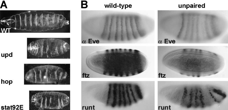
Embryonic effects of JAK pathway mutations. (A) Representative embryonic cuticle phenotypes are shown from wild-type, hopc111 germ-line mutant clone-derived (GLC) embryos, stat92EP1681 GLC embryos, and Df(1)os1A (upd mutant) embryos. The defects seen are similar for all three mutations and are described in the text. (B) The effects of loss of upd on the expression of three pair–rule genes is shown. Wild-type embryos (left) and Df(1)os1A/ Y embryos (right) are compared at cellular blastoderm for presence of Even-skipped (Eve) protein, fushi tarazu (ftz) RNA, and runt RNA. Each shows reduced expression of the fifth stripe in the upd mutant embryos (middle). Variable reductions in other stripes of expression are described in the text (not shown). In all pictures, anterior is to the left. Cuticles are shown in ventral views, whereas stainings are shown in lateral views.

Molecular map of the upd region. The map indicates the position of phages from the chromosomal walk, in kilobases. The positions of alleles that delimit the extent of the upd locus are shown beneath the chromosome, with bold lines indicating deleted DNA. Broken lines indicate uncertainties in the exact position of breakpoints. The position of transcription units is indicated at bottom. The 1-kb transcript has not been precisely mapped and falls within the indicated area. The 2.5-kb RNA is expressed only during late embryogenesis (after 13 hr), and the 1-kb RNA is present maternally, and again late in embryogenesis. The upd candidate transcript, 2.2 kb, is expressed during early zygotic stages (also see Fig. 3). CREB is shown for reference (porc is off the map at right). Proximal is to the right. Identification of single site lesions in two upd alleles, updYM55 and updYC43, confirms the assignment of these mutations to the same complementation group. Because these mutations fail to complement os alleles and only affect the protein encoded by the 2.2-kb transcript (Fig. 4), we conclude that os and upd are separable functions of the same locus. The breakpoints of os4-51-1, osc18, and os109 (all os−, upd+) map at least 13 kb from the 2.2-kb transcript, suggesting that the os phenotype may derive from the disruption of an enhancer that lies far from the 3′ end of the upd transcript.
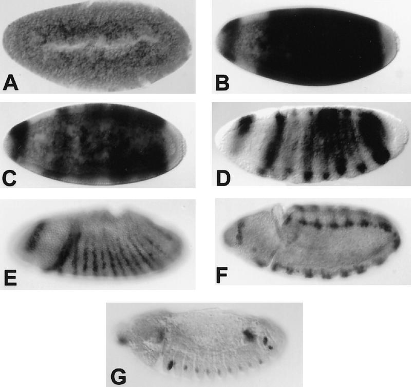
Embryonic expression of the upd transcript. Embryonic expression of the upd RNA is highly dynamic. No maternal product can be detected above background (A), but before cellularization, the RNA is expressed broadly throughout the trunk of the embryo and in a dorsal crescent in the head (B). As cellularization proceeds, expression resolves transiently into seven stripes (C,D). During early gastrulation, 14 stripes of expression appear (E). Later expression is largely restricted to the tracheal pits (F,G). Anterior is to the left and views are primarily lateral.
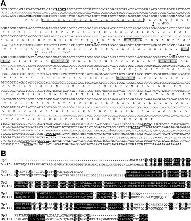
Sequence of upd. (A) The sequence of the upd cDNA (λKZ-GR) is shown. The conceptual ORF is shown below the DNA sequence. The presumed signal sequence is denoted by the open box, putative amino-linked glycosylation signals are indicated by the shaded boxes, and AU-rich sequences implicated in mRNA stability are underlined and bold. Intron positions are indicated by inverted open triangles. (B) The predicted protein sequence of D. melanogaster (Dm) Upd is aligned with the predicted protein sequence of the D. ananassae (Da) Om(1E). Amino acid identities are shown in black boxes.
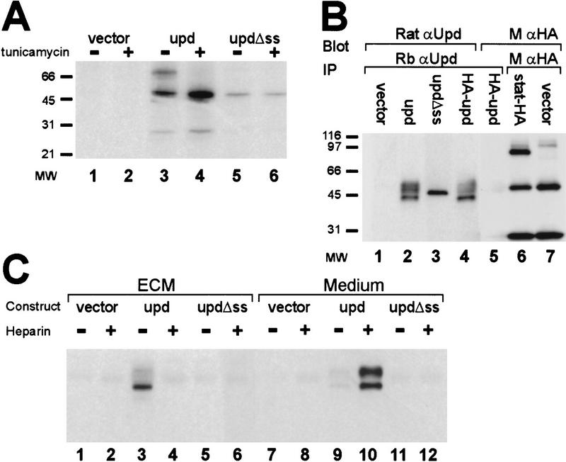
Modification and localization of Upd. (A) The size of Upd protein was determined in the presence and absence of tunicamycin. Transfected constructs are empty vector (lanes 1,2), upd cDNA (lanes 3,4), and an amino-terminal truncation of upd that removes the predicted signal sequence (lanes 5,6). (B) The possible cleavage of Upd at the signal sequence was investigated by use of an amino-terminal HA epitope tag. Cells were transfected with empty vector (lane 1), upd cDNA (lane 2), amino-terminal truncation of upd that removes the signal sequence (lane 3), and upd cDNA tagged with a HA epitope at the amino terminus (lanes 4,5). Protein was immunoprecipitated with rabbit α-upd, electrophoresed, blotted, and detected with rat α-upd (lanes 1–4) or with α-HA (lane 5). A control shows HA antibody detection of HA-tagged Stat92E protein (lane 6) HA expression vector background (lane 7). (C) Cells were transfected with vector alone (lanes 1,2,7,8), upd cDNA (lanes 3,4,9,10), or upd missing the signal sequence (Δss) (lanes 5,6,11, 12). Upd protein was recovered from either the ECM (lanes 1–6) or the medium (lanes 7–12), in the presence or absence of heparin, by immunoprecipitation with rabbit α-upd and detection with rat α-upd.

Activation of Hop by Upd. The level of tyrosine phosphorylation on Hop is shown for Cl.8 cells transfected with upd or cocultured (CC) with S2 cells transfected with upd. Lane 1 shows the endogenous levels of Hop protein (middle), tyrosine-phosphorylated Hop (top), and Upd protein (bottom) in Cl.8 cells that were transfected with vector alone. When transfected with full-length Upd, Hop phosphorylation is stimulated (lane 2), whereas deletion of the Upd signal sequence eliminates Hop activation (lane 3). Similarly, S2 cells transfected with vector alone (lane 4) fail to activate Hop, whereas Cl.8 cells cocultured with S2 cells transfected with full-length Upd (lane 5) display activation of Hop.

Model for JAK pathway activity in embryogenesis. We propose that Upd is the ligand for stimulation of the JAK pathway in early embryonic development. Upd protein is produced in a restricted set of embryonic cells, in which it is glycosylated and secreted, and diffusion is restricted by association with the ECM. Through binding of Upd to a yet unidentified receptor, the Hop JAK is stimulated, resulting in phosphorylation of Stat92E. Ultimately, transcription of specific pair–rule genes, such as eve, is activated (see text for details).
References
-
- Ashburner M. Drosophila: A laboratory manual. Cold Spring Harbor, NY: Cold Spring Harbor Laboratory Press; 1989.
-
- Binari R, Perrimon N. Stripe-specific regulation of pair-rule genes by hopscotch, a putative Jak family tyrosine kinase in Drosophila. Genes & Dev. 1994;8:300–312. - PubMed
-
- Brown NH, Kafatos FC. Functional cDNA libraries from Drosophila embryos. J Mol Biol. 1988;203:425–437. - PubMed
Publication types
MeSH terms
Substances
Associated data
- Actions
Grants and funding
LinkOut - more resources
Full Text Sources
Other Literature Sources
Molecular Biology Databases
Research Materials