. 1998 Nov;66(11):5196-201.
doi: 10.1128/IAI.66.11.5196-5201.1998.

Lipoprotein release by bacteria: potential factor in bacterial pathogenesis

Affiliations

Lipoprotein release by bacteria: potential factor in bacterial pathogenesis

H Zhang et al. Infect Immun. 1998 Nov.

Abstract

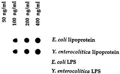
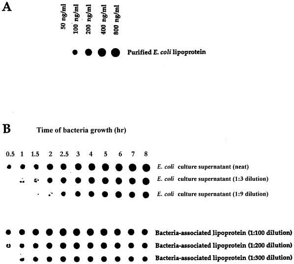
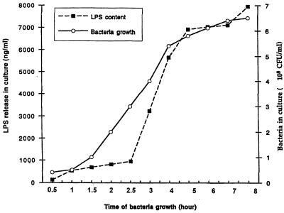
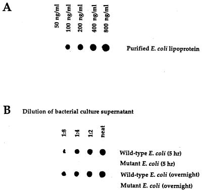
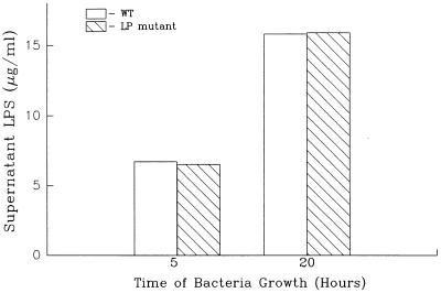
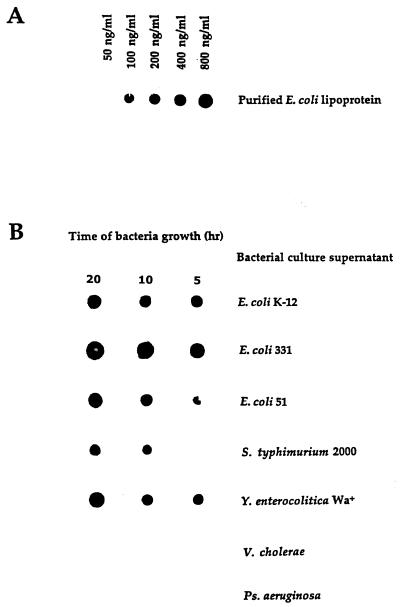
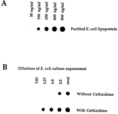
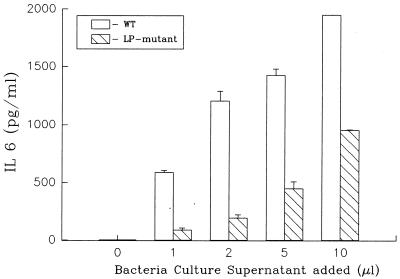
Lipoprotein (LP) is a major component of the outer membrane of bacteria in the family Enterobacteriaceae. LP induces proinflammatory cytokine production in macrophages and lethal shock in LPS-responsive and -nonresponsive mice. In this study, the release of LP from growing bacteria was investigated by immuno-dot blot analysis. An immuno-dot blot assay that could detect LP at levels as low as 100 ng/ml was developed. By using this assay, significant levels of LP were detected in culture supernatants of growing Escherichia coli cells. During mid-logarithmic growth, approximately 1 to 1.5 microgram of LP per ml was detected in culture supernatants from E. coli. In contrast, these culture supernatants contained 5 to 6 microgram/ml of lipopolysaccharide (LPS). LP release was not unique to E. coli. Salmonella typhimurium, Yersinia enterocolitica, and two pathogenic E. coli strains also released LP during in vitro growth. Treatment of bacteria with the antibiotic ceftazidime significantly enhanced LP release. Culture supernatants from 5-h cultures of E. coli were shown to induce in vitro production of interleukin-6 (IL-6) by macrophages obtained from LPS-nonresponsive C3H/HeJ mice. In contrast, culture supernatants from an E. coli LP-deletion mutant were significantly less efficient at inducing IL-6 production in C3H/HeJ macrophages. These results suggest, for the first time, that LP is released from growing bacteria and that this released LP may play an important role in the induction of cytokine production and pathologic changes associated with gram-negative bacterial infections.


Figures

FIG. 1
FIG. 1
Detection of LP by immuno-dot blot analysis. Purified E. coli LP and LPS in BHI broth were loaded onto nitrocellulose membranes and processed as described in Materials and Methods. Data are from one experiment that was representative of three separate experiments.
FIG. 2
FIG. 2
LP can be detected in culture supernatants obtained from growing E. coli. E. coli K-12 was grown in BHI medium. At various times, 5-ml samples were collected and assessed for one of the following: (i) number of bacteria (see Fig. 3), (ii) amount of LPS by the LAL assay (see Fig. 3), or (iii) amount of LP by the dot blot assay (Fig. 1). (A) Dot blot assay involving purified LP in BHI medium, was performed at the same time as the dot blot assay in panel B. (B) Dot blot assay of the amount of LP present in culture supernatants obtained at the times indicated. Culture supernatants were assessed undiluted or after being diluted 1:3 or 1:9 in PBS. Data are from one experiment that was representative of five separate experiments.
FIG. 3
FIG. 3
Bacterial growth and LPS release by E. coli K-12. Bacterial growth and LPS contents of bacterial culture supernatants at different times after culture initiation were determined as described in the legend to Fig. 2 and in Materials and Methods. Data are from one experiment that was representative of two separate experiments.
FIG. 4
FIG. 4
LP is not detectable in culture supernatants obtained from an E. coli LP deletion mutant. E. coli K-12 and an E. coli mutant (K-12 strain JE5055) that had a deletion of the lpp gene were grown, and the culture supernatant was assessed as described in the legend to Fig. 2. Data are from one experiment that was representative of two separate experiments.
FIG. 5
FIG. 5
An E. coli LP deletion mutant releases normal levels of LPS during growth. The LPS contents of culture supernatants obtained from wild-type E. coli and an E. coli LP deletion mutant were assessed as described in the legend to Fig. 3. There was no statistical difference (P > 0.05) between the amounts of LPS contained in culture supernatant obtained from wild-type E. coli and the LP deletion mutant. Data are from one experiment that was representative of three separate experiments.
FIG. 6
FIG. 6
LP is present in culture supernatants obtained from different bacteria from the family Enterobacteriaceae. The LP content in culture supernatants obtained from different bacteria was assessed as described in the legend to Fig. 2. Data are from one experiment that was representative of three separate experiments.
FIG. 7
FIG. 7
E. coli treated with ceftazidime shows an enhanced release of LP into culture supernatants. Culture supernatants obtained from untreated E. coli and E. coli treated with ceftazidime were assessed for levels of LP. The culture conditions and ceftazidime treatment are described in Materials and Methods. Data are from one experiment that was representative of three separate experiments.
FIG. 8
FIG. 8
Supernatants obtained from an E. coli LP deletion mutant are less efficient at inducing IL-6 production in macrophages than are supernatants obtained from wild-type E. coli. Peritoneal exudate macrophages were obtained from C3H/HeJ mice and exposed to different concentrations of culture supernatant obtained from wild-type E. coli and an E. coli LP deletion mutant. The supernatants obtained from wild-type and mutant E. coli had equivalent levels of LPS (wild-type, 6.4 μg/ml; mutant, 6.1 μg/ml). Macrophage culture supernatants were collected after 8 h of culture and assessed for IL-6 content by enzyme-linked immunosorbent assay as described in Materials and Methods. Levels of IL-6 in supernatant obtained from macrophages stimulated with any of the different concentrations of culture supernatant obtained from the E. coli LP deletion mutant were statistically different (P < 0.01) from levels of IL-6 in supernatant obtained from macrophages stimulated with culture supernatant obtained from wild-type E. coli. Data are from one experiment that was representative of three separate experiments.

Similar articles

Cited by

References

    1. Abernathy R S, Spink W W. Studies with Brucella endotoxin in humans: significance of susceptibility to endotoxin in the pathogenesis of brucellosis. J Clin Invest. 1958;37:219–231. - PMC - PubMed
    1. Andersen B M, Solberg O. The endotoxin-liberating effect of antibiotics on meningococci in vitro. Acta Pathol Microbiol Scand Sect B. 1980;88:231–236. - PubMed
    1. Berkowitz F E, Vallabh P, Altman D I, Diamantes F, VanWyk H J, Stroucken J M. Jarisch-Herxheimer reaction in meningococcal meningitis. Am J Dis Child. 1983;137:599–603. - PubMed
    1. Bessler W G, Resch K, Hancock E, Hantke K. Induction of lymphocyte-proliferation and membrane changes by lipoprotein derivatives of the lipoprotein from the outer membrane of E. coli. Z Immunitaetsforsch. 1977;153:11–19. - PubMed
    1. Bessler W G, Ottenbreit B P. Studies on the mitogenic principal of lipoprotein from the outer membrane of E. coli. Biochem Biophys Res Commun. 1977;76:239–246. - PubMed

LinkOut - more resources