Comparative Study
. 1998 Nov;66(11):5252-9.
doi: 10.1128/IAI.66.11.5252-5259.1998.

Disruption of an internal membrane-spanning region in Shiga toxin 1 reduces cytotoxicity

Affiliations
Comparative Study

Disruption of an internal membrane-spanning region in Shiga toxin 1 reduces cytotoxicity

M L Suhan et al. Infect Immun. 1998 Nov.

Abstract

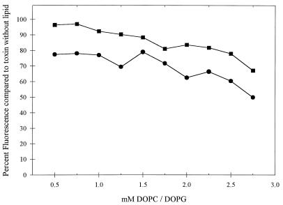
Shiga toxin type 1 (Stx1) belongs to the Shiga family of bipartite AB toxins that inactivate eukaryotic 60S ribosomes. The A subunit of Stxs are N-glycosidases that share structural and functional features in their catalytic center and in an internal hydrophobic region that shows strong transmembrane propensity. Both features are conserved in ricin and other ribosomal inactivating proteins. During eukaryotic cell intoxication, holotoxin likely moves retrograde from the Golgi apparatus to the endoplasmic reticulum. The hydrophobic region, spanning residues I224 through N241 in the Stx1 A subunit (Stx1A), was hypothesized to participate in toxin translocation across internal target cell membranes. The TMpred computer program was used to design a series of site-specific mutations in this hydrophobic region that disrupt transmembrane propensity to various degrees. Mutations were synthesized by PCR overlap extension and confirmed by DNA sequencing. Mutants StxAF226Y, A231D, G234E, and A231D-G234E and wild-type Stx1A were expressed in Escherichia coli SY327 and purified by dye-ligand affinity chromatography. All of the mutant toxins were similar to wild-type Stx1A in enzymatic activity, as determined by inhibition of cell-free protein synthesis, and in susceptibility to trypsin digestion. Purified mutant or wild-type Stx1A combined with Stx1B subunits in vitro to form a holotoxin, as determined by native polyacrylamide gel electrophoresis immunoblotting. StxA mutant A231D-G234E, predicted to abolish transmembrane propensity, was 225-fold less cytotoxic to cultured Vero cells than were the wild-type toxin and the other mutant toxins which retained some transmembrane potential. Furthermore, compared to wild-type Stx1A, A231D-G234E Stx1A was less able to interact with synthetic lipid vesicles, as determined by analysis of tryptophan fluorescence for each toxin in the presence of increasing concentrations of lipid membrane vesicles. These results provide evidence that this conserved internal hydrophobic motif contributes to Stx1 translocation in eukaryotic cells.


Figures

FIG. 1
FIG. 1
Inhibition of protein synthesis by wild-type or mutant Stx1As. Rabbit reticulocyte lysates were preincubated with periplasmic extracts containing wild-type Stx1A (○) or F226Y (▵), A231D (▿), G234E (◊), or A231D-G234E (□) mutant Stx1A at the dilutions indicated. Background activity (omitted mRNA) was subtracted from each value, and percent protein synthesis was determined by comparison to the control reaction preincubated with buffer only. Datum points are the means in one representative experiment ± the SE.
FIG. 2
FIG. 2
Immunoblot of wild-type (wt) or mutant Stx1A following trypsin digestion. Periplasmic extracts of mutant and wild-type Stx1As were incubated at 37°C for 15 min with no trypsin (lanes 2, 6, 10, 14, and 18) or with trypsin at 0.05 (lanes 3, 7, 11, 15, and 19), 0.5 (lanes 4, 8, 12, 16, and 20), or 5 ng/ml (lanes 5, 9, 13, 17, and 21) ng/ml. Lanes: 1, pUC19ΔH (vector-only control); 2 to 5, wild-type Stx1A; 6 to 9, F226Y Stx1A; 10 to 13, A231D Stx1A; 14 to 17, G234E Stx1A; 18 to 21, A231D-G234E Stx1A (AG). The locations of Stx1A and the two primary trypsin degradation products (★) are shown on the left. The positions of the following molecular size standards are indicated on the right (from top to bottom: 95.5, 55, 43, 29, 18.4, and 12.4 kDa).
FIG. 3
FIG. 3
Wild-type and mutant holotoxins in native-PAGE immunoblots with stacking and resolving gels (indicated at the left) developed with anti-Stx1A serum (A) or an anti-Stx1B monoclonal antibody (B). Lanes: 1, Stx1B; 2, wild-type Stx1A; 3, wild-type Stx1A associated with Stx1B; 4, G234E Stx1A; 5, G234E Stx1A plus Stx1B; 6, A231D Stx1A; 7, A231D Stx1A plus Stx1B; 8, F226Y Stx1A; 9, F226Y Stx1A plus Stx1B; 10, A231D-G234E Stx1A; 11, A231D-G234E Stx1A plus Stx1B. The positions of the holotoxin and Stx1B and Stx1A monomers are indicated at the right.
FIG. 4
FIG. 4
Cytotoxicity of wild-type and mutant holotoxins. Holotoxins or Stx1As, at the concentrations indicated, were incubated with Vero cells for 72 h. Cell viability was determined by microscopic analysis and expressed as a percentage. The datum points are the means ± the SE of one representative experiment. Holotoxins are indicated by shaded symbols, and Stx1As are indicated by open symbols as follows: wild type, • and ○; F226Y, ▴ and ▵; A231D, ▾ and ▿; G234E, ⧫ and ◊; A231D-G234E alone, ■ and □.
FIG. 5
FIG. 5
Interaction of wild-type Stx1A and A231D-G234E Stx1A with synthetic lipid vesicles. Purified wild-type (•) or A231D-G234E (■) A subunits at 18 μg/ml were combined with increasing concentrations of 70% DOPC–30% DOPG (mol/mol) vesicles. Fluorescence intensities were obtained at an excitation wavelength of 290 nm and an emmission wavelength of 323 nm and expressed as a percentage of that of the toxin without lipid vesicles. The results of one representative experiment are shown.

Similar articles

Cited by

References

    1. Acheson D W K, Donohue-Rolfe A, Keusch G T. The family of Shiga and Shiga-like toxins. In: Alouf J E, Freer J H, editors. Sourcebook of bacterial toxins. 1st ed. Vol. 1. London, England: Academic Press, Inc.; 1991. pp. 415–433.
    1. Austin P R, Hovde C J. Purification of recombinant Shiga-like toxin type I B subunit. Protein Expr Purif. 1995;6:771–779. - PubMed
    1. Austin P R, Jablonski P E, Bohach G A, Dunker A K, Hovde C J. Evidence that the A2 fragment of Shiga-like toxin type I is required for holotoxin integrity. Infect Immun. 1994;62:1768–1775. - PMC - PubMed
    1. Burgess B J, Roberts L M. Proteolytic cleavage at arginine residues within the hydrophilic disulphide loop of the Escherichia coli Shiga-like toxin I A subunit is not essential for cytotoxicity. Mol Microbiol. 1993;10:171–179. - PubMed
    1. Calderwood S B, Auclair F, Donohue-Rolfe A, Keusch G T, Mekalanos J J. Nucleotide sequence of the Shiga-like toxin genes of Escherichia coli. Proc Natl Acad Sci USA. 1987;84:4364–4368. - PMC - PubMed

Publication types

LinkOut - more resources