Comparative Study
. 1998 Nov;66(11):5372-8.
doi: 10.1128/IAI.66.11.5372-5378.1998.

Mechanisms involved in the pathogenesis of sepsis are not necessarily reflected by in vitro cell activation studies

Affiliations
Comparative Study

Mechanisms involved in the pathogenesis of sepsis are not necessarily reflected by in vitro cell activation studies

C R Amura et al. Infect Immun. 1998 Nov.

Abstract

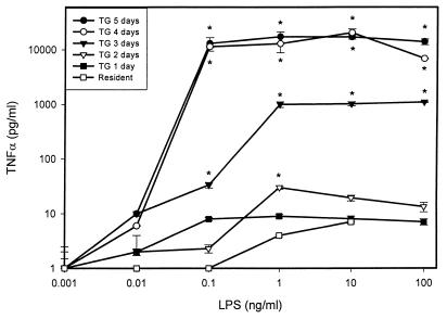
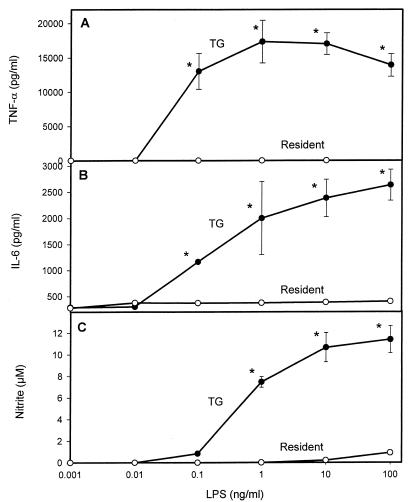
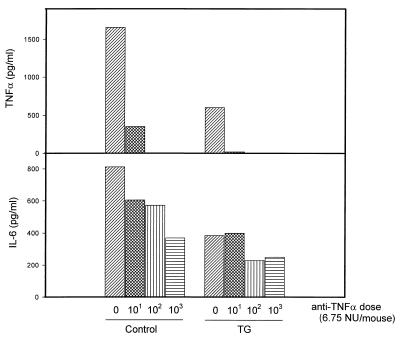
It is thought that lipopolysaccharide (LPS) from gram-negative bacteria contributes significantly to the pathogenesis of septic shock. In vitro studies to address the mechanisms involved in this process have often investigated human monocytes or mouse macrophages, since these cells produce many of the mediators found in septic patients. Targeting of these mediators, especially tumor necrosis factor alpha (TNF-alpha), has been pursued as a means of reducing mortality in sepsis. Two experimental approaches were designed to test the assumption that in vitro studies with macrophages accurately predict in vivo mechanisms of LPS pathogenesis. In the first approach, advantage was taken of the fact that on consecutive days after injection of thioglycolate into mice, increased numbers of macrophages could be harvested from the peritoneum. These cells manifested markedly enhanced levels of in vitro TNF-alpha, interleukin 6 (IL-6), and nitric oxide production in response to LPS. In D-galactosamine-sensitized mice, however, thioglycolate treatment significantly decreased mortality due to LPS, as well as levels of circulating TNF-alpha and IL-6. Anti-TNF-alpha treatment confirmed this cytokine's role in the observed lethality. In a second experimental approach, we compared the mouse macrophage-stimulating potencies of different LPS preparations with their lethalities to mice. In these studies, the in vitro macrophage-stimulating profiles presented by rough-LPS and smooth-LPS preparations were the reverse of their relative lethal potencies in vivo. In conclusion, peritoneal macrophages appear not to be the major cells responsible for the overall host response during endotoxic shock. These findings underscore the importance of verifying the correlation of in vivo systems with in vitro systems when attributing specific functions to a cell type.


Figures

FIG. 1
FIG. 1
LPS-stimulated TNF-α production by mouse macrophages harvested at different days after thioglycolate (TG) injection. Peritoneal exudate was harvested at various times following thioglycolate administration and treated in vitro with increasing concentrations of R-LPS. Supernatants were assessed for the presence of TNF-α after 18 h. TNF-α was quantitated as the extent of cytotoxicity for the fibroblast cell line L929 as described in Materials and Methods. Results represent averages of triplicate determinations ± standard errors of the means (SEM) from one representative experiment, repeated four times under equivalent conditions. ∗, P < 0.05 (by Student’s t test) with respect to the group treated with LPS alone.
FIG. 2
FIG. 2
Effects of thioglycolate on mediator secretion by LPS-stimulated mouse macrophages. Cells were harvested at 0 (resident) or 5 (TG) days after thioglycolate injection. TNF-α (A), IL-6 (B), and nitric oxide (C) production after 18 h of R-LPS stimulation was analyzed in supernatants as described in Materials and Methods. ∗, P < 0.05 (by Student’s t test) in comparison to the day 0 group.
FIG. 3
FIG. 3
LPS-induced mortality in thioglycolate (TG)-pretreated mice. Mice were pretreated either with thioglycolate or with saline. Five days later, all animals received graded doses of R-LPS and 20 mg of d-GalN/g of body weight. Lethality was recorded 14 h after R-LPS challenge in d-GalN-sensitized mice. Cumulative data were depicted according to Reed and Muench’s method (31). ∗, statistically significant (P < 0.05 by Fisher’s exact test) compared with the respective R-LPS dose in thioglycolate-treated animals. The number of mice in each group is given in parentheses.
FIG. 4
FIG. 4
Effects of thioglycolate (TG) pretreatment on circulating TNF-α (A) or IL-6 (B) levels induced by LPS in d-GalN-sensitized mice. Mice were treated as described in the text, and cytokine levels in serum collected 1.5 h after R-LPS challenge were determined by ELISA. Cytokine levels in sera collected 4 h after stimulation were below the detection limit. Results from one representative experiment, repeated five times, are depicted.
FIG. 5
FIG. 5
Effects of anti-TNF-α Ab treatment on LPS-induced mortality in thioglycolate (TG)- versus saline-pretreated, d-GalN-sensitized mice (n = 8 per group). Various amounts of anti-TNF-α neutralizing Abs were administered to mice 3 h before the administration of d-GalN plus R-LPS (5 μg/kg). Mortality was recorded after 14 h, and cumulative data were depicted according to the method described by Reed and Muench (31). ∗, P < 0.05 (by Fisher’s exact test).
FIG. 6
FIG. 6
Effect of anti TNF-α Ab on TNF-α and IL-6 production induced by LPS in d-GalN-sensitized mice pretreated with thioglycolate (TG) or saline (control). Mice were treated as described in the text, and cytokine levels in serum were evaluated by ELISA. Results from one representative experiment, repeated five times with similar results, are depicted.
FIG. 7
FIG. 7
(A) In vitro TNF-α response induced by S-LPS or R-LPS (Ra-LPS) in peritoneal-exudate macrophages. TNF-α was quantitated by cytotoxicity toward L929 cells, as described above. Results are averages of triplicate determinations ± SEM from one representative experiment (partially reproduced from reference 45) with permission of the publisher). (B) (Left) Mortality rates induced by different preparations of LPS (S-LPS and Ra-LPS) in d-GalN-sensitized mice. Animal mortality was recorded 14 h after challenge, and cumulative data were depicted according to the method described by Reed and Muench (31). ∗, P < 0.05 by Fisher’s exact test. (Right) Mortality rates induced by S-LPS and Ra-LPS in normal mice, 24 h after LPS challenge.

Similar articles

Cited by

References

    1. Abraham E, Wunderink R, Silverman H, Perl T M, Nasraway S, Levy H, Bone R, Wenzel R P, Balk R, Allred R, et al. Efficacy and safety of monoclonal antibodies to tumor necrosis factor alpha in patients with sepsis syndrome. A randomized, controlled, double-blind, multicenter clinical trial. TNF-alpha Mab sepsis group. JAMA. 1997;273:934–941. - PubMed
    1. Amura C R, Chen L C, Hirohashi N, Lei M G, Morrison D C. Two functionally independent pathways for the LPS-dependent activation of mouse peritoneal macrophages. J Immunol. 1997;159:5079–5083. - PubMed
    1. Amura C R, Fontan P A, Sanjuan N, Sordelli D O. The effect of treatment with interleukin-1 and tumor necrosis factor on Pseudomonas aeruginosa lung infection in a granulocytopenic mouse model. Clin Immunol Immunopathol. 1994;73:261–266. - PubMed
    1. Beutler B, Milsark I W, Cerami A. Passive immunization against cachectin/tumor necrosis factor protects mice from lethal effects of endotoxin. Science. 1985;229:869–871. - PubMed
    1. Bone R C. Sepsis syndrome. New insights into its pathogenesis and treatment. Infect Dis Clin N Am. 1991;5:793–805. - PubMed

Publication types

MeSH terms