

CD4(+) T-lymphocyte and immunoglobulin G2 responses in calves immunized with Anaplasma marginale outer membranes and protected against homologous challenge
- PMID: 9784551
- PMCID: PMC108677
- DOI: 10.1128/IAI.66.11.5406-5413.1998
CD4(+) T-lymphocyte and immunoglobulin G2 responses in calves immunized with Anaplasma marginale outer membranes and protected against homologous challenge
Abstract
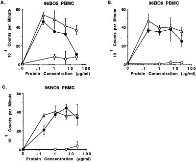
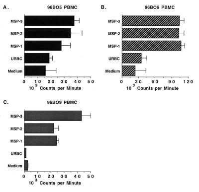
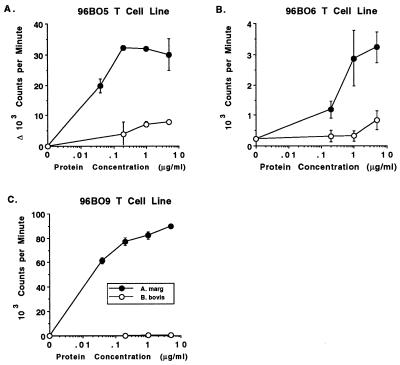
Protective immunity against the ehrlichial pathogen Anaplasma marginale has been hypothesized to require induction of immunoglobulin G2 (IgG2) antibody against outer membrane protein epitopes and coordinated activation of macrophages for phagocytosis and killing. In the present study, cell-mediated immune responses, including induction of IgG isotype switching, were characterized in calves immunized with purified outer membranes of the Florida strain of A. marginale. Importantly, these calves were subsequently shown to be protected upon experimental challenge with the Florida strain, and calves which developed the highest IgG2 titers were completely protected against infection. Peripheral blood mononuclear cells (PBMC) obtained after immunization proliferated strongly in response to both whole A. marginale homogenates and purified outer membranes, and this responsiveness persisted until the time of challenge. Responding cells were shown to be CD4(+) T cells, and CD4(+) T-cell lines cultured for 2 to 4 weeks also proliferated specifically in response to A. marginale and produced high titers of gamma interferon. The helper T-cell response included recognition of conserved epitopes, as PBMC proliferation was stimulated by the homologous Florida strain, four genetically distinct A. marginale strains, and Anaplasma ovis. The outer membrane proteins stimulating the PBMC responses in protected calves included major surface proteins (MSPs) MSP-1, MSP-2, and MSP-3, which were previously shown to induce partial protection against infection. These studies demonstrate, for the first time, potent helper T-cell responses in cattle protectively immunized with outer membranes against A. marginale challenge and identify three MSPs that are recognized by immune T cells. These experiments provide the basis for subsequent identification of the helper T-cell epitopes on MSP-1, MSP-2, and MSP-3 that are needed to evoke anamnestic antibody and effector T-cell responses elicited by protein or nucleic acid immunization.
Figures

Similar articles
-
The repertoire of Anaplasma marginale antigens recognized by CD4(+) T-lymphocyte clones from protectively immunized cattle is diverse and includes major surface protein 2 (MSP-2) and MSP-3.Infect Immun. 1998 Nov;66(11):5414-22. doi: 10.1128/IAI.66.11.5414-5422.1998. Infect Immun. 1998. PMID: 9784552 Free PMC article.
-
CD4(+) T lymphocytes from calves immunized with Anaplasma marginale major surface protein 1 (MSP1), a heteromeric complex of MSP1a and MSP1b, preferentially recognize the MSP1a carboxyl terminus that is conserved among strains.Infect Immun. 2001 Nov;69(11):6853-62. doi: 10.1128/IAI.69.11.6853-6862.2001. Infect Immun. 2001. PMID: 11598059 Free PMC article.
-
Immunogenicity of Anaplasma marginale type IV secretion system proteins in a protective outer membrane vaccine.Infect Immun. 2007 May;75(5):2333-42. doi: 10.1128/IAI.00061-07. Epub 2007 Mar 5. Infect Immun. 2007. PMID: 17339347 Free PMC article.
-
Strategies to interrupt the development of Anaplasma marginale in its tick vector. The effect of bovine-derived antibodies.Ann N Y Acad Sci. 1996 Jul 23;791:157-65. doi: 10.1111/j.1749-6632.1996.tb53522.x. Ann N Y Acad Sci. 1996. PMID: 8784497 Review.
-
Antigenic variation in the persistence and transmission of the ehrlichia Anaplasma marginale.Microbes Infect. 2000 Feb;2(2):167-76. doi: 10.1016/s1286-4579(00)00271-9. Microbes Infect. 2000. PMID: 10742689 Review.
Cited by
-
Protective immunity induced by immunization with a live, cultured Anaplasma marginale strain.Vaccine. 2013 Aug 2;31(35):3617-22. doi: 10.1016/j.vaccine.2013.04.069. Epub 2013 May 9. Vaccine. 2013. PMID: 23664994 Free PMC article.
-
Epitope-based vaccines with the Anaplasma marginale MSP1a functional motif induce a balanced humoral and cellular immune response in mice.PLoS One. 2013 Apr 8;8(4):e60311. doi: 10.1371/journal.pone.0060311. Print 2013. PLoS One. 2013. PMID: 23579784 Free PMC article.
-
A hybrid protein containing MSP1a repeats and Omp7, Omp8 and Omp9 epitopes protect immunized BALB/c mice against anaplasmosis.Vet Res. 2018 Jan 19;49(1):6. doi: 10.1186/s13567-018-0503-4. Vet Res. 2018. PMID: 29351812 Free PMC article.
-
Immune Response to Tick-Borne Hemoparasites: Host Adaptive Immune Response Mechanisms as Potential Targets for Therapies and Vaccines.Int J Mol Sci. 2020 Nov 20;21(22):8813. doi: 10.3390/ijms21228813. Int J Mol Sci. 2020. PMID: 33233869 Free PMC article. Review.
-
A review of bovine anaplasmosis (Anaplasma marginale) with emphasis on epidemiology and diagnostic testing.J Vet Diagn Invest. 2025 Jul;37(4):517-538. doi: 10.1177/10406387251324180. Epub 2025 Mar 28. J Vet Diagn Invest. 2025. PMID: 40156087 Free PMC article. Review.
References
-
- Adler H, Peterhans E, Nicolet J, Jungi T W. Inducible l-arginine-dependent nitric oxide synthase activity in bone marrow-derived macrophages. Biochem Biophys Res Commun. 1994;198:510–515. - PubMed
-
- Alderink F J, Dietrich R. Anaplasmosis in Texas: epidemiologic and economic data from a questionnaire survey. In: Hidalgo R J, Jones E W, editors. Proceedings of the Seventh National Anaplasmosis Conference. Mississippi State, Miss: Mississippi State University Press; 1981. pp. 27–44.
Publication types
MeSH terms
Substances
LinkOut - more resources
Full Text Sources
Medical
Research Materials
