

The repertoire of Anaplasma marginale antigens recognized by CD4(+) T-lymphocyte clones from protectively immunized cattle is diverse and includes major surface protein 2 (MSP-2) and MSP-3
- PMID: 9784552
- PMCID: PMC108678
- DOI: 10.1128/IAI.66.11.5414-5422.1998
The repertoire of Anaplasma marginale antigens recognized by CD4(+) T-lymphocyte clones from protectively immunized cattle is diverse and includes major surface protein 2 (MSP-2) and MSP-3
Abstract
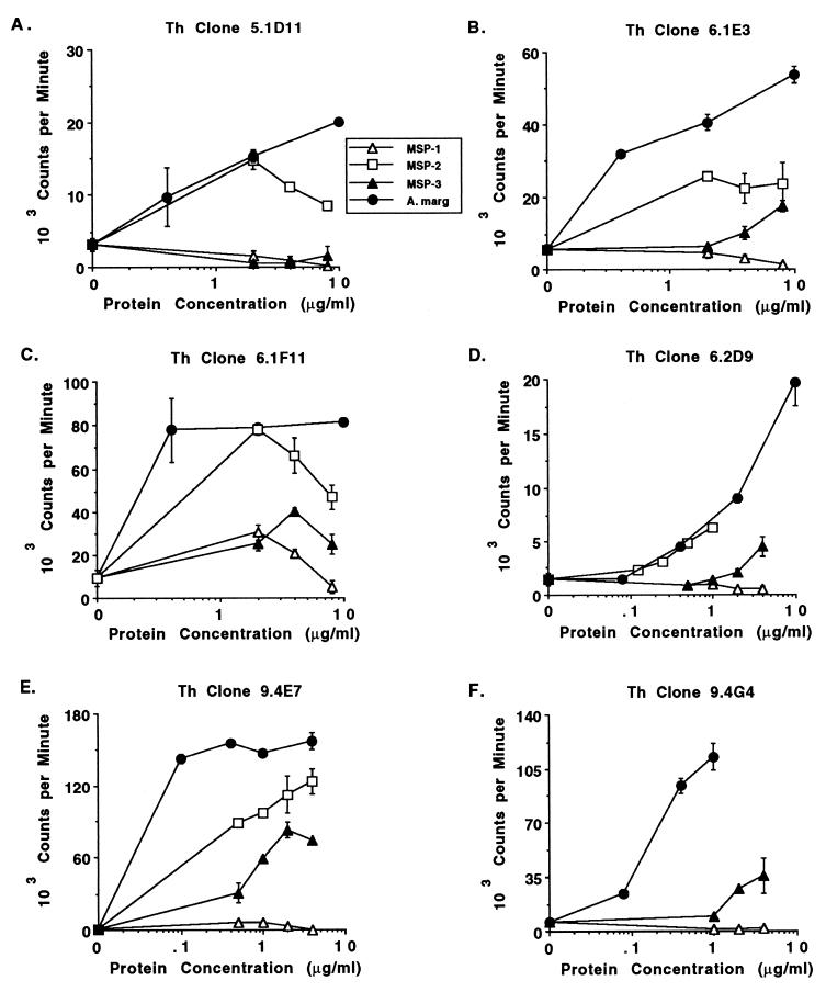
Major surface proteins of Anaplasma marginale are vaccine candidates. We recently demonstrated that immunization of calves with outer membranes of the Florida strain of A. marginale resulted in protective immunity that correlated with a memory CD4(+) T-lymphocyte response specific for major surface protein 1 (MSP-1), MSP-2, and MSP-3 (W. C. Brown, V. Shkap, D. Zhu, T. C. McGuire, W. Tuo, T. F. McElwain, and G. H. Palmer, Infect. Immun. 66:5406-5413, 1998). As immunogens, these proteins have been shown to induce complete or partial protection against homologous challenge. To further define the T helper (Th) cell response to these and other A. marginale antigens and to determine conservation of Th cell epitopes among genetically distinct A. marginale strains, Th cell clones obtained prior to challenge from three immunized calves were characterized for antigen-specific responses. Nine distinct antigenic profiles were defined by 11 Th cell clones derived by stimulation with the Florida strain. Several clones responded to MSP-2, MSP-3, or both. All of these MSP-2- or MSP-3-specific clones and the majority of other clones that did not respond to MSPs recognized all bovine blood-passaged strains of A. marginale. These results demonstrate conservation of certain Th cell epitopes between MSP-2 and MSP-3 and show that Th cell epitopes in MSP-2, MSP-3, and undefined antigens are conserved among strains of A. marginale. Of seven clones that responded to the blood-passaged Virginia strain, two did not recognize antigen prepared from this strain cultured in tick cells, suggesting differences in the antigenic composition between these stages. Analysis of the cytokines expressed by the Th cells revealed that all clones expressed gamma interferon and tumor necrosis factor alpha, and most coexpressed interleukin-4. Our results provide a rationale for identifying Th cell epitopes conserved among different strains of A. marginale for inclusion in a nucleic acid or recombinant protein vaccine.
Figures
References
-
- Adler H, Peterhans E, Nicolet J, Jungi T W. Inducible l-arginine-dependent nitric oxide synthase activity in bone marrow-derived macrophages. Biochem Biophys Res Commun. 1994;198:510–515. - PubMed
Publication types
MeSH terms
Substances
LinkOut - more resources
Full Text Sources
Other Literature Sources
Medical
Research Materials
