. 1998 Nov;66(11):5423-32.
doi: 10.1128/IAI.66.11.5423-5432.1998.

DNA and a CpG oligonucleotide derived from Babesia bovis are mitogenic for bovine B cells

Affiliations

DNA and a CpG oligonucleotide derived from Babesia bovis are mitogenic for bovine B cells

W C Brown et al. Infect Immun. 1998 Nov.

Abstract

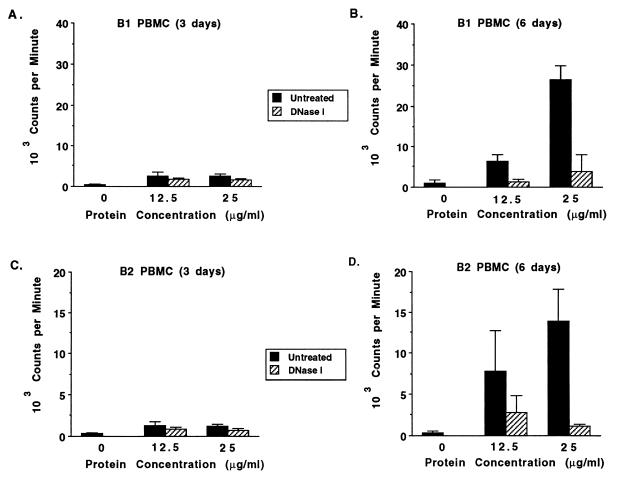
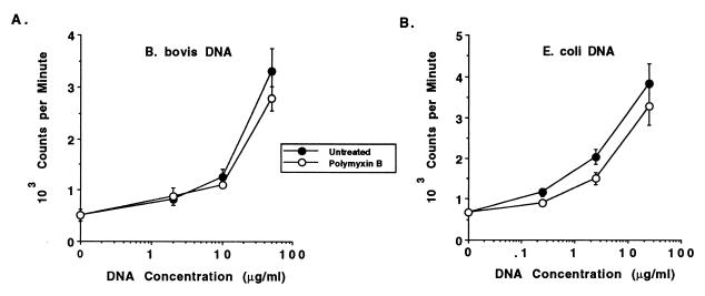
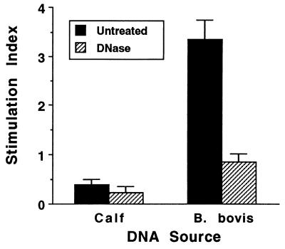
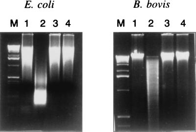
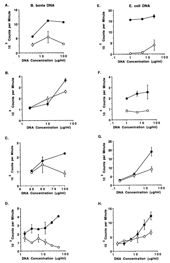
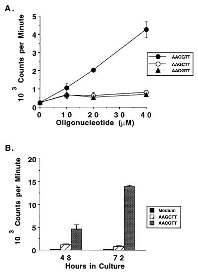
DNAs from bacteria and variety of nonvertebrate organisms, including nematodes, mollusks, yeasts, and insects, cause polyclonal activation of murine B lymphocytes. Similar studies have not been reported for bovine B cells, and to date no studies have reported mitogenic properties of protozoal DNA for any species. However, we and others have observed that protozoal parasite antigens can induce the proliferation of lymphocytes from nonexposed donors. Extending these studies, we now show that the mitogenic property of protozoal antigen preparations is in part attributable to parasite DNA and that Babesia bovis DNA is directly mitogenic for bovine B cells. DNase treatment of B. bovis extracts abrogated B. bovis-induced proliferation of peripheral blood mononuclear cells from nonexposed cattle. Like DNAs from other organisms that were mitogenic for murine B cells, B. bovis DNA is largely nonmethylated and induced a dose-dependent proliferation of bovine B cells, which was reduced upon methylation. Furthermore, B. bovis and E. coli DNAs enhanced immunoglobulin secretion by cultured B cells, inducing moderate increases in immunoglobulin G1 and stronger increases in immunoglobulin G2. Because certain nonmethylated CpG motifs present in bacterial DNA are known to stimulate proliferation of murine and human B cells, an 11-kb fragment of B. bovis DNA was analyzed for CG dinucleotide content and for the presence of known immunostimulatory sequences (ISS) centered on a CG motif. The frequency of CG dinucleotides was approximately one-half of the expected frequency, and several CpG hexameric sequences with known activity for murine B cells were identified. An oligodeoxynucleotide containing one of these ISS (AACGTT), which is present within the rhoptry-associated protein-1 (rap-1) open reading frame, was shown to stimulate B-cell proliferation. These ISS may be involved in host immune modulation during protozoal infection and may be useful as vaccine adjuvants.


Figures

FIG. 1
FIG. 1
Nonspecific stimulation by unfractionated B. bovis merozoite antigen is DNase sensitive. PBMC from two calves never exposed to babesial parasites were cultured for 3 (A and C) or 6 (B and D) days with medium alone (0) or 12.5 or 25 μg of B. bovis CM antigen per ml that was untreated (solid bars) or DNase I treated (striped bars), and proliferation was determined. Results are expressed as the mean cpm ± 1 SD of triplicate cultures.
FIG. 2
FIG. 2
Dose-dependent proliferation of B cells to DNA prepared from B. bovis or E. coli is not inhibited by polymyxin B. Negatively selected B cells were cultured for 3 days with medium or 2, 10, or 50 μg of B. bovis DNA per ml (A) or 0.25, 2.5, or 25 μg of E. coli DNA per ml (B) without (solid circles) or with (open circles) 10 μg of polymyxin B sulfate per ml. Results are presented as the mean cpm ± 1 SD of duplicate cultures.
FIG. 3
FIG. 3
Stimulation of B-cell proliferation by B. bovis DNA is abrogated by DNase treatment. Negatively selected B cells were cultured in duplicate wells for 3 days with medium or 50 μg of either calf thymus DNA or B. bovis DNA per ml that was untreated (solid bars) or treated (striped bars) with DNase I. Results are presented as the SI and represent the mean ± 1 SD of three independent experiments.
FIG. 4
FIG. 4
Resistance of methylated B. bovis DNA to cleavage by HpaII. DNAs prepared from E. coli or B. bovis were treated with SssI methylase and compared with untreated DNA for sensitivity to HpaII digestion. DNA was visualized after electrophoresis on ethidium bromide-stained agarose gels. Lanes: M, 1-kb markers; 1, untreated DNA; 2, untreated DNA incubated with HpaII; 3, methylated DNA; 4, methylated DNA incubated with HpaII.
FIG. 5
FIG. 5
Effect of methylating B. bovis and E. coli DNA with CpG methylase on stimulating B-cell proliferation. DNA was extracted from either B. bovis (A to D) or E. coli (E to H) and treated with SssI methylase as described in the text. Untreated (solid circles) and methylated (open circles) DNAs were then assayed for dose-dependent proliferation with 2 × 10⁵ B cells purified by positive (panels A, D, and H) or negative (panels B, C, E, F, and G) selection and cultured for 3 days with 0.15 to 100 μg of B. bovis DNA or 0.15 to 50 μg of E. coli DNA per ml. Results are presented as the mean cpm ± 1 SD of duplicate or triplicate cultures.
FIG. 6
FIG. 6
Dose dependency and time course of the proliferative response of B cells to the CpG phosphodiester oligonucleotide derived from B. bovis. B cells purified by negative selection were tested for proliferation in a 3-day assay with medium or 10, 20, and 40 μM concentrations of either the B. bovis RAP-1-derived ISS oligonucleotide TAAAAACGTTAC (solid circles) or the control oligonucleotides TAAAAAGCTTAC (open circles) and TAAAAAQGTTAC (solid triangles) (A). In a different experiment, negatively selected B cells were cultured for either 48 or 72 h and radiolabeled for the last 6 h of culture with medium (solid bars) or 40 μM TAAAAACGTTAC (gray bars) or TAAAAAGCTTAC (striped bars). The CG dinucleotide is indicated with an underscore (B).

Similar articles

Cited by

References

    1. Ballas Z K, Rasmussen W L, Krieg A M. Induction of NK activity in murine and human cells by CpG motifs in oligodeoxynucleotides and bacterial DNA. J Immunol. 1996;157:1840–1845. - PubMed
    1. Bate C A W, Taverne J, Playfair J H L. Malarial parasites induce TNF production by macrophages. Immunology. 1988;64:227–231. - PMC - PubMed
    1. Bate C A W, Taverne J, Playfair J H L. Soluble malarial antigens are toxic and induce the production of tumor necrosis factor in vivo. Immunology. 1989;66:600–605. - PMC - PubMed
    1. Bird A P. CpG islands as gene markers in the vertebrate nucleus. Trends Genet. 1987;3:342–347.
    1. Branda R F, Moore A L, Mathews L, McCormack J J, Zon G. Immune stimulation by an antisense oligomer complementary to the rev gene of HIV-1. Biochem Pharmacol. 1993;45:2037–2043. - PubMed

Publication types

MeSH terms