Comparative Study
. 1998 Nov;66(11):5565-9.
doi: 10.1128/IAI.66.11.5565-5569.1998.

Structural and antigenic characteristics of Streptococcus sobrinus glucan binding proteins

Affiliations
Comparative Study

Structural and antigenic characteristics of Streptococcus sobrinus glucan binding proteins

D J Smith et al. Infect Immun. 1998 Nov.

Abstract

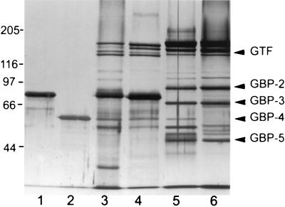
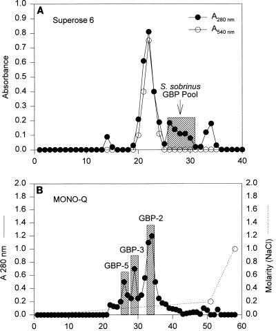
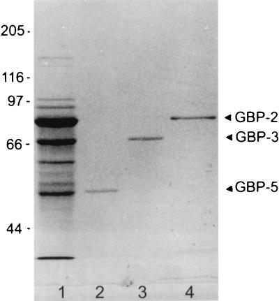
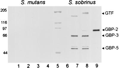
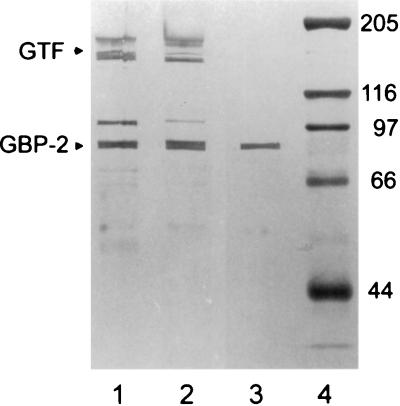
Three purified glucan binding proteins (GBP-2, GBP-3, and GBP-5) from Streptococcus sobrinus 6715 were compared structurally by mass spectroscopy of tryptic fragments and antigenically by Western blot analysis with rat antisera to each GBP or to peptides containing putative glucan binding epitopes of mutans streptococcal glucosyltransferases. Structural and antigenic analyses indicated that GBP-3 and GBP-5 are very similar but that both are essentially unrelated to GBP-2. None of these S. sobrinus GBPs appeared to have a strong antigenic relationship with GBPs from Streptococcus mutans. Thus, S. sobrinus GBP-2 and GBP-3 appear to be distinct proteins with potentially different functions. S. sobrinus GBP-5 may be a proteolytic fragment of GBP-3, or, alternatively, the genes coding for these proteins may be closely related.


Figures

FIG. 1
FIG. 1
Silver-stained SDS–7% polyacrylamide gel of guanidine eluates and purified GBPs from mutans streptococci. Lanes 1 and 2 contain enriched preparations of S. mutans GBP-A (0.5 μg [lane 1]) and GBP-B (0.4 μg [lane 2]). Lanes 3 to 6 contain GBP mixtures from S. mutans Ingbritt, S. mutans SJ, S. sobrinus 6715, and S. sobrinus DS, respectively. These GBP mixtures were obtained by 6 M guanidine elution of GBPs from Sephadex after incubation with culture supernatant, as described in the text. The relative migration positions of S. sobrinus GBP-2 to GBP-5 and GTF are indicated by the labeled arrows on the right side of the gel. The molecular masses (in kilodaltons) of coelectrophoresed standards are indicated on the left side of the gel.
FIG. 2
FIG. 2
Purification of S. sobrinus 6715 GBPs. (A) A280 elution profile of GBPs eluted from Sephadex G100 by 3 M guanidine-HCl and separated with Superose 6 in 6 M guanidine-HCl. GTF enzymatic activity (release of total reducing sugars) is illustrated by the open circles, which indicate A540 after the Somogyi assay. (B) Elution profile of GBPs in the shaded Superose 6 peak (arrow [A]) after ion-exchange chromatography in a Mono-Q 5/5 column. The NaCl gradient used to elute the GBPs is indicated by the dotted line connecting the open circles. The molecular mass characteristics of GBP-2, GBP-3, and GBP-5 are given in Table 1.
FIG. 3
FIG. 3
Coomassie blue-stained SDS–7% polyacrylamide gel of chromatographically purified GBPs from S. sobrinus 6715. In lane 1 is the GBP pool (2 μg) from Superose 6 gel filtration (Figure 2A). In lanes 2, 3, and 4 are the GBP-5-, GBP-3-, and GBP-2-containing pools, respectively, from Mono-Q ion-exchange chromatography (Figure 2B). Lanes 2, 3, and 4 contain 0.1 μg (each) of protein. The migration positions of the purified S. sobrinus GBPs are indicated by the labeled arrows on the right side of the gel. No other components were detected in the GBP-2 (lane 4) and GBP-5 (lane 2) pools, although a trace of GBP-2 can be detected in the GBP-3 pool (lane 3). The estimated molecular masses (in kilodaltons) of coelectrophoresed standards are indicated on the left side of the gel.
FIG. 4
FIG. 4
Western blot of S. mutans SJ GBPs (left) and S. sobrinus 6715 GBPs (right) eluted from Sephadex G100 after electrophoresis on an SDS–7% polyacrylamide gel. Blots were developed for IgG in sera (1:200) from rats injected with the enriched GBP pools shown in Fig. 3. Antisera to GBP-5, GBP-3, or GBP-2 were added to lanes 2 to 4 and 7 to 9, respectively. An antiserum to a sham-immunized rat was added to lanes 1 and 6. The molecular masses of protein standards (lane 5) are indicated (in kilodaltons) on the left of the blot.
FIG. 5
FIG. 5
Western blot of transblotted SDS–7% polyacrylamide gels developed for IgG in serum (1:50) from a rat injected with the GLU peptide construct. Lanes 1 and 2 contain previously electrophoresed S. sobrinus 6715 and S. sobrinus DS guanidine eluates, respectively. Lane 3 contains previously electrophoresed enriched S. sobrinus GBP-2 (0.6 μg). Lane 4 contains molecular standards whose estimated masses (in kilodaltons) are indicated to the right of the blot. The stained S. sobrinus components migrating between the 205- and 116-kDa positions reflect the migration of GTF (see Fig. 1). The stained component migrating to approximately 100 kDa is likely to be degraded GTF.

Similar articles

Cited by

References

    1. Abo H, Matsumura T, Kodama T, Ohta H, Fukui K, Kato K, Kagawa H. Peptide sequences for sucrose splitting and glucan binding within Streptococcus sobrinus glucosyltransferase (water-insoluble glucan synthetase) J Bacteriol. 1991;173:989–996. - PMC - PubMed
    1. Banas J A, Russell R R B, Ferretti J S. Sequence analysis of the gene for the glucan-binding protein of Streptococcus mutans Ingbritt. Infect Immun. 1990;58:667–673. - PMC - PubMed
    1. Gilmore K S, Russell R R B, Ferretti J J. Analysis of the Streptococcus downei gtfS gene, which specifies a glucosyltransferase that synthesizes soluble glucans. Infect Immun. 1990;58:2452–2458. - PMC - PubMed
    1. Hanada N, Kuramitsu H K. Isolation and characterization of the Streptococcus mutans gtfD gene, coding for primer-dependent soluble glucan synthesis. Infect Immun. 1989;57:2079–2085. - PMC - PubMed
    1. Lis M, Shiroza T, Kuramitsu H K. Role of the C-terminal direct repeating units of the Streptococcus mutans glucosyltransferase-S in glucan binding. Appl Environ Microbiol. 1995;61:2040–2042. - PMC - PubMed

Publication types