. 1998 Nov;66(11):5570-5.
doi: 10.1128/IAI.66.11.5570-5575.1998.

A protein (M9) associated with monoclonal antibody-mediated agglutination of Mycoplasma gallisepticum is a member of the pMGA family

Affiliations

A protein (M9) associated with monoclonal antibody-mediated agglutination of Mycoplasma gallisepticum is a member of the pMGA family

L Liu et al. Infect Immun. 1998 Nov.

Abstract

A 62-kDa cell surface antigen (M9) of Mycoplasma gallisepticum PG31 that mediates antibody-induced agglutination of the organism was purified and subjected to N-terminal amino-acid sequencing. A 999-bp region of the cDNA encoding the M9 protein was generated by reverse transcription-PCR, and its nucleotide sequence was determined. PCR primers based on this sequence were used to screen a genomic DNA library of PG31. A full-length M9 protein-encoding gene was isolated and sequenced, revealing 96% nucleotide identity with the pMGA1.1 gene of M. gallisepticum S6. Sequence analyses of the M9 gene and flanking open reading frames that encode other pMGA family members suggest that a tandemly repeated GAA sequence may influence pMGA gene expression.


Figures
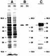
FIG. 1
FIG. 1
Separation and analysis of M9 by SDS-PAGE and electroblotting. (A) SDS-PAGE-resolved, Ponceau S-stained blot of whole-cell lysate of M. gallisepticum PG31 (a) and the supernatant retrieved after centrifugation of this whole-cell lysate (b). (B) The blot from panel A was destained and probed with MAb G9. (C) The immunoaffinity- and gel-purified protein sample was concentrated, subjected to SDS-PAGE, blotted onto a PVDF membrane, and stained with Ponceau S. One of four identical lanes is shown, each of which contained 10 μg of M9 protein. The M9 protein bands (indicated by the arrow) were excised for sequencing. (A small amount of albumin originating from the ascitic fluid appears as a band above the M9 protein band.) Molecular size markers (in kilodaltons; Diversified Biotech, Boston, Mass.) are for, from top to bottom, phosphorylase b, bovine serum albumin (68 kDa, panel C only), glutamate dehydrogenase, ovalbumin, lactate dehydrogenase, carbonic anhydrase, and lactoglobulin.
FIG. 2
FIG. 2
Schematic diagrams of M. gallisepticum DNA fragments containing the M9 gene. (A) Composite diagram (top line) of the chromosomal region containing the M9 gene (ORF-B) based on sequence analysis of the clones VSP 2 and VSP 3 (middle shaded lines) and locations of transcribed sequences identified by RT-PCR (bottom filled line). The locations of primer binding sites used for RT-PCR analysis are shown with arrows indicating directionality. (B) Schematic diagram of the insert in clone VSP 1 showing the locations of orf-C, -D, and -E. Positions of TAA stop codons in orf-A and -C are indicated.
FIG. 3
FIG. 3
Nucleotide sequence of the M9 gene and predicted amino acid sequence. Double-underlined amino acids agree with sequence data obtained from peptide fragments. Underlined nucleotides indicate positions of oligonucleotide primers (indicated in parentheses) used for RT-PCR and library screening. Arrows mark the sequence determined from RT-PCR product analysis as well as from the genomic clones. An asterisk denotes the stop codon.
FIG. 4
FIG. 4
Alignment of the M9 protein’s predicted amino acid sequence with those of the products of related ORFs from strain PG31 and with that of the pMGA1.1 protein of strain S6. Alignment was performed with the CLUSTAL W (19) program of the MacVector software package. Regions of dark shading and light boxed areas indicate amino acid identity and similarity, respectively. Amino acids that are different are unshaded. Gaps indicated by dashes were added to obtain the best alignment.

Similar articles

Cited by

References

    1. Aebersold R. Internal amino acid sequence analysis of proteins after in situ protease digestion on nitrocellulose. In: Matsudaira P, editor. A practical guide to protein and peptide purification for microsequencing. 2nd ed. San Diego, Calif: Academic Press, Inc.; 1993. pp. 102–124.
    1. Amaravadi L, King M W. A rapid and efficient, nonradioactive method for screening recombinant DNA libraries. BioTechniques. 1994;16:98–103. - PubMed
    1. Baseggio N, Glew M D, Markham P F, Whithear K G, Browning G F. Size and genomic location of the pMGA multigene family of Mycoplasma gallisepticum. Microbiology. 1996;142:1429–1435. - PubMed
    1. Don R H, Cox P T, Wainwright B J, Baker K, Mattick J S. Touchdown PCR to circumvent spurious priming during gene amplification. Nucleic Acids Res. 1991;19:4008. - PMC - PubMed
    1. Dybvig K, Clark C D, Aliperti G, Schlesinger M J. A chicken repetitive DNA sequence that is highly sensitive to single-strand specific endonucleases. Nucleic Acids Res. 1983;11:8495–8508. - PMC - PubMed

Publication types

Substances

Associated data

LinkOut - more resources