. 1998 Oct;77(10):1779-90.
doi: 10.1177/00220345980770100501.

Autoregulation of periodontal ligament cell phenotype and functions by transforming growth factor-beta1

Affiliations

Autoregulation of periodontal ligament cell phenotype and functions by transforming growth factor-beta1

T A Brady et al. J Dent Res. 1998 Oct.

Abstract

During orthodontic tooth movement, mechanical forces acting on periodontal ligament (PDL) cells induce the synthesis of mediators which alter the growth, differentiation, and secretory functions of cells of the PDL. Since the cells of the PDL represent a heterogeneous population, we examined mechanically stress-induced cytokine profiles in three separate clones of human osteoblast-like PDL cells. Of the four pro-inflammatory cytokines investigated, only IL-6 and TGF-beta1 were up-regulated in response to mechanical stress. However, the expression of other pro-inflammatory cytokines such as IL-1 beta, TNF-alpha, or IL-8 was not observed. To understand the consequences of the increase in TGF-beta1 expression following mechanical stress, we examined the effect of TGF-beta1 on PDL cell phenotype and functions. TGF-beta1 was mitogenic to PDL cells at concentrations between 0.4 and 10 ng/mL. Furthermore, TGF-beta1 down-regulated the osteoblast-like phenotype of PDL cells, i.e., alkaline phosphatase activity, calcium phosphate nodule formation, expression of osteocalcin, and TGF-beta1, in a dose-dependent manner. Although initially TGF-beta1 induced expression of type I collagen mRNA, prolonged exposure to TGF-beta1 down-regulated the ability of PDL cells to express type I collagen mRNA. Our results further show that, within 4 hrs, exogenously applied TGF-beta1 down-regulated IL-6 expression in a dose-dependent manner, and this inhibition was sustained over a six-day period. In summary, the data suggest that mechanically stress-induced TGF-beta1 expression may be a physiological mechanism to induce mitogenesis in PDL cells while down-regulating its osteoblast-like features and simultaneously reducing the IL-6-induced bone resorption.


Figures

Figure 1
Figure 1
Characterization of various PDL cell lines. (A) Alkaline phosphatase activity in PDL cells, PL-1, PL-4, and PL-124 (104 cells/well), cultured for 7 days in vitro. (B) Generation of cAMP in PDL cells activated with PGE2, for 10 min. (C) Calcium phosphate nodule formation in PDL cells grown for 28 days in vitro. (D) Expression of mRNA for TGF-β1, osteocalcin, and type I-α1 collagen in PL-1, PL-4, and PL-124 cells as assessed by RT/PCR. The data in (A), (B), and (C) represent the mean and standard error of the mean (± SEM) of quadruplicate values. Bands of PCR products in (D) represent one out of three separate experiments on PL-1, PL-4, and PL-124 cells.
Figure 2
Figure 2
Effect of mechanical stress on PDL cells. (A) PL-1 or PL-124 cells were subjected to either 0 hr (lanes 1, 3, 5, and 7), 1 hr (lanes 2 and 4) or 24 hrs (lanes 6 and 8) of mechanical stress. Subsequently, cells were incubated for 4 hrs; RNA was extracted and analyzed by RT/PCR for the presence of mRNA for cytokines. Gingival fibroblasts activated with recombinant human IL-1β for 4 hrs were used as positive controls in these experiments (lane 9). Alternatively, PL-1 or PL-124 unstressed control cells (PL-1c, PL-124c) or cells subjected to mechanically induced tension (PL-1ms, PL-124ms) were incubated for 6 hrs at 37°C, after a one-hour stress regimen (B) or a 24-hour stress regimen (C). Thereafter, the supernatants were harvested and analyzed by ELISA for the presence of cytokines. The data in (B) and (C) represent the mean ± SEM of triplicate values. The data in all panels represent one out of three separate experiments.
Figure 3
Figure 3
Effects of various concentrations of TGF-β1 on PDL cell growth. PL-4 cells (5 × 10³/well) grown in 96-well plates were incubated in the absence or presence of various concentrations of TGF-β1 for 3, 6, 9, 12, or 28 days. The cell growth was assessed spectrophotometrically following crystal violet staining and solubilization of cells. The data represent the mean ± SEM of quadruplicate values from one of three separate experiments.
Figure 4
Figure 4
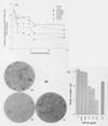
Effects of various concentrations of TGF-β1 on the alkaline phosphatase activity and calcium phosphate nodule formation in PDL cells. (A) PL-4 cells (5 × 10³/well) grown in 96-well plates were incubated in the absence or presence of various concentrations of TGF-β1 for 3, 6, 9, 12, or 28 days. Alkaline phosphatase activity was assessed spectrophotometrically in the presence of p-nitrophenyl phosphate in digitonin-permeabilized cells. All experiments were done in quadruplicate. The SEM at each point was less than 2% of the mean. (B) Localization of alkaline phosphatase activity in PL-4 cells following TGF-β1 treatment. PL-4 cells were incubated in the continuous presence of 0 ng/mL (a) or 10 ng/mL (b) TGF-β1 for 28 days. Alternatively, PL-4 cells were incubated in the presence of TGF-β1 for 14 days, washed, and replenished with medium devoid of TGF-β1 and examined on day 28 (c). All cells were examined for the presence of alkaline phosphatase activity by histochemical staining as described in “ Methods”. The photographs represent one of three separate experiments performed in duplicates. (C) PL-4 cells were incubated with various concentrations of TGF-β1 for 9 days (solid bars). To examine the effect of removal of TGF-β1, we grew PL-4 cells in 10 ng/mL TGF-β1 for 14 days, then washed them and grew them again in TCM devoid of TGF-β1 for an additional 14 days (hatched bars). The numbers of nodules were counted after von Kossa staining of cells. Each point represents the means ± SEM of triplicate values.
Figure 5
Figure 5
Effect of TGF-β1 on the expression of TGF-β1 mRNA in PDL cells (5 × 10⁴/well) were incubated in the absence or presence of various concentrations of TGF-β1 for 0.16, 1, 3, or 6 days. Subsequently, RNA was extracted, and the expression of TGF-β1 mRNA was examined by RT/PCR. The histograms represent the mean of triplicate luminescence values of PCR products on agarose gels in one of three separate experiments performed on PL-4 or PL-124 cells with similar results. The SEM were within 5% of the mean in Figs. 5 to 8.
Figure 6
Figure 6
Effect of TGF-β1 on the expression of osteocalcin mRNA in PDL cells. PL-4 cells (5 × 10⁴/well) were incubated in the absence or presence of various concentrations of TGF-β1 for 0.16, 1, 3, or 6 days. Subsequentlv, RNA was extracted, and the expression of osteocalcin-specific mRNA was assessed by RT/PCR. The histograms depict the mean of triplicate luminescence values of PCR products on agarose gels shown in the photographs. The data represent one of three separate experiments performed on PL-4 or PL-124 cells with similar results.
Figure 7
Figure 7
Effect of TGF-β1 on type I-α1 collagen mRNA expression in PDL cells. PL-4 cells (5 × 10⁴/well) were incubated in the absence or presence of various concentrations of TGF-β1 for 0.16, 1, 3, or 6 days. Subsequently, RNA was extracted, and the expression of type I-α1 collagen mRNA was examined by RT/PCR. The histograms represent the mean of triplicate luminescence values of PCR products in one of three separate experiments performed on PL-4 or PL-124 cells with similar results.
Figure 8
Figure 8
Effect of TGF-β1 on IL-6 mRNA expression in PDL cells. PL-4 cells (5 × 10⁴/well) were incubated in the absence or presence of various concentrations of TGF-β1 for 0.16,1, 3, or 6 days. Subsequently, RNA was extracted, and the expression of IL-6 mRNA was examined by RT/PCR. The histograms represent the mean of the triplicate luminescence values of PCR products in one of three separate experiments performed on PL-4 or PL-124 cells with similar results.

References

    1. Arceo N, Sauk JJ, Moehring J, Foster RA, Somerman MJ. Human periodontal cells initiate mineral like nodules in vitro. J Periodont Res. 1991;62:499–503. - PubMed
    1. Banes AJ, Gilbert J, Taylor D, Monbureau O. A new vacuum-operated stress-providing instrument that applies static or variable duration cyclic tension or compression to cells in vitro. J Cell Sci. 1985;75:35–42. - PubMed
    1. Binderman I, Zor U, Kaye AM, Shimshoni Z, Harell A, Somjen D. The transduction of mechanical force into biochemical events in bone cells may involve activation of phospholipase A2. Calcif Tissue Int. 1988;42:261–266. - PubMed
    1. Bonewald LF, Mundy GR. Role of transforming growth factor-β on bone remodeling. Clin Orthop Rel Res. 1990;250:261–276. - PubMed
    1. Breen EC, Ignotz RA, McCabe L, Stein JL, Stein GS, Lian JB. TGF beta alters growth and differentiation related gene expression in proliferating osteoblasts in vitro, preventing development of the mature bone phenotype. J Cell Physiol. 1994;160:323–335. - PubMed

Publication types

MeSH terms

LinkOut - more resources