. 1998 Oct 19;143(2):319-31.
doi: 10.1083/jcb.143.2.319.

The membrane transport factor TAP/p115 cycles between the Golgi and earlier secretory compartments and contains distinct domains required for its localization and function

Affiliations

The membrane transport factor TAP/p115 cycles between the Golgi and earlier secretory compartments and contains distinct domains required for its localization and function

D S Nelson et al. J Cell Biol. .

Abstract

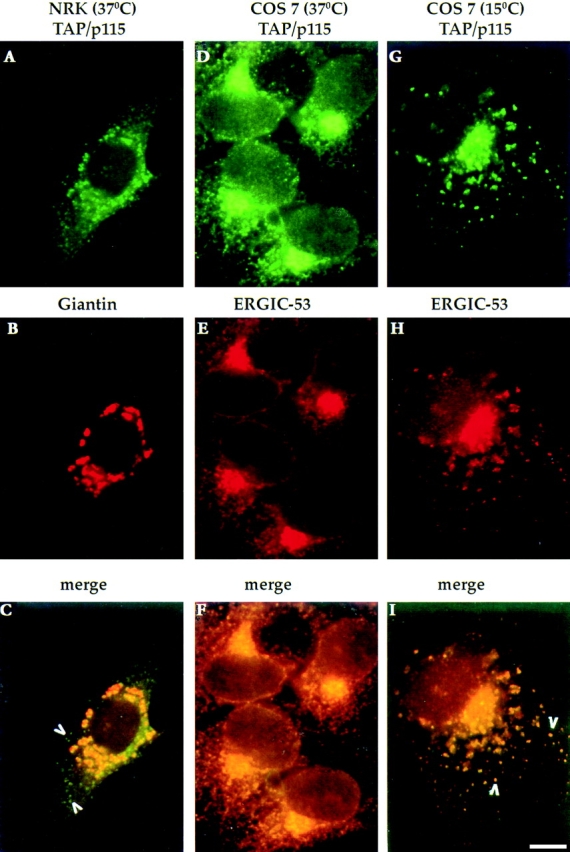
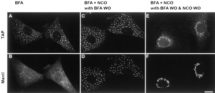
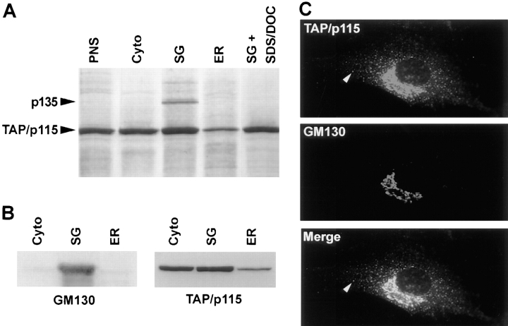
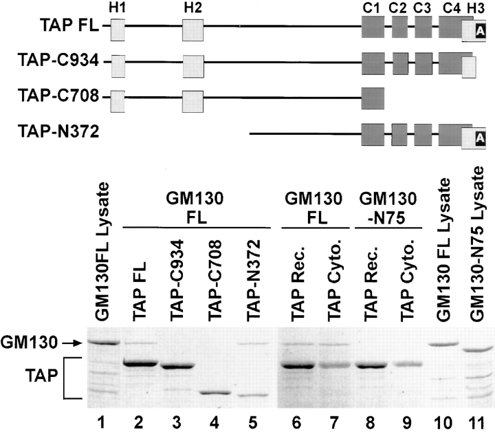
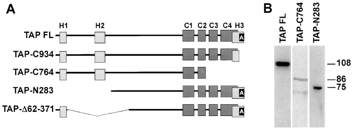
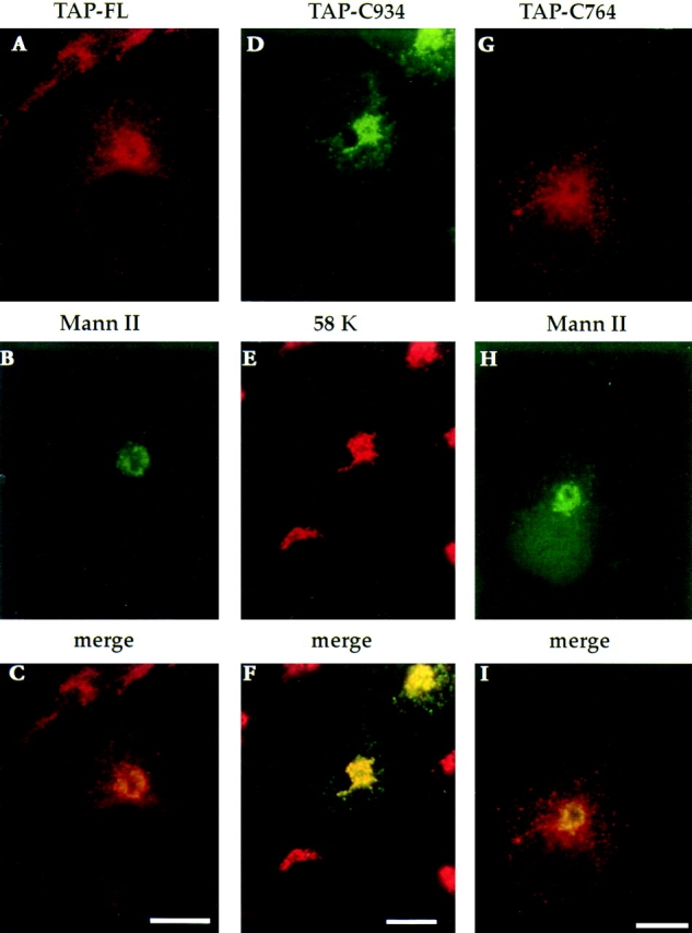
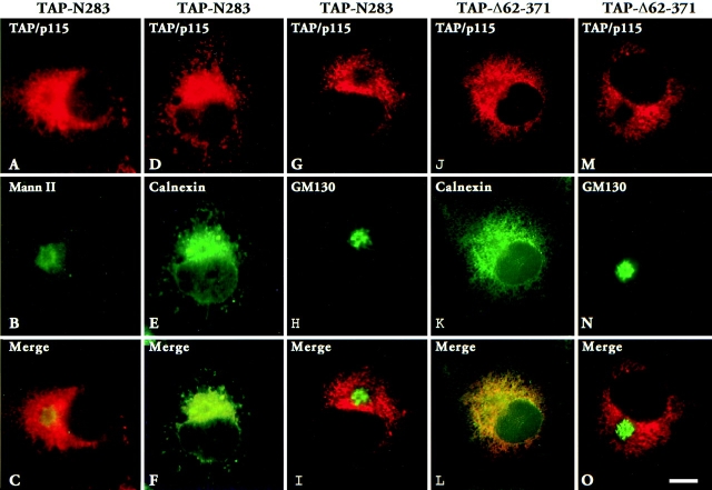
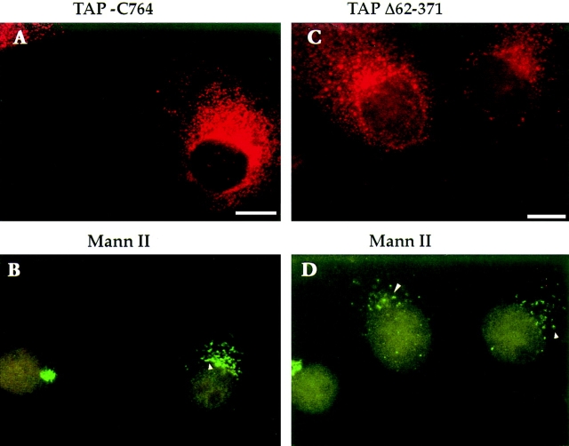
The mammalian protein TAP/p115 and its yeast homologue Uso1p have an essential role in membrane traffic (Nakajima et al., 1991; Waters et al., 1992; Sztul et al., 1993; Rabouille et al.; 1995). To inquire into the site and mechanism of TAP/p115 action, we aimed to localize it and to identify domains required for its function. We show that in interphase cells, TAP/p115 localizes predominantly to the Golgi and to peripheral structures that represent vesicular tubular clusters (VTCs) involved in ER to Golgi transport. Using BFA/ nocodazole treatments we confirm that TAP/p115 is present on ER to Golgi transport intermediates. TAP/ p115 redistributes to peripheral structures containing ERGIC-53 during a 15 degreesC treatment, suggesting that it is a cycling protein. Within the Golgi, TAP/p115 is associated with pleiomorphic structures on the cis side of the cis-Golgi cisterna and the cis-most cisterna, but is not detected in more distal compartments of the Golgi. TAP/p115 binds the cis-Golgi protein GM130, and the COOH-terminal acidic domain of TAP/p115 is required for this interaction. TAP/p115 interaction with GM130 occurs only in the Golgi and is not required for TAP/p115 association with peripheral VTCs. To examine whether interaction with GM130 is required to recruit TAP/p115 to the Golgi, TAP/p115 mutants lacking the acidic domain were expressed and localized in transfected cells. Mutants lacking the GM130-binding domain showed normal Golgi localization, indicating that TAP/p115 is recruited to the Golgi independently of its ability to bind GM130. Such mutants were also able to associate with peripheral VTCs. Interestingly, TAP/p115 mutants containing the GM130-binding domain but lacking portions of the NH2-terminal region were restricted from the Golgi and localized to the ER. The COOH-terminal domain required for GM130 binding and the NH2-terminal region required for Golgi localization appear functionally relevant since expression of TAP/p115 mutants lacking either of these domains leads to loss of normal Golgi morphology.


Figures

Figure 1
Figure 1
TAP/p115 is a cycling protein. NRK or COS-7 cells were incubated at 37° or at 15°C for 2 h, fixed, and then processed for IF. In NRK cells, TAP/p115 (A) colocalizes with the Golgi marker giantin (B) in a perinuclear region. TAP/p115 is also present in peripheral punctate structures that do not contain giantin (arrowheads in C). In COS-7 cells incubated at 37°C, TAP/ p115 (D) and ERGIC-53 (E) colocalize in a juxtanuclear region and in peripheral punctate structures (F). In COS-7 cells incubated at 15°C, TAP/p115 (G) and ERGIC-53 (H) colocalize in dispersed punctate structures (arrowheads in I). Bar, 10 μm.
Figure 2
Figure 2
Peripheral structures containing TAP/p115 are transport intermediates. NRK cells were treated with 5 μg/ml BFA for 30 min, fixed, and processed for IF. TAP/p115 (A) is present in punctate structures, whereas mannosidase II (B) is localized to the ER. Cells like in A and B were treated with 10 μg/ml nocodazole for 90 min. Cells were then incubated in medium without BFA but containing nocodazole (BFA + NCO with BFA WO) for 30 min, fixed, and processed for IF. Mannosidase II (D) redistributed from the ER into punctate structures containing TAP/p115 (C). Cells like in C and D were subjected to nocodazole wash-out (BFA + NCO with BFA WO & NCO WO) for 45 min, fixed, and then processed for IF. TAP/p115 (E) and mannosidase II (F) colocalized in a morphologically normal Golgi complexes. Bar, 10 μm.
Figure 3
Figure 3
TAP/p115 associates with vesicular tubular structures and cis-Golgi membranes. Immunogold localization of TAP/p115 (arrowheads) in pancreatic acinar cells. Gc, Golgi complex; n, nucleus. Bar, 0.2 μm.
Figure 4
Figure 4
TAP/p115 binds GM130 in the Golgi. (A) TAP/p115 was immunoprecipitated from PNS (lane PNS), cytosolic (lane cyto), stacked Golgi (lane SG), and ER (lane ER) fractions solubilized with Triton X-100 or from the SG fraction solubilized with Triton X-100/SDS/DOC (lane SG + SDS/DOC). A silver-stained gel is shown. p135 coimmunoprecipitates with TAP/ p115 only from the SG fraction. (B) Rat liver fractions were analyzed by immunoblot for the presence of GM130 and TAP/p115. GM130 was detected predominantly in the SG fraction whereas TAP/p115 was also present in cytosolic and ER fractions. (C) NRK cells were processed for IF using anti-TAP/p115 and anti-GM130 antibodies. TAP/p115 and GM130 colocalize in the Golgi region, but GM130 is not detected in peripheral punctate structures containing TAP/ p115 (arrowheads).
Figure 5
Figure 5
The acidic COOH terminus of TAP/p115 and the basic NH2 terminus of GM130 are required for binding. Diagram of analyzed constructs. Light gray rectangles, Uso1p homology (H1, H2, and H3) regions. Dark gray rectangles, coiled-coil domains (C1–C4). Black rectangle, the acidic domain (A). (Lanes 1–5) Recombinant TAP/p115 constructs were incubated with recombinant full-length GM130 (20% of input is loaded in lane 1). Complexes were recovered by immunoprecipitation with anti-TAP/ p115 antibodies. Coomassie blue–stained gel is shown. Full-length TAP/p115 (lane 2) and TAP-N372 (lane 5) bound full-length GM130, whereas mutants lacking the COOH-terminal acidic domain, TAP-C934 (lane 3) and TAP-C708 (lane 4) did not bind GM130. (Lanes 6–11) Recombinant full-length TAP/p115 or cytosolic TAP/p115 were incubated with recombinant full-length GM130 (20% of input is loaded in lane 10) or GM130-N75 (20% of input is loaded in lane 11). Complexes were recovered by immunoprecipitation with anti-TAP/p115 antibodies. Coomassie blue–stained gel is shown. Recombinant (lane 6) and cytosolic (lane 7) TAP/p115 bound GM130, but neither bound GM130-N75 (lanes 8 and 9). The results indicate that the COOH-terminal acidic domain of TAP/p115 and the NH2-terminal basic domain of GM130 are required for binding.
Figure 6
Figure 6
TAP/p115 mutants used in transfection studies. (A) Diagram of analyzed constructs. Light gray rectangles, Uso1p homology (H1, H2, and H3) regions. Dark gray rectangles, coiled-coil domains (C1–C4). Black rectangle, acidic domain (A). (B) Lysates of COS-7 cells tranfected with specific TAP/p115 constructs were analyzed by immunoblotting. Recombinant proteins of the appropriate molecular weight (∼108 kD for TAP-FL; ∼86 kD for TAP-C764, and ∼75 kD for TAP-N283) were detected.
Figure 7
Figure 7
TAP/p115 mutants lacking the GM130-binding domain localize to the Golgi. Full-length TAP/p115 (A–C), TAP-C934 (D–F), and TAP-C764 (G–I) were transiently expressed in COS-7 cells. Cells were fixed and processed for IF using anti-TAP/ p115 and either anti–mannosidase II or anti-58K antibodies. Recombinant proteins were concentrated in the Golgi region and colocalized with the Golgi markers. Bars, 10 μm.
Figure 8
Figure 8
TAP/p115 mutants with NH2-terminal deletions do not localize to the Golgi. TAP-N283 (A–I) or TAP-Δ62–371 (J–O) were transiently expressed in COS-7 cells. Cells were fixed and processed for IF using anti-TAP/p115 and either anti–mannosidase II, anti-calnexin, or anti-GM130 antibodies. Mutant TAP/p115 did not concentrate with mannosidase II in the Golgi (C), but colocalized with calnexin in the ER (F and L). Mutants did not cause the redistribution of GM130 from the Golgi to the ER (I and O). Bar, 10 μm.
Figure 9
Figure 9
Expression of TAP/p115 mutants leads to loss of characteristic Golgi morphology. TAP-C764 (A and B) and TAP-Δ62–371 (C and D) were transiently expressed in COS-7 cells, and 63 h after transfection cells were fixed and processed for IF using anti–mannosidase II and anti-TAP/p115 antibodies. In cells expressing high levels of mutant TAP/ p115 (A and C), mannosidase II is found in punctate structures clustered in the perinuclear region (arrowheads in B and D). Bar, 10 μm.

Similar articles

Cited by

References

    1. Ansorge W. Fast and sensitive detection of protein and DNA bands by treatment with potassium permanganate. J Biochem Biophys Methods. 1985;11:13–20. - PubMed
    1. Bajjalieh SM, Scheller RH. The biochemistry of neurotransmitter secretion. J Biol Chem. 1995;270:1971–1974. - PubMed
    1. Balch WE, McCaffery JM, Plutner H, Farquhar MG. Vesicular stomatitis virus glycoprotein is sorted and concentrated during export from the endoplasmic reticulum. Cell. 1994;76:841–852. - PubMed
    1. Banfield DK, Lewis MJ, Rabouille C, Warren G, Pelham HR. Localization of Sed5, a putative vesicle targetingmolecule, to the cis-Golgi network involves both its transmembrane and cytoplasmic domains. J Cell Biol. 1994;127:357–371. - PMC - PubMed
    1. Bannykh SI, Balch W. Membrane dynamics at the endoplasmic reticulum–Golgi interface. J Cell Biol. 1997;138:1–4. - PMC - PubMed

Publication types

MeSH terms