. 1998 Oct 19;143(2):359-73.
doi: 10.1083/jcb.143.2.359.

Mitochondrial fusion in yeast requires the transmembrane GTPase Fzo1p

Affiliations

Mitochondrial fusion in yeast requires the transmembrane GTPase Fzo1p

G J Hermann et al. J Cell Biol. .

Abstract

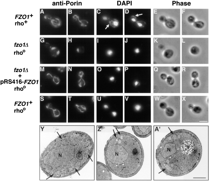
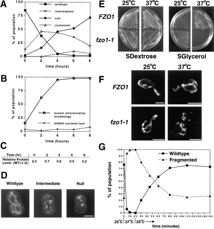
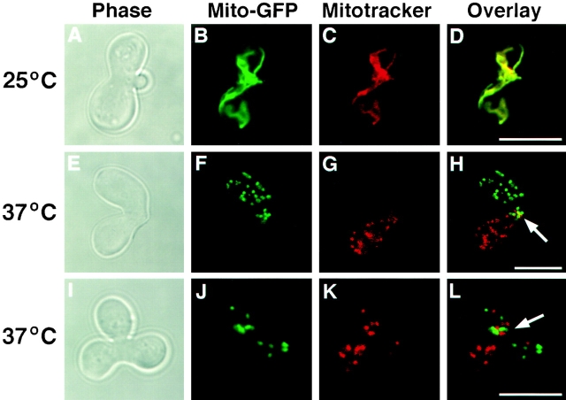
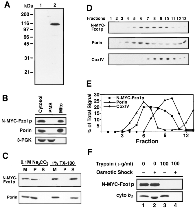
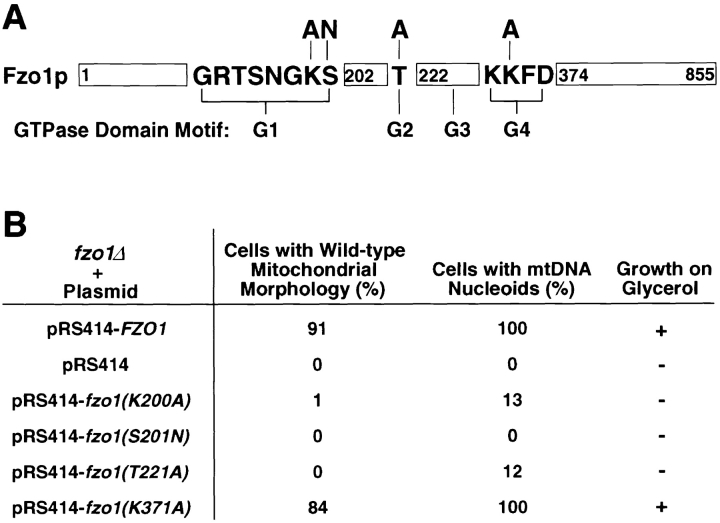
Membrane fusion is required to establish the morphology and cellular distribution of the mitochondrial compartment. In Drosophila, mutations in the fuzzy onions (fzo) GTPase block a developmentally regulated mitochondrial fusion event during spermatogenesis. Here we report that the yeast orthologue of fuzzy onions, Fzo1p, plays a direct and conserved role in mitochondrial fusion. A conditional fzo1 mutation causes the mitochondrial reticulum to fragment and blocks mitochondrial fusion during yeast mating. Fzo1p is a mitochondrial integral membrane protein with its GTPase domain exposed to the cytoplasm. Point mutations that alter conserved residues in the GTPase domain do not affect Fzo1p localization but disrupt mitochondrial fusion. Suborganellar fractionation suggests that Fzo1p spans the outer and is tightly associated with the inner mitochondrial membrane. This topology may be required to coordinate the behavior of the two mitochondrial membranes during the fusion reaction. We propose that the fuzzy onions family of transmembrane GTPases act as molecular switches to regulate a key step in mitochondrial membrane docking and/or fusion.


Figures

Figure 1
Figure 1
S. cerevisiae FZO1 is required for mitochondrial function. (A) Domain structure of three Fzo family members showing the position of the predicted GTPase (GTPase), heptad repeat (Hep), and transmembrane (TM) domains. Amino acid identities between the entire protein sequences and the predicted GTPase domains of the S. cerevisiae Fzo1p, S. pombe Fzo1p (the newest family member), and D. melanogaster Fzo family members are indicated. GenBank/EMBL/DDBJ accession numbers are as follows: S. cerevisiae (Z36048), S. pombe (ALO23533), and D. melanogaster (U95821). (B) fzo1Δ cells exhibit growth defects on fermentable and nonfermentable carbon sources. Two fzo1Δ (top, JSY1809 and JSY1810) and two wild-type (bottom, JSY1811 and JSY1812) strains from a single tetrad were grown on YPDextrose medium for 2 d at 30°C (left) or YPGlycerol medium for 5 d at 30°C (right).
Figure 2
Figure 2
fzo1Δ cells exhibit defective mitochondrial morphology and lose their mtDNA. (A–X) Indirect immunofluorescence images of yeast cells stained with anti-porin antiserum to visualize mitochondrial membranes (leftmost columns), and DAPI to visualize nuclei and mtDNA (middle columns; white arrows in C and D indicate mtDNA nucleoids). Phase-contrast images of the same cells are shown in the rightmost columns. (A–F) Normal mitochondrial morphology and distribution of mtDNA nucleoids in a wild-type rho+ strain (JSY1812). (G–L) Mutant fzo1Δ rhoo cells (JSY1810) contained spherical or slightly elongated mitochondrial structures that lack DAPI-stained mtDNA. (M–R) Wild-type mitochondrial morphology, but not mtDNA, was restored in an fzo1Δ rhoo mutant strain (JSY2579) upon reintroduction of the wild-type FZO1 gene. (S–X) Mitochondrial morphology is normal in a wild-type rhoo strain (JSY2555) that lacks mtDNA. (Y–A′) Transmission electron micrographs of wild-type and fzo1Δ cells. (Y) Mitochondrial profiles in wild-type cells (JSY1812) are dispersed equally throughout the cell cortex and display numerous cristae (black arrows). (Z and A′) Mitochondrial profiles in fzo1Δ cells (JSY1810) are clustered together near the cell periphery and contain fewer cristae. N, nuclei; V, vacuoles; rho +, containing mtDNA; rhoo, lacking mtDNA. Bars: (A–X) 5 μm; (Y–A′) 1μm.
Figure 3
Figure 3
Depletion or loss of Fzo1p function leads to defects in mitochondrial morphology and mtDNA maintenance. (A–D) Depletion of Fzo1p causes defects in mitochondrial morphology that precede the loss of mtDNA nucleoids. fzo1Δ cells containing pGAL1-N-3XMYC-FZO1 (JSY2273) were shifted from raffinose medium, which allows nearly wild-type levels of Fzo1p expression, to dextrose medium, which represses expression from the GAL1 promoter. (A) Mitochondrial morphology scored by DiOC6 staining after shift to depletion medium (n = 100): mitochondrial morphologies were classified as wild type (branched tubular network), intermediate (tubular condensed network), null (spherical or partially elongated structures), and unstained (nonrespiring mitochondria do not accumulate DiOC6). (B) Distribution of mtDNA nucleoids scored by DAPI staining during the Fzo1p depletion shown in A (n = 200). The mutant mitochondrial morphology class included the intermediate, null, and unstained categories scored in A. (C) N-3XMYC-Fzo1p levels during the depletion experiment. Protein extracts prepared from equivalent numbers of cells were analyzed by Western blotting with anti-MYC antiserum. N-3XMYC-Fzo1 protein levels during the depletion were normalized to the level of N-3XMYC-Fzo1p expressed from the wild-type FZO1 promoter (WT = 1.0; JSY2034). (D) Representative wild-type, intermediate, and null mitochondrial morphologies visualized with a matrix-targeted form of the GFP (mito-GFP). (E and F) The temperature-sensitive fzo1-1 mutation causes rapid and reversible fragmentation of the mitochondrial network. (E) fzo1-1 cells cannot grow on a nonfermentable carbon source at 37°C. fzo1Δ strains containing pRS414-FZO1 (JSY2926) or pRS414-fzo1-1 (JSY2793) were grown on SDextrose media (left panels) for 2 d at 25° or 37°C and SGlycerol media (right panels) for 6 d at 25° or 37°C. (F) Mitochondrial morphology in FZO1 (JSY2793) and fzo1-1(JSY2802) cells grown at 25°C and shifted to 37°C for 10 min. Mitochondrial compartments were visualized with mito-GFP. Representative cells are shown. (G) Log-phase fzo1-1 cells (JSY2804) grown at 25°C (t = 0) were shifted to 37°C. After 20 min at 37°C, the cells were returned to 25°C. Mitochondrial morphology was quantified at the indicated times using mito-GFP (n ≥ 200). A representative experiment is shown. Bar, 2.5 μm.
Figure 4
Figure 4
FZO1 is required for mitochondrial fusion during mating. fzo1-1 cells of opposite mating type (JSY2802 and JSY2804) were labeled with mito-GFP or Mitotracker red and mated at 25° (A–D) and 37°C (E–H and I–L). Confocal microscopy was used to score the distribution of mito-GFP (green in B, F, and J) and Mitotracker (red in C, G, and K) in serial optical sections (representative single optical section are shown). Fusion and mixing of mitochondrial contents (yellow in D) was evaluated in merged mito-GFP and Mitotracker red images (D, H, and L). Zygote morphology was visualized by phase-contrast microscopy (A, E, and I). The zygote bud is on the right in A and on the top in I. White arrows, regions where parental mitochondrial membranes have intermixed. Bar, 5 μm.
Figure 5
Figure 5
Fzo1p localizes to the mitochondrial network. (A–D) fzo1Δ cells (JSY2394) expressing the N-9XMYC-Fzo1 protein were stained with anti-MYC antiserum (A and C) and DAPI (B and D). The N-9XMYC-Fzo1p staining was completely coincident with mitochondria as marked by mtDNA nucleoids (compare white arrows in A and C with B and D). (E and F) Anti-MYC serum did not stain fzo1Δ cells (JSY2392) expressing the Fzo1p lacking the MYC tag. Bar, 5 μm.
Figure 6
Figure 6
Fzo1p is a mitochondrial integral membrane protein with its GTPase domain exposed to the cytoplasm. (A) Total cell extracts from fzo1Δ cells containing either the wild-type pRS414-FZO1 (lane 1; JSY2392) or pRS414-N-9XMYC- FZO1 plasmids (lane 2; JSY2394) analyzed by Western blotting with an anti-MYC antiserum. The same fzo1Δ strain harboring pRS414-N-9XMYC-FZO1 (JSY2394) was used for all of the experiments described in 6B-F. (B) N-9XMYC-Fzo1p cofractionated with the mitochondrial protein porin. Protein extracts from equivalent numbers of cells were fractionated by differential centrifugation, separated by SDS-PAGE, and analyzed by Western blotting with anti-MYC, anti-porin, and anti–3-PGK serum. Cytosol, postnuclear cytoplasmic extract; Mito, crude mitochondrial pellet; PMS, supernatant depleted of mitochondria. (C) N-9XMYC-Fzo1p is an integral membrane protein. Purified mitochondria (M) were treated to solubilize peripheral membrane proteins (0.1 M Na2CO3) or integral membrane proteins (1% Triton X-100), separated into pellet (P) and supernatant (S) fractions, and analyzed by SDS-PAGE and Western blotting. The release of soluble cytochrome b 2 and the peripheral F1β ATPase subunit into the supernatant fraction after 0.1M Na2CO3 treatment was confirmed by Western blotting (data not shown). (D and E) N-9XMYC-Fzo1p fractionates in an intermediate density mitochondrial membrane fraction. Fractions from a 30–50% sucrose step gradient (top, fraction 1) analyzed by Western blotting with anti-MYC, anti-porin, and anti–CoxIV serum. (E) The percentage of total N-MYC-Fzo1p, porin, and CoxIV per fraction. (F) The GTPase domain of Fzo1p is exposed on the cytoplasmic surface of mitochondria. Untreated (lane 1), trypsin-treated (lanes 3 and 4, 100 μg/ml), and osmotically shocked (lanes 2 and 4) mitochondria were analyzed by SDS-PAGE and Western blotting with anti-MYC and anti-cytochrome b 2 serum.
Figure 7
Figure 7
GTPase domain mutations block Fzo1p function. (A) Schematic representation of Fzo1p illustrating the location and sequence of the GTPase domain motifs (G1–G4; not to scale) with mutations changing conserved residues indicated. The mutant amino acids are depicted above the original residues. (B) Mutations in the G1 and G2 motifs disrupt the function of Fzo1p. Glycerol growth, mitochondrial morphology (anti-porin, n = 400), and mtDNA nucleoid distribution (DAPI staining, n ≥ 100) analyzed in fzo1Δ cells (JSY2354) containing wild-type FZO1 (JSY2392) or mutated fzo1 genes (JSY2355-2358) carried on low copy plasmids (pRS414).

References

    1. Adari H, Lowy DR, Willumsen BM, Der CJ, McCormick F. Guanosine triphosphatase activating protein (GAP) interacts with the p21 raseffector binding domain. Science. 1988;240:518–521. - PubMed
    1. Azpiroz R, Butow RA. Patterns of mitochondrial sorting in yeast zygotes. Mol Biol Cell. 1993;4:21–36. - PMC - PubMed
    1. Bakeeva LE, Chentsov YS, Skulachev VP. Mitochondrial framework (reticulum mitochondriale) in rat diaphragm muscle. Biochim Biophys Acta. 1978;501:349–369. - PubMed
    1. Bakeeva LE, Chentsov YS, Skulachev VP. Ontogenesis of mitochondrial reticulum in rat diaphragm muscle. Eur J Cell Biol. 1981;25:175–181. - PubMed
    1. Baudin A, Ozier-Kalogeropoulos O, Denouel A, Lacroute F, Cullin C. A simple and efficient method for direct gene deletion in Saccharomyces cerevisiae. . Nucleic Acids Res. 1993;21:3329–3330. - PMC - PubMed

Publication types

MeSH terms