. 1998 Oct 19;143(2):391-401.
doi: 10.1083/jcb.143.2.391.

A single gene product, claudin-1 or -2, reconstitutes tight junction strands and recruits occludin in fibroblasts

Affiliations

A single gene product, claudin-1 or -2, reconstitutes tight junction strands and recruits occludin in fibroblasts

M Furuse et al. J Cell Biol. .

Abstract

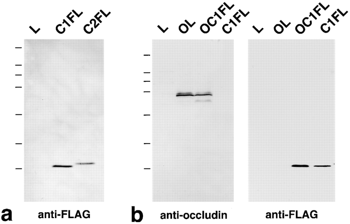
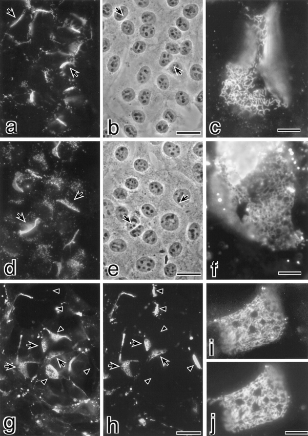
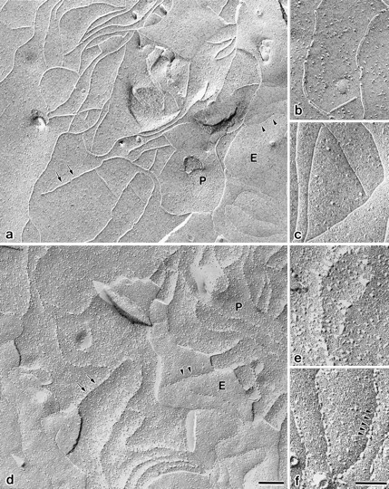
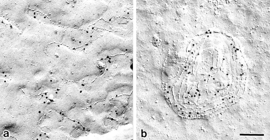
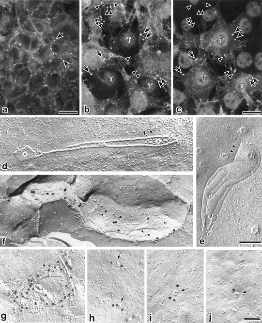
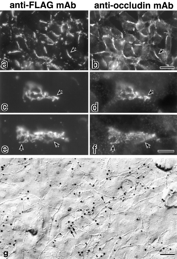
Three integral membrane proteins, clau- din-1, -2, and occludin, are known to be components of tight junction (TJ) strands. To examine their ability to form TJ strands, their cDNAs were introduced into mouse L fibroblasts lacking TJs. Immunofluorescence microscopy revealed that both FLAG-tagged claudin-1 and -2 were highly concentrated at cell contact sites as planes through a homophilic interaction. In freeze-fracture replicas of these contact sites, well-developed networks of strands were identified that were similar to TJ strand networks in situ and were specifically labeled with anti-FLAG mAb. In glutaraldehyde-fixed samples, claudin-1-induced strands were largely associated with the protoplasmic (P) face as mostly continuous structures, whereas claudin-2-induced strands were discontinuous at the P face with complementary grooves at the extracellular (E) face which were occupied by chains of particles. Although occludin was also concentrated at cell contact sites as dots through its homophilic interaction, freeze-fracture replicas identified only a small number of short strands that were labeled with anti-occludin mAb. However, when occludin was cotransfected with claudin-1, it was concentrated at cell contact sites as planes to be incorporated into well- developed claudin-1-based strands. These findings suggested that claudin-1 and -2 are mainly responsible for TJ strand formation, and that occludin is an accessory protein in some function of TJ strands.


Figures

Figure 1
Figure 1
Stable L transfectants used in this study. Total lysates from the same number of parental L cells and stable transfectants were separated by SDS-PAGE and immunoblotted with anti-FLAG mAb or anti-occludin mAb. (a) Expression of FLAG-tagged claudin-1 and -2 in stable L transfectants in parental L cells (L), L transfectants expressing FLAG–claudin-1 (C1FL), and L transfectants expressing FLAG–claudin-2 (C2FL). FLAG– claudin-1 and FLAG–claudin-2 with expected molecular masses were detected. (b) Expression of occludin and/or FLAG–claudin-1 in parental L cells (L), L transfectants expressing occludin (OL), L transfectants coexpressing occludin and FLAG–claudin-1 (OC1FL), and L transfectants expressing FLAG–claudin-1 (C1FL). Occludin and FLAG–claudin-1 with expected molecular masses were detected. Bars indicate molecular masses of 200, 116, 97, 66, 45, 31, and 21 kD, respectively, from the top.
Figure 2
Figure 2
Concentration of claudin-1 and -2 at cell contact planes of stable L transfectants. (a–f) Stable L transfectants expressing FLAG–claudin-1 (C1FL cells) (a–c) or FLAG–claudin-2 (C2FL cells) (d–f) were immunofluorescently stained with anti-FLAG mAb (a, c, d, and f). (b and e) Phase-contrast images. Both FLAG–claudin-1 and -2 were highly concentrated at cell–cell borders as planes (arrows). At higher magnification, face or oblique images of these cell contact planes revealed the concentration of FLAG–claudin-1 (c) and FLAG–claudin-2 (f) in a network pattern. (g–j) Homophilic interaction of claudin-1 and -2. Stable L transfectants expressing GFP-tagged claudin-1 (C1GL cells) were cocultured with C1FL cells. Subcellular distributions of GFP–claudin-1 and FLAG–claudin-1 were visualized by GFP fluorescence (g and i) and immunofluorescence with anti-FLAG mAb (h and j), respectively. Both GFP–claudin-1 (g) and FLAG–claudin-1 (h) were concentrated at cell contact sites as planes between adjacent C1GL and C1FL cells (arrows), and either was detected at cell contact sites as planes between C1GL cells or C1FL cells (arrowheads). At higher magnification, the network pattern of GFP–claudin-1 concentration at the contact planes between adjacent C1GL and C1FL cells (i) was identical to that of FLAG–claudin-1 concentration (j). Bars: (a, b, d, and e) 20 μm; (c and f) 5 μm; (g and h) 20 μm; (i and j) 5 μm.
Figure 3
Figure 3
Freeze-fracture images of cell contact planes of stable L transfectants expressing FLAG–claudin-1 (C1FL cells) (a–c) and FLAG–claudin-2 (C2FL cells) (d–f). Cells were fixed with glutaraldehyde and then processed for freeze-fracture. At low magnification, large networks of strands/grooves were frequently observed. In a and d, the fracture plane jumped from one membrane (E face) (E) to another (P face) (P) maintaining the continuity of network pattern of strands (arrows) and grooves (arrowheads). These features were similar to those of TJ networks observed in situ. At higher magnification, in C1FL cells (b and c) strand particles were largely associated with the P face to form mostly continuous strands with intervening spaces of various widths (b), leaving complementary continuous grooves that were occupied by only a small number of particles on the E face (c). In sharp contrast, in C2FL cells (e and f), strands on the P face were fairly discontinuous (e), and on the E face intramembranous particles (arrowheads) formed chains that occupied the grooves (f). Bars: (a and d) 0.2 μm; (b, c, e, and f) 0.1 μm.
Figure 4
Figure 4
Immunolabeling of freeze-fracture replicas of L transfectants expressing FLAG–claudin-1 (a) or FLAG–claudin-2 (b) with anti-FLAG mAb. Cells were quickly frozen without chemical fixation, and then processed for freeze-fracture. In both cell lines, induced strands were specifically labeled with anti-FLAG mAb (10-nm gold particles). Note that without chemical fixation the difference in the extent of the association of strand particles with the P face was not clear between these two transfectants. Bar, 0.2 μm.
Figure 5
Figure 5
Concentration of occludin at cell contact sites of stable L transfectants expressing occludin (OL cells). (a) OL cells were immunofluorescently stained with anti-occludin mAb. In OL cells, occludin was concentrated at cell–cell borders in a punctate pattern (arrows). (b and c) Homophilic interaction of occludin. Stable L transfectants expressing HA-tagged occludin (OHL cells) and those expressing T7-tagged occludin (OTL cells) were cocultured, and subcellular distributions of HA-occludin and T7-occludin were detected by immunofluorescence with anti-HA pAb (b) and anti-T7 mAb (c), respectively. Three OTL cells (T) can be seen surrounded by many OHL cells. Both HA-occludin (b) and T7-occludin (c) were concentrated at cell contact sites as dots between adjacent OTL and OHL cells (arrows), whereas either was detected at cell contact dots between OTL cells or OHL cells (arrowheads). (d and e) OL cells were fixed with glutaraldehyde and then processed for freeze-fracture. A small number of short TJ strand-like structures (arrows) were observed on the P face (d), and corresponding grooves (arrowheads) were detected on the E face (e). Strand particles were largely associated with the P face, and occasionally associated with gap junctions (asterisks). (f–j) Cells were quickly frozen without chemical fixation and processed for freeze-fracture, and then replicas were labeled with anti-occludin mAb (10-nm gold particles). In addition to the short strands on the P face (f and g), a large number of dimple-like structures (arrows) were specifically labeled (h–j). Asterisk, gap junction. Bars: (a) 20 μm; (b and c) 10 μm; (d and e) 0.1 μm; (f–j) 0.1 μm.
Figure 6
Figure 6
Cotransfection of FLAG–claudin-1 and occludin into L cells. (a–f) Stable transfectants coexpressing both molecules (OC1FL cells) were doubly stained with anti-FLAG mAb (a, c, and e) and anti-occludin mAb (b, d, and f). Similarly to C1FL cells, FLAG–claudin-1 was concentrated at cell contact sites as planes (a) in a network pattern (c and e). In contrast to OL cells in which occludin was concentrated at cell–cell borders in a punctate pattern (refer to Fig. 5 a), occludin was coconcentrated with FLAG–claudin-1 at cell contact sites as planes (arrow) in OC1FL cells (b). As shown in c–f (arrows), the pattern of occludin concentration (d and f) was included in the network pattern of claudin-1 concentration (c and e). (g) Double immunolabeling of freeze-fracture replicas of OC1FL cells with anti-FLAG mAb and anti-occludin pAb. Cells were quickly frozen without chemical fixation and processed for freeze-fracture, and then replicas were doubly labeled with anti-FLAG mAb (arrowheads, 10-nm gold particles) and anti-occludin pAb (arrows, 15-nm gold particles). Both 10-nm and 15-nm gold particles were admixed along the strands, indicating that FLAG–claudin-1 and occludin were copolymerized into strands. Bars: (a and b) 20 μm; (c–f) 5 μm; (g) 0.15 μm.

References

    1. Anderson JM, Van Itallie CM. Tight junctions and the molecular basis for regulation of paracellular permeability. Am J Physiol. 1995;269:G467–G475. - PubMed
    1. Ando-Akatsuka Y, Saitou M, Hirase T, Kishi M, Sakakibara A, Itoh M, Yonemura S, Furuse M, Tsukita Sh. Interspecies diversity of the occludin sequence: cDNA cloning of human, mouse, dog, and rat-kangaroo homologues. J Cell Biol. 1996;133:43–47. - PMC - PubMed
    1. Balda MS, González-Mariscal L, Matter K, Cereijido M, Anderson JM. Assembly of the tight junction: The role of diacylglycerol. J Cell Biol. 1993;123:293–302. - PMC - PubMed
    1. Balda MS, Whitney JA, Flores C, González S, Cereijido M, Matter K. Functional dissociation of paracellular permeability and transepithelial electrical resistance and disruption of the apical-basolateral intramembrane diffusion barrier by expression of a mutant tight junction membrane protein. J Cell Biol. 1996;134:1031–1049. - PMC - PubMed
    1. Chen Y-H, Merzdorf C, Paul DL, Goodenough DA. COOH terminus of occludin is required for tight junction barrier function in early Xenopusembryos. J Cell Biol. 1997;138:891–899. - PMC - PubMed

Publication types