. 1998 Oct 19;143(2):429-42.
doi: 10.1083/jcb.143.2.429.

Layilin, a novel talin-binding transmembrane protein homologous with C-type lectins, is localized in membrane ruffles

Affiliations

Layilin, a novel talin-binding transmembrane protein homologous with C-type lectins, is localized in membrane ruffles

M L Borowsky et al. J Cell Biol. .

Abstract

Changes in cell morphology and motility are mediated by the actin cytoskeleton. Recent advances in our understanding of the regulators of microfilament structure and dynamics have shed light on how these changes are controlled, and efforts continue to define all the structural and signaling components involved in these processes. The actin cytoskeleton-associated protein talin binds to integrins, vinculin, and actin. We report a new binding partner for talin that we have named layilin, which contains homology with C-type lectins, is present in numerous cell lines and tissue extracts, and is expressed on the cell surface. Layilin colocalizes with talin in membrane ruffles, and is recruited to membrane ruffles in cells induced to migrate in in vitro wounding experiments and in peripheral ruffles in spreading cells. A ten-amino acid motif in the layilin cytoplasmic domain is sufficient for talin binding. We have identified a short region within talin's amino-terminal 435 amino acids capable of binding to layilin in vitro. This region overlaps a binding site for focal adhesion kinase.


Figures

Figure 7
Figure 7
Endogenous layilin is in membrane ruffles. Cells are shown stained with peptide-depleted anti-layilin antiserum (a), affinity-purified anti-layilin antiserum (d and g), phalloidin (c and f), and anti-talin (i). b, e, and h are double exposures of a and c, d and f, and g and i. Layilin antibodies stain membrane ruffles (arrowheads) which also contain talin (i) and actin (f). The talin monoclonal stains both ruffles (h and i, arrowheads) and focal contacts (h and i, arrows), while the layilin antiserum stains ruffles but not focal contacts (g and h). Bar, 10 μm.
Figure 1
Figure 1
Northern blot analysis of CHO total and poly-A+ RNA. 5 μg of CHO total RNA or 1 μg of CHO poly-A+ RNA were analyzed for expression of a gene encoding a candidate talin-interacting protein. The single detectable transcript is highly enriched in the poly-A+ RNA. The lane showing total RNA was exposed for 15 d and that showing poly-A+ RNA for 2 d. The positions of the 18S (1874 nt) and 28S (4718 nt) ribosomal RNAs are indicated for size reference.
Figure 2
Figure 2
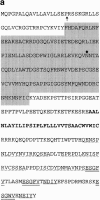
(a) The deduced layilin amino acid sequence is shown. The predicted signal sequence cleavage site is indicated with an arrow. A grey box highlights the C-type lectin homology. A single potential N-linked glycosylation site is marked with a diamond. The proposed transmembrane domain is shown in boldface. Three copies of a 16–18 amino acid repeat (layilin homology 1, LH1) are shown with a broken underline. Three copies of a penta-amino acid repeat (LH2) are double-underlined, and two copies of a tetra-amino acid repeat (LH3) are underlined. The cDNA encoding this polypeptide has been submitted to GenBank with accession number AF093673. (b) Alignment of the carbohydrate-recognition domains from rat mannose-binding protein A, dog E-selectin, and hamster layilin. Shown at the bottom is the CRD consensus sequence derived by Drickamer (1993): single-letter amino acid code indicates absolutely conserved residues; other abbreviations: O, any oxygen-containing amino acid; Z, either E or Q; θ, aliphatic; Φ, aromatic; Ω, aliphatic or aromatic residue (Drickamer, 1993). The alignment was performed using the GCG program PILEUP, and was then manually adjusted. The five amino acid insertions found in layilin and E-selectin are underlined, a 7–amino acid insertion unique to layilin is double-underlined, a second 7–amino acid insertion is shown with a broken underline.
Figure 2
Figure 2
(a) The deduced layilin amino acid sequence is shown. The predicted signal sequence cleavage site is indicated with an arrow. A grey box highlights the C-type lectin homology. A single potential N-linked glycosylation site is marked with a diamond. The proposed transmembrane domain is shown in boldface. Three copies of a 16–18 amino acid repeat (layilin homology 1, LH1) are shown with a broken underline. Three copies of a penta-amino acid repeat (LH2) are double-underlined, and two copies of a tetra-amino acid repeat (LH3) are underlined. The cDNA encoding this polypeptide has been submitted to GenBank with accession number AF093673. (b) Alignment of the carbohydrate-recognition domains from rat mannose-binding protein A, dog E-selectin, and hamster layilin. Shown at the bottom is the CRD consensus sequence derived by Drickamer (1993): single-letter amino acid code indicates absolutely conserved residues; other abbreviations: O, any oxygen-containing amino acid; Z, either E or Q; θ, aliphatic; Φ, aromatic; Ω, aliphatic or aromatic residue (Drickamer, 1993). The alignment was performed using the GCG program PILEUP, and was then manually adjusted. The five amino acid insertions found in layilin and E-selectin are underlined, a 7–amino acid insertion unique to layilin is double-underlined, a second 7–amino acid insertion is shown with a broken underline.
Figure 3
Figure 3
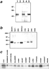
Expression of layilin protein in cells and tissues. (a) A rabbit polyclonal antiserum was raised against a synthetic peptide comprising the COOH-terminal 20 amino acids of layilin and used to Western blot a lysate of CHO cells. Lane 1, immune serum; lane 2, immune serum preincubated with 1 mg/ml layilin peptide; lane 3, immune serum; lane 4, immune serum after depletion over immobilized layilin peptide; lane 5, affinity-purified antibody eluted from the immobilized layilin peptide. The position and size in kilodaltons of molecular weight standards are shown. (b) Western blot with affinity-purified anti-layilin antiserum of mouse, rat, monkey, hamster, and human cell lines. Specifically reacting bands in rat cell lines migrate slightly faster than do the prevalent immunoreactive species in monkey, mouse and hamster. NRK, normal rat kidney; REF, rat embryo fibroblast. The position and size in kD of molecular weight standards are shown. 5 μg of lysate were loaded per lane. (c) Organs were dissected from a healthy adult female mouse, homogenized, and lysed in gel-loading buffer. 10 μg of each lysate were loaded per lane, and were blotted for layilin. Total CHO protein is included to indicate the position of layilin.
Figure 4
Figure 4
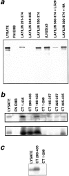
(a) GST fusion proteins containing portions of the layilin cytoplasmic domain were immobilized on agarose and mixed with a CHO cell detergent lysate. Each fusion protein contains the layilin amino acids indicated, except (LH23)×3, which has three copies of the amino acid motif spanning layilin amino acids 243–252. After washing, the agarose beads were boiled in gel-loading buffer, and the released material was analyzed by Western blotting for talin. The lanes containing proteins bound by GST fusions of fibronectin EIIIB (FN EIIIB) or layilin are overloaded approximately 13-fold relative to the total lysate lane. (b) Glutathione agarose preloaded with GST fusions containing either FN EIIIB or the indicated amino acids of chicken talin was incubated with a CHO cell detergent lysate, washed, and boiled in gel-loading buffer. Proteins released from the beads were detected by Western blotting for focal adhesion kinase (FAK, top) or layilin (bottom). The lanes containing proteins bound by GST fusions to chicken talin and fibronectin EIIIB are overloaded approximately 25-fold relative to the total lysate lane. (c) Glutathione agarose preloaded with GST fusions containing various fragments of chicken talin was incubated with a detergent lysate of NIL8 cells expressing HA-tagged layilin, washed, and boiled in gel-loading buffer. Proteins released from the beads were detected by Western blotting for the HA-epitope tag. Lanes containing material bound to GST fusions are overloaded approximately 20-fold relative to the total lysate lane.
Figure 5
Figure 5
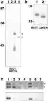
Layilin is a glycoprotein expressed on the cell surface. (a) CHO cells were surface-labeled with biotin, lysed in RIPA buffer, immunoprecipitated with either peptide-depleted (lanes 1 and 2) or affinity-purified (lanes 3 and 4) anti-layilin antiserum, and labeled bands were detected with HRP-streptavidin. Samples in lanes 2 and 4 were treated with PNGase F to remove N-linked carbohydrates. The position of the layilin band is marked with a solid arrowhead before PNGase F treatment, and with an open arrowhead after PNGase F treatment. The position and size in kD of molecular mass standards are shown. (b) A detergent lysate of CHO cells was treated with (lane 2) or without (lane 1) PNGase F to remove N-linked sugars, Western-blotted, and probed with affinity-purified anti-layilin antiserum. Note that essentially all the layilin present in the lysate is PNGase F-sensitive. The position and size in kD of molecular weight standards are shown. (c) Layilin is predominantly found on the cell surface. CHO cells were surface-labeled with biotin (lanes 2–4) or mock surface-labeled without biotin (lanes 5–7), lysed, and passed over an avidin column to remove labeled material. The first three fractions eluted from each column (lanes 2–4 and 5–7) were analyzed by Western blotting for β1-integrin and layilin. When assayed for β1-integrin (top), the lysate (lane 1) contains two bands: the upper band (arrow) represents mature β1-integrin; the lower band (asterisk) is an intracellular precursor. Biotin selectively labels the mature (top) band without affecting the precursor form of β1 (compare lanes 2 and 5, top), indicating the reagent does not have access to the cells' interior. The same fractions (bottom) reveal that most layilin, like mature β1-integrin, is biotin-labeled (compare lanes 2 and 5, bottom). All lanes were loaded with equivalent fractions of the cell lysate.
Figure 6
Figure 6
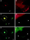
Localization of HA-tagged layilin in spreading (a, c, and e) or migrating (b, d, and f) NIL8 hamster cells. Cells spreading on a fibronectin matrix (a, c, and e) contain peripheral ruffles that stain for both HA-layilin (a) and phalloidin (e). Yellow ruffles in the double exposure (c) show the extent of overlap of staining. Migrating NIL8 cells also contain layilin-rich ruffles at their leading edges (b, d, and f). Cells shown are stained with 12CA5 in the presence of 1 mg/ml LC20 (b), 12CA5 in the presence of 1 mg/ml HA peptide (d), and the same cell as in d double-stained with affinity-purified anti-layilin antiserum (f). Bar, 10 μm.

References

    1. Albigès-Rizo C, Frachet P, Block MR. Down regulation of talin alters cell adhesion and the processing of the α5β1 integrin. J Cell Sci. 1995;108:3317–3329. - PubMed
    1. Algrain M, Turunen O, Vaheri A, Louvard D, Arpin M. Ezrin contains cytoskeleton and membrane binding domains accounting for its proposed role as a membrane-cytoskeleton linker. J Cell Biol. 1993;120:129–139. - PMC - PubMed
    1. Alwine JC, Kemp DJ, Stark GR. Method for detection of RNAs in agarose gels by transfer to diazobenzyloxymethyl-paper and hybridization with DNA probes. Proc Natl Acad Sci USA. 1977;74:5350–5354. - PMC - PubMed
    1. Anderson RA, Marchesi VT. Regulation of the association of membrane skeletal protein 4.1 with glycophorin by a polyphosphoinositide. Nature. 1985;318:295–298. - PubMed
    1. Beckerle MC. The adhesion plaque protein, talin, is phosphorylated in vivo in chick embryo fibroblasts exposed to a tumor-promoting phorbol ester. Cell Regul. 1990;1:227–236. - PMC - PubMed

Publication types

MeSH terms