. 1998 Oct 19;143(2):469-86.
doi: 10.1083/jcb.143.2.469.

Onset of keratin 17 expression coincides with the definition of major epithelial lineages during skin development

Affiliations

Onset of keratin 17 expression coincides with the definition of major epithelial lineages during skin development

K M McGowan et al. J Cell Biol. .

Abstract

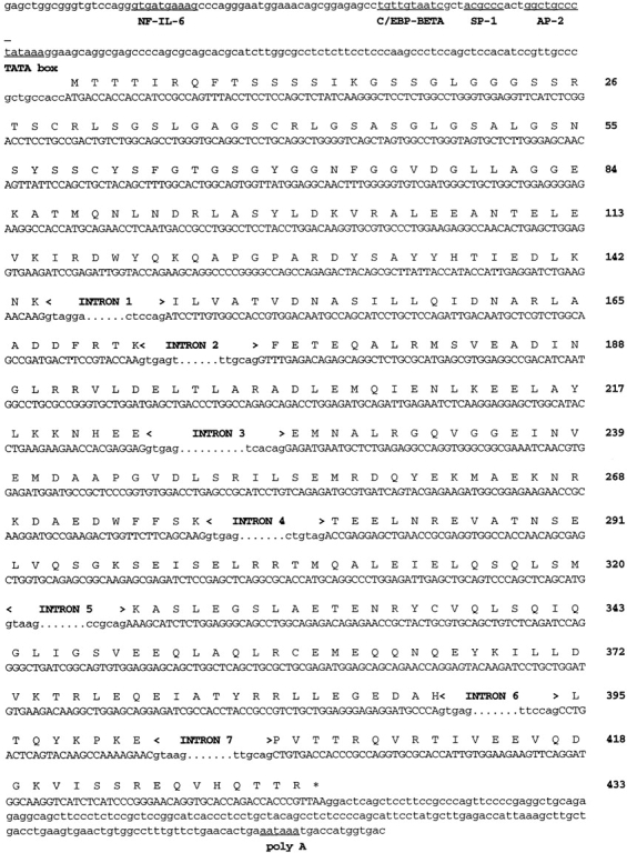
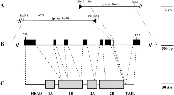
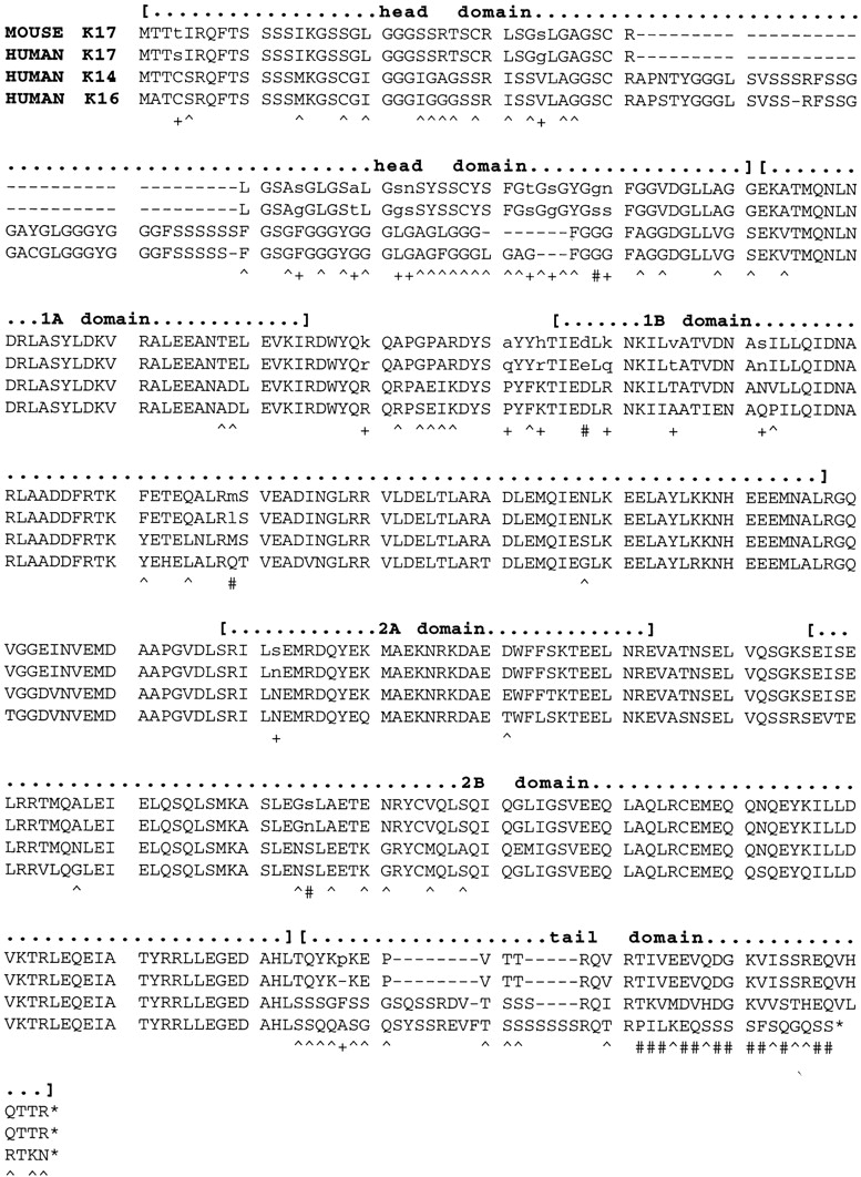
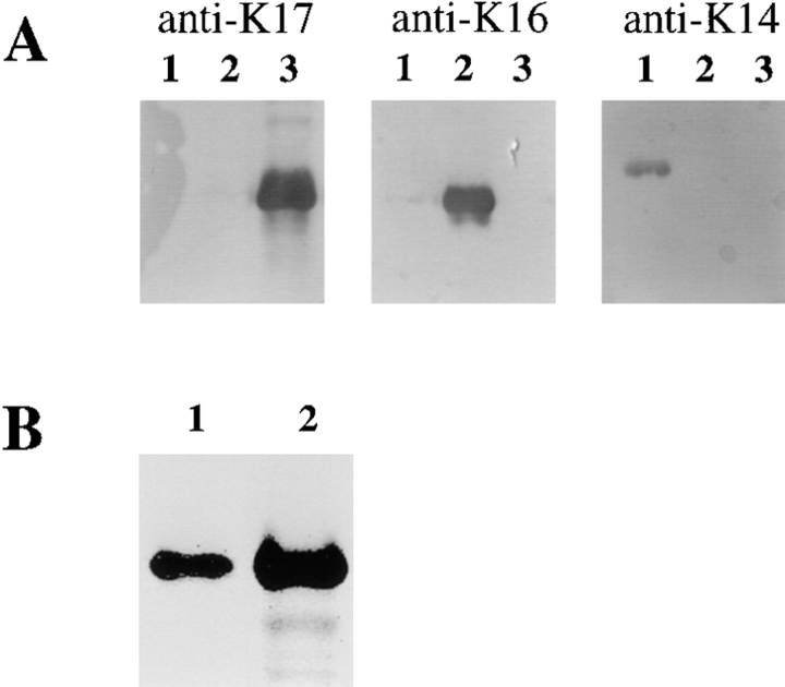
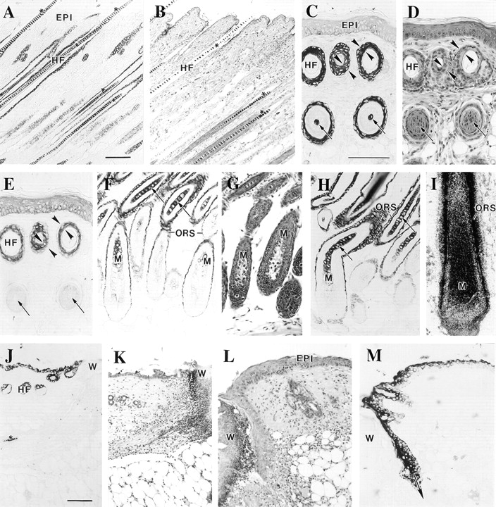
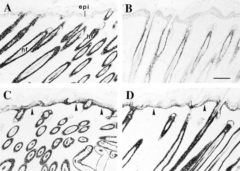
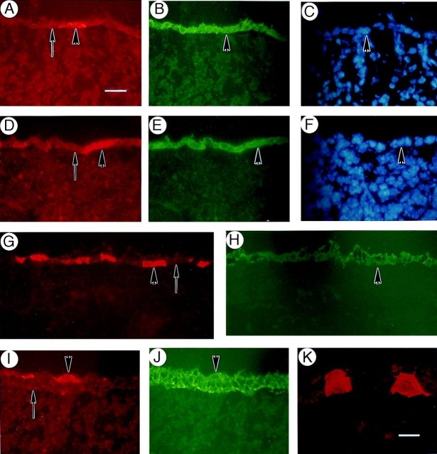
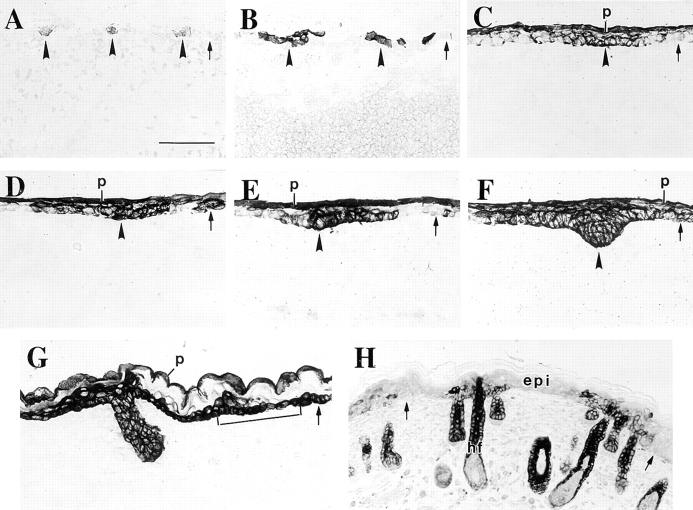
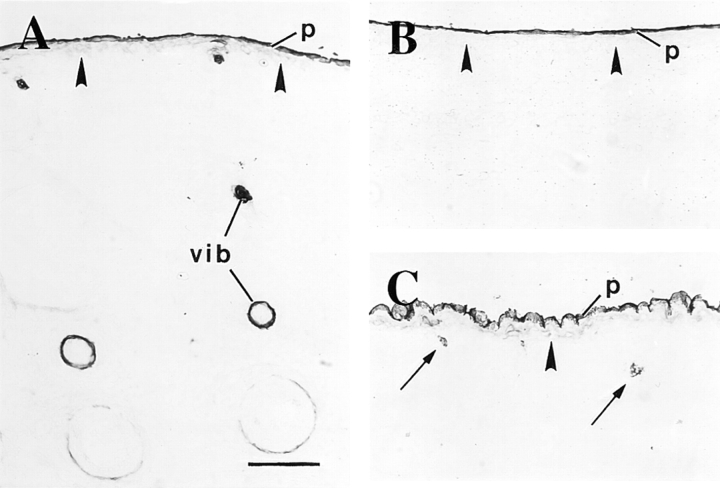
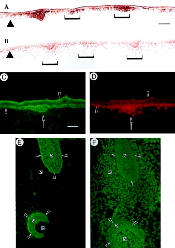
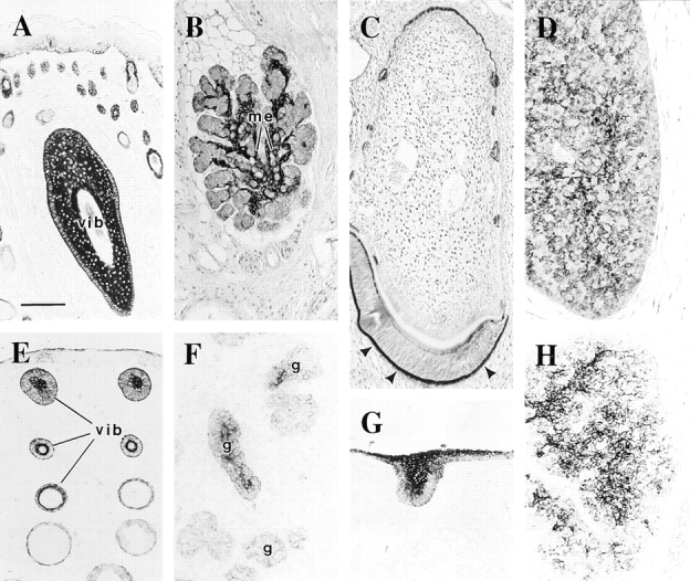
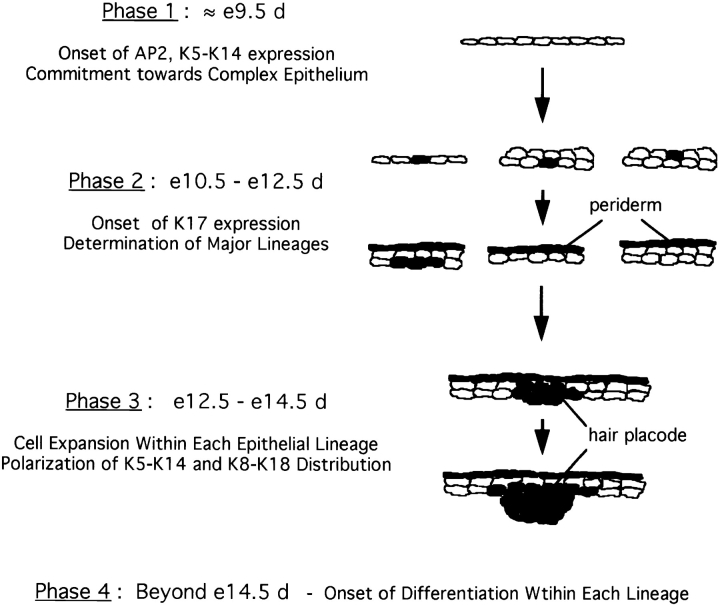
The type I keratin 17 (K17) shows a peculiar localization in human epithelial appendages including hair follicles, which undergo a growth cycle throughout adult life. Additionally K17 is induced, along with K6 and K16, early after acute injury to human skin. To gain further insights into its potential function(s), we cloned the mouse K17 gene and investigated its expression during skin development. Synthesis of K17 protein first occurs in a subset of epithelial cells within the single-layered, undifferentiated ectoderm of embryonic day 10.5 mouse fetuses. In the ensuing 48 h, K17-expressing cells give rise to placodes, the precursors of ectoderm-derived appendages (hair, glands, and tooth), and to periderm. During early development, there is a spatial correspondence in the distribution of K17 and that of lymphoid-enhancer factor (lef-1), a DNA-bending protein involved in inductive epithelial-mesenchymal interactions. We demonstrate that ectopic lef-1 expression induces K17 protein in the skin of adult transgenic mice. The pattern of K17 gene expression during development has direct implications for the morphogenesis of skin epithelia, and points to the existence of a molecular relationship between development and wound repair.


Figures

Figure 2
Figure 2
Nucleotide sequence of exons and flanking regions for the mouse K17 gene. Sequences corresponding to protein coding strand for exons 1–8 are shown in uppercase letter, and the predicted amino acid encoded (one-letter code) is indicated directly above. Amino acids are numbered at right starting with the initial methionine. The 5′ boundary of exon 1 has not been determined. Sequences corresponding to intron boundaries are shown as lowercase letters. In the 5′ upstream sequence, the canonical TATA sequence (−95 bp from the ATG translation start) is underlined, as are potential binding sites for known transcription factors (Boulikas, 1994). In the 3′ sequence, the canonical polyA signal sequence (AATAAA) is underlined. This sequence can be accessed at GenBank (GenBank/EMBL/DDBJ accession number applied for).
Figure 1
Figure 1
Relationship between genomic and protein domain structure for mouse K17. From the two overlapping phage clones designated 16-8 and 16-9 shown in A, a full-length type I keratin gene was reconstructed as shown in B. DNA sequencing showed that this gene contains an open reading frame identical to that of a near full-length (this study) and partial (Knapp et al., 1987) cDNA clones for mouse K17, and highly related to a genomic clone for human K17 (Troyanovsky et al., 1992). The structure of the mouse K17 gene is identical to that of the human orthologous gene and all other type I keratin genes (Troyanovsky et al., 1992), in that it contains eight exons (black boxes in B) and seven intervening introns comprised within ∼5 kilobases of sequence. (C) K17 is predicted to show the tripartite domain structure common to all IF proteins with a nonhelical (HEAD) domain at the NH2 terminus, a central domain featuring extended regions of heptad repeats (1A, 1B, 2A, and 2B), and another nonhelical (TAIL) domain at the COOH terminus. There is no obvious relationship beween the genomic and protein domain structures.
Figure 3
Figure 3
Alignment of the predicted amino acid sequences for mouse K17 (this study) and human K17 (Troyanovsky et al., 1992), K14 (Marchuk et al., 1984), and K16 (Paladini et al., 1995). This alignment was produced using the DNASIS v.3.5 software (Hitachi Software Engineering Inc., Tokyo, Japan), using default parameters. The boundaries of the major domains shared by all IF proteins (Fuchs and Weber, 1994) are indicated with brackets: a nonhelical head domain at the NH2 terminus; the α-helical subdomains 1A, 1B, 2A, and 2B characteristic of the central rod domain; and the nonhelical tail domain at the COOH terminus. Asterisks, nonsense stop codons. Underneath the aligned sequences, the symbols + and # identify residues that are different between mouse and human K17; #, the subset for which mouse K17 is identical to either K14 or K16; ^, residues that are conserved between mouse and human K17 but different in both K14 and K16.
Figure 4
Figure 4
Validation of the specificity of a rabbit antiserum raised against a conjugated 12-mer peptide corresponding to the COOH terminus of mouse and human K17. (A) 50 ng of FPLC-purified human recombinant K14 (lane 1), K16 (lane 2), and K17 (lane 3) were prepared as described (Wawersik et al., 1997), electrophoresed in triplicate, and transferred on nitrocellulose membranes. These were reacted with the monoclonal antibody LL001 directed against K14 (Purkis et al., 1990), a rabbit polyclonal antiserum directed against K16 (Takahashi et al., 1994), and the newly produced antiserum against K17 (this study). (B) Immunoblot analysis of electrophoresed total skin proteins (20 μg) prepared from the back of an adult mouse (lane 2). A single band comigrating with purified human recombinant K17 (25 ng; lane 1), with an apparent M r of 48 kD, reacts with the polyclonal anti-K17 peptide antiserum made for this study.
Figure 5
Figure 5
Immunolocalization of K17 protein in sections prepared from adult mouse skin. Tissues were fixed, paraffin embedded, and 5-μm sections were processed for hematoxylin-eosin staining, for K17 of K6 immunohistochemistry using a peroxidase-based detection method, or for in situ hybridization as described (Carroll et al., 1997). A (K17 stain) and B (H & E), consecutive cross sections of intact trunk skin showing anagen phase hair follicles (HF). C (K17 stain), D (H & E), and E (K6 stain) represent consecutive cross sections of adult tail skin where on can observe profiles of hair follicles at two different levels. In contrast to K6, K17 immunostaining occurs in both levels, indicating a more extensive distribution along the longitudinal axis of the follicle. Furthermore, K17 staining is present in all the ORS layers whereas K6 is polarized to the innermost layers, as indicated by the opposing arrowheads in C, D, and E. Finally, the arrows in these frames point to the occurrence of K17 staining in the hair shaft in the lower segment of the follicle. F (K17 stain), G (H & E), and H (K17 stain) document the occurrence of K17 immunoreactivity in the matrix epithelial cells (M) and early differentiating trichocytes (arrows). This staining is clearly distinct from that specific to the ORS compartment. (I) Localization of K17 mRNA by in situ hybridization using a 3′ noncoding probe. In this large vibrissae follicle, a strong hybridization occurs for both the hair matrix (M) and hair epithelial cells as well as the ORS. J (K17 stain), K, L (H & E), and M (K17 stain) illustrate the induction of K17 immunoreactivity in wound edge epidermis at 8 h (J and K) and 48 h (L and M) after injury. W, wound site. Bars: (A and B) 100 μm; (C–I) 100 μm; (J–M) 100 μm.
Figure 11
Figure 11
Immunolocalization of keratins in the skin of transgenic mice that ectopically express lef-1 in the basal layer of epidermis. Skin tissue samples were fixed in Bouin's, paraffin embedded, and then sections were immunostained for either K17 or K6 using a peroxidase-based detection method. (A) Trunk skin from a 11-d-old nontransgenic littermate control stained for K17. As in Fig. 5, the hair follicle (hf) ORS is strongly immunostained, whereas the interfollicular epidermis (epi) is negative. (B–D) Trunk skin from a 11-d-old transgenic animal stained for K6 (B) and K17 (C and D). Consistent with the normal histology (data not shown), K6 protein occurs in the innermost layers of the hair follicle ORS, whereas the epidermis is negative. In contrast, the basal layer of transgenic epidermis shows a strong signal for K17 (arrowheads in C and D), as does the hair follicle ORS. Arrowhead, dermo-epidermal interface. Bar, 100 μm.
Figure 6
Figure 6
Distribution of K17 protein an early stage of mouse skin development. Triple- immunofluorescence staining was performed on sections prepared from fresh-frozen e12.5-d mouse embryos to assess the context in which K17 expression first occurs in the embryonic ectoderm. (A–C) Portion of the upper dorsum showing single-layer ectoderm as determined by the vital Hoechst stain for DNA (C). A single cell is seen to express K17 (arrowhead in A), whereas K14 expression is uniformly established at that stage (B). (D–F) Another example of single-layer ectoderm (Hoechst stain in F). Two adjacent cells are seen to express K17 (arrowhead in D), whereas uniform K8–K18 expression is maintained at that stage (E). (G and H) Portion of the lower dorsum showing two-layered ectoderm. In this example, K17 induction occurs in the bottom layer of the ectoderm (arrowhead in G), which is otherwise uniformly stained for K14 (H). (J and K) Portion of the ventral surface showing three-layered ectoderm. In this case, K17 induction occurs in the top layer of the ectoderm (arrowhead in I), whereas again the staining for K14 is uniform (J). (K) At higher magnification, the filamentous nature of the anti-K17 staining is readily apparent in individual epithelial cells. In all panels, the arrow depicts the interface between the surface epithelium and the underlying mesenchyme. Bars: (A–J) 20 μm; (K) 10 μm.
Figure 7
Figure 7
Distribution of K17 protein during skin development. Sections were prepared from fresh-frozen e10.5-, e12.5-, e14.5-, and e16.5-d mouse embryos and from newborn mice and immunostained for K17 using a peroxidase-based detection method. Examples were chosen so as to represent the continuum of events characterizing skin morphogenesis. K17 is first induced in the single layer ectoderm in e10.5-d embryos (arrowheads in A). At e12.5 d, small clusters of K17-positive cells occur at periodic intervals in the cross sectioned ectoderm (arrowheads in B). These clusters subsequently evolve into placodes and then primary hair germs, as suggested by the pattern of K17-staining in frames C–G (open arrowheads). At e14.5 d, the periderm (p) is also distinctly stained for K17 (C–E). Moreover, beginning at e14.5 d (F) and firmly established by e16.5 d (bracket in G), the basal layer of embryonic epidermis expresses K17. Between e16.5 d and birth (H), the staining for K17 in the basal layer as well as the periderm is gradually lost. At birth, the skin features an adult pattern of K17 expression, in that the latter is largely restricted to the hair follicles. In all frames, the small arrow depicts the interface beween the surface epithelium and the underlying mesenchyme. See text for further explanation. Bars: (A–G) 50 μm; (H) 82 μm.
Figure 8
Figure 8
Distribution of K6 isoform proteins during skin development. Sections were prepared from fresh-frozen mouse embryos and immunostained for K6 protein using a peroxidase-based detection method. K6 staining is first detected in e14.5-d embryos, where it is restricted to a subset of cells within the vibrissae (vib in A) and the periderm (p; shown are whisker pads in A and trunk skin in B). At e16.5 d, K6 staining is detected in a small subset of epithelial cells within primary hair germs (arrows in C), coinciding with the onset of differentiation-specific gene expression (see text). Solid arrowhead, interface between the surface epithelium and the underlying mesenchyme. Bar, 100 μm.
Figure 9
Figure 9
Colocalization of K17 and lef-1 during skin development. Sections were prepared from fresh-frozen mouse embryos at specific developmental stages and immunostained for K17 and for the transcription factor lef-1, either on consecutive (A and B and E and F) or the same sections (C and D, double immunofluorescence). (A and B) Upper dorsum of a e12.5-d embryo stained for K17 (A) and lef-1 (B). Brackets denote placodes at various stages of their formation resulting from epithelio–mesenchymal interactions. A strong, epithelium-restricted staining for K17 is seen in these placodes, while the signal for lef-1 is confined to the nucleus in both the epithelial cells and the proximal mesenchymal cells. (C and D) Upper dorsum of a e12.5-d embryo subjected to double immunofluorescence for K17 (C) and lef-1 (D). K17 is expressed strongly in the placode epithelial cells denoted by an arrow and in the periderm (inverted arrowhead), and sporadically in the embryonic basal layer. Although excluded from the periderm, a strong nuclear staining for lef-1 is detected in the placode epithelial cells and in the mesenchymal cells proximal to it. In addition, embryonic basal cells that express K17 strongly are also immunopositive for lef-1. The upward arrowheads in A–D depict the interface between the epithelium and the underlying mesenchyme. (E and F) Consecutive sections through vibrissae follicles (v) from an e14.5-d embryo subjected to immunofluorescence for K17 (E) and lef-1 (F). Although K17 immunoreactivity is restricted to the epithelial cells, the signal for lef-1 is particularly strong in the nucleus of mesenchymal cells (m) surrounding the epithelium. Arrowheads, interface between the epithelium and the mesenchyme. Bars: (A and B) 70 μm; (C–F) 20 μm.
Figure 10
Figure 10
Distribution of K17 protein in adult- and embryonic-stage epithelia known to develop in a lef-1–dependent manner. Sections were prepared from either paraffin-embedded (A–D) or fresh-frozen e14.5-d whole embryos (E–H), and immunostained for K17 using a peroxidase-based detection method. A (adult) and E (embryo), whisker pads; vib, a tangentially sectioned vibrissae follicle in A. B (adult) and F (embryo), submaxillary gland; me, myoepithelium that envelops the glandular epithelial tissue. C (adult) and G (embryo), tooth. Arrowhead, enamel organ. D (adult) and H (embryo), thymus. Bar, 100 μm.
Figure 12
Figure 12
A model for early skin morphogenesis. This model relates the induction of K17 expression in developing mouse skin to other molecular events as described by Byrne et al. (1994). For the sake of simplicity, this model focuses on the events involving keratinocytes and ignores the other cell types. Shading, presence of K17 protein. (Phase 1) The onset of K5–K14 expression in the single-layer embryonic ectoderm, beginning at ∼e9.5 d, reflects a commitment towards the formation of a complex epithelium. Induction of K5–K14 occurs concomitant with, but independently of, stratification to a two-layered epithelium. (Phase 2) The onset of K17 expression, starting at ∼e10.5 d, reflects the determination of the major lineages in skin epithelia. Specifically, we postulate that K17 is induced in the subset of K8–K18, K5–K14 expressing epithelial cells that are responding to the mesenchymal cues that induce appendage formation, and in cells that will give rise to periderm. As is the case for K5–K14 approximately a day earlier, onset of K17 expression may occur in single-layer ectoderm or alternatively, in either layer of a two-layer-thick epithelium, depending on the region of the body. (Phase 3) Cell expansion and onset of epithelial polarization. Between e12.5 and e14.5 d, the epithelial sheet thickens considerably and clusters of K17-expressing epithelial cells extend into the underlying mesenchyme at sites of strong lef-1 transcription factor expression. Consistent with the lack of differentiation-specific gene expression, we propose that this time window corresponds primarily to a cell expansion phase. (Phase 4) Onset of cell type-specific differentiation. Starting at e14.5 d as previously reported, terminal differentiation is initiated within the various lineages as manifested through K1–K10 expression in embryonic epidermis and hair-specific keratin gene expression in hair germs. The periderm is destined to be lost via shedding beginning at e16.5 d.

References

    1. Bessho Y, Nakanishi S, Nawa H. Glutamate receptor agonists enhance the expression of BDNF mRNA in cultured cerebellar granule cells. Mol Brain Res. 1993;18:201–208. - PubMed
    1. Bonnekoh B, Huerkamp C, Wevers A, Geisel J, Sebok B, Bange FC, Greenhalgh DA, Bottger EC, Krieg T, Mahrle G. Up-regulation of keratin 17 expression in human HaCaT keratinocytes by interferon-gamma. J Invest Dermatol. 1995;104:58–61. - PubMed
    1. Boulikas T. A compilation and classification of DNA binding sites for protein transcription factors from vertebrates. Crit Rev Eukaryot Gene Expr. 1994;4:117–322. - PubMed
    1. Bousquet O, Coulombe PA. Cytoskeleton: missing links found? . Curr Biol. 1996;6:1563–1566. - PubMed
    1. Brock J, McCluskey J, Baribault H, Martin P. Perfect wound healing in the keratin 8 deficient mouse embryo. Cell Motil Cytoskeleton. 1996;35:358–366. - PubMed

Publication types

MeSH terms

Associated data