. 1998 Oct 19;143(2):523-32.
doi: 10.1083/jcb.143.2.523.

The nonreceptor protein tyrosine phosphatase PTP1B binds to the cytoplasmic domain of N-cadherin and regulates the cadherin-actin linkage

Affiliations

The nonreceptor protein tyrosine phosphatase PTP1B binds to the cytoplasmic domain of N-cadherin and regulates the cadherin-actin linkage

J Balsamo et al. J Cell Biol. .

Abstract

Cadherin-mediated adhesion depends on the association of its cytoplasmic domain with the actin-containing cytoskeleton. This interaction is mediated by a group of cytoplasmic proteins: alpha-and beta- or gamma- catenin. Phosphorylation of beta-catenin on tyrosine residues plays a role in controlling this association and, therefore, cadherin function. Previous work from our laboratory suggested that a nonreceptor protein tyrosine phosphatase, bound to the cytoplasmic domain of N-cadherin, is responsible for removing tyrosine-bound phosphate residues from beta-catenin, thus maintaining the cadherin-actin connection (). Here we report the molecular cloning of the cadherin-associated tyrosine phosphatase and identify it as PTP1B. To definitively establish a causal relationship between the function of cadherin-bound PTP1B and cadherin-mediated adhesion, we tested the effect of expressing a catalytically inactive form of PTP1B in L cells constitutively expressing N-cadherin. We find that expression of the catalytically inactive PTP1B results in reduced cadherin-mediated adhesion. Furthermore, cadherin is uncoupled from its association with actin, and beta-catenin shows increased phosphorylation on tyrosine residues when compared with parental cells or cells transfected with the wild-type PTP1B. Both the transfected wild-type and the mutant PTP1B are found associated with N-cadherin, and recombinant mutant PTP1B binds to N-cadherin in vitro, indicating that the catalytically inactive form acts as a dominant negative, displacing endogenous PTP1B, and rendering cadherin nonfunctional. Our results demonstrate a role for PTP1B in regulating cadherin-mediated cell adhesion.


Figures
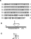
Figure 1
Figure 1
Chick PTP1B cDNA and protein. (A) Comparison of the amino acid sequence of chick PTP1B (GenBank/EMBL/DDBJ accession number U86410) and its human homologue PTP1B (GenBank/EMBL/DDBJ accession number M31724). Alignments were done with Geneworks software (Intelligenetics, Campbell, CA). Boxes, identical residues; underline, unique chicken sequence used to raise a polyclonal antibody. (B) Recombinant chick PTP1B was constructed with a c-myc or HA tag in frame at the NH2 terminus of the full-length PTP1B sequence. Thick underline, consensus PTP domain. The position of the chicken specific sequence is indicated (amino acids 347–356). Catalytically inactive PTP1B was constructed by site-directed mutagenesis in which cysteine 215, within the consensus PTP catalytic domain, was changed to serine. (C) A Triton X-100 homogenate of embryonic chick neural retinas was fractionated by SDS-PAGE, transferred to a PVDF membrane, and then immunoblotted with the chicken-specific antibody (chk) or the anti-PTP1B antibody from TL. Left, numbers indicate the migration of the molecular mass standards.
Figure 2
Figure 2
Distribution of chick PTP1B in neural retina and association with N-cadherin in retina and N-cadherin–expressing L cells. (A) Homogenates of embryonic chick neural retina were fractionated into ER-enriched (M1) and plasma membrane-enriched (M2) fractions. Each were separated by SDS-PAGE, transferred to PVDF, and then immunoblotted with the anti-chkPTP1B antibody. (B) Chick neural retina or N-cadherin– expressing L cells were homogenized in neutral detergent, immunoprecipitated with the anti–N-cadherin antibody NCD-2 (NCD) or control rat IgG (Co). The precipitates were separated by SDS-PAGE, transferred to PVDF, and then immunoblotted with anti-chkPTP1B antibody. Left, numbers indicate the migration of the molecular mass standards.
Figure 4
Figure 4
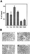
Expression of mutant PTP1B inhibits N-cadherin–mediated adhesion. (A) Adhesion assays. Cells were harvested using 0.1% trypsin (GIBCO BRL) in buffer containing 1 mM Ca2+. Single cells were plated on 96-well plates coated with anti–N-cadherin antibody NCD-2. Cells adhering to the substrate were measured after 45 min by staining with crystal violet. Co, LN cells; Cov, LN cells transfected with vector lacking insert; Wt, LN cells transfected with the wild-type PTP1B; Mut, LN cells transfected with the mutant PTP1B. Adhesion is plotted as percent of control values. Each point is the average of three measurements. The results presented are from one experiment representative of many. (B) Phase–contrast micrographs showing the phenotype of control LN cells (panel a), LN cells transfected with vector alone (panel b), LN cells transfected with mutant PTP1B (panel c), and LN cells tranfected with wild-type PTP1B (panel d).
Figure 3
Figure 3
Expression of N-cadherin and chick PTP1B in transfected cells as detected by RT-PCR and immunoblot. (A) RT-PCR was performed using primers specific to tagged chkPTP1B, N-cadherin, and actin to ensure that comparable amounts of material were used. (B) Expression of chick PTP1B peptide in transfected cells. Cell lysates were fractionated by SDS-PAGE, transferred to PVDF membrane, and then immunoblotted with the indicated antibodies. Co, parental N-cadherin–expressing cells; Wt, LN cells transfected with wild-type PTP1B; Mut, LN cells transfected with mutant PTP1B. Left, numbers indicate the migration of the molecular mass standards.
Figure 5
Figure 5
β-catenin shows increased levels of tyrosine-phosphate in LN cells transfected with mutant PTP1B. LN cells transfected with either wild-type (Wt) or mutant (Mut) PTP1B were lysed in neutral detergent, immunoprecipitated with anti–N-cadherin antibody NCD-2, and then separated into bound and free fractions. (A) The NCD-2–bound fractions were separated by SDS-PAGE and immunoblotted with anti–β-catenin antibody. (B) The NCD-2–free fractions were immunoprecipitated with anti–β-catenin antibody, separated by SDS-PAGE, and then immunoblotted with anti–β-catenin antibody or antiphosphotyrosine antibody PY20. Left, numbers indicate the migration of the molecular mass standards.
Figure 6
Figure 6
The association of N-cadherin with actin is disrupted in cells transfected with mutant PTP1B. LN cells were transfected with wild-type (Wt), or mutant (Mut) chkPTP1B or with vector alone and immunoprecipitated with anti–N-cadherin antibody NCD-2. The immunoprecipitates were fractionated by SDS-PAGE and immunoblotted with (A) anti-actin antibody or (B) NCD-2. Left, numbers indicate the migration of the molecular mass standards.
Figure 7
Figure 7
ChkPTP1B associates with N-cadherin in transfected LN cells. LN cells transfected with HA-tagged chick PTP1B were homogenized in neutral detergent-containing buffer and immunoprecipitated with anti–N-cadherin antibody NCD-2. The immunoprecipitates were separated by SDS-PAGE and immunoblotted with (A) anti-HA antibody, (B) anti–N-cadherin antibody NCD-2, and (C) anti-phosphotyrosine antibody PY20. Co, LN cells transfected with vector alone; Wt, LN cells transfected with the wild-type PTP1B; Mut, LN cells transfected with the mutant PTP1B. Left, numbers indicate the migration of the molecular mass standards.
Figure 8
Figure 8
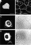
Cellular distribution of chkPTP1B in LN cells. LN cells transfected with vector containing GFP alone, wild-type GFP–chkPTP1B, or mutant GFP–chkPTP1B were immunostained with anti-GFP antibody and analyzed by laser confocal microscopy. LN cells expressing GFP alone show diffuse GFP (A). The same cells stained with anti–β-catenin antibody show the characteristic morphology of L cells expressing N-cadherin with β-catenin localized at sites of cell–cell contact (B). LN cells expressing either wild-type or mutant GFP–chkPTP1B show extensive perinuclear (ER) localization (C and E). Wild-type GFP–chkPTP1B is also localized to areas of cell-cell contact (C and D [phase]). LN cells expressing mutant GFP–chkPTP1B show little or no fluorescence at the cell periphery (E and F [phase]) and lack the well-defined borders seen in control cells and cells expressing wild-type GFP–chkPTP1B. Bar, 20 μm.
Figure 9
Figure 9
Association of GFP–chkPTP1B fusion protein with N-cadherin. LN cells transfected with GFP– chkPTP1B were homogenized in neutral detergent-containing buffer and immunoprecipitated with anti–N-cadherin antibody NCD-2. The immunoprecipitates were separated by SDS-PAGE and immunoblotted with the indicated antibodies. (A) Anti-GFP antibody, (B) anti-PTP1B antibody. Co, LN cells transfected with vector alone; Wt, LN cells transfected with the wild-type PTP1B; Mut, LN cells transfected with the mutant PTP1B. Asterisk, position of IgG heavy chain. Left, numbers indicate the migration of the molecular mass standards. Arrows, position of GFP–PTP1B at ∼70 kD and endogenous PTP1B at ∼40 kD.
Figure 10
Figure 10
In vitro association of PTP1B with N-cadherin. Phosphorylated, catalytically inactive, GST– c-myc-chkPTP1B fusion protein was purified on a glutathione–Sepharose affinity column. One aliquot was treated with 0.2 U recombinant protein tyrosine phosphatase (Yersinia enterolitica, Calbiochem-Novabiochem, La Jolla, CA) and the reaction stopped with 4 mM vanadate. Purified N-cadherin was incubated with control or dephosphorylated PTP1B for 20 min at room temperature. Putative complexes were precipitated with anti–c-myc antibody and separated by SDS-PAGE. PTP1B-bound N-cadherin was detected with the anti–N-cadherin antibody NCD-2. Phosphorylated and dephosphorylated c-myc PTP1B were detected with anti–c-myc antibody. The molecular mass of the bacterial PTP1B fusion protein, 50 kD, represents the entire ORF. Asterisk at ∼55 kD, position of immunoglobulin heavy chain derived from the anti–c-myc antibody. Left, migration of N-cadherin and PTP1B. Right, numbers indicate the migration of the molecular mass standards.

Similar articles

Cited by

References

    1. Arregui, C.O., J. Balsamo, and J. Lilien. 1998. Impaired integrin-mediated adhesion and signaling in fibroblasts expressing a dominant-negative mutant PTP1B. J. Cell Biol. 143:In press. - PMC - PubMed
    1. Balsamo J, Thiboldeaux R, Swaminathan N, Lilien J. Antibodies to the retina N-acetylgalactosaminylphosphotransferase modulate N-cadherin–mediated adhesion and uncouple the N-cadherin transferase complex from the actin-containing cytoskeleton. J Cell Biol. 1991;113:429–436. - PMC - PubMed
    1. Balsamo J, Ernst H, Zanin MK, Hoffman S, Lilien J. The interaction of the retina cell surface N-acetylgalactosaminylphosphotransferase with an endogenous proteoglycan ligand results in inhibition of cadherin-mediated adhesion. J Cell Biol. 1995;129:1391–1401. - PMC - PubMed
    1. Balsamo J, Leung T, Ernst H, Zanin MK, Hoffman S, Lilien J. Regulated binding of a PTP1B-like phosphatase to N-cadherin: control of cadherin mediated adhesion by dephosphorylation of β-catenin. J Cell Biol. 1996;134:801–813. - PMC - PubMed
    1. Bandyopadhyay, D., A. Kusari, K.A. Kenner, F. Liu, J. Chernoff, T.A. Gustafson, and J. Kusari. 1997. Protein-tyrosine phosphatase 1B complexes with the insulin receptor in vivo and is tyrosine-phosphorylated in the presence of insulin. J. Biol. Chem 272:1639–1645. - PubMed

Publication types

MeSH terms

Associated data