. 1998 Nov 1;18(21):8625-36.
doi: 10.1523/JNEUROSCI.18-21-08625.1998.

The epithelial inward rectifier channel Kir7.1 displays unusual K+ permeation properties

Affiliations

The epithelial inward rectifier channel Kir7.1 displays unusual K+ permeation properties

F Döring et al. J Neurosci. .

Abstract

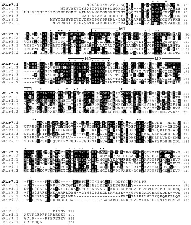
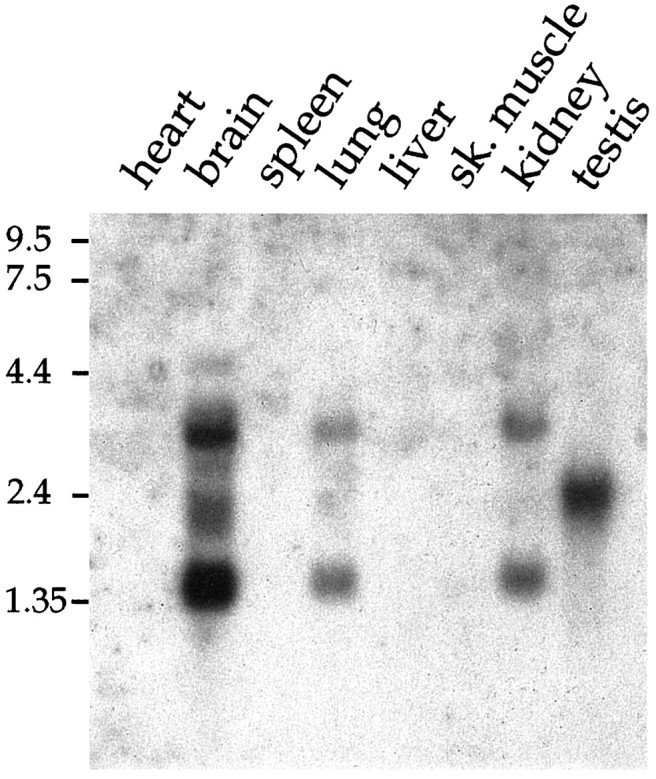
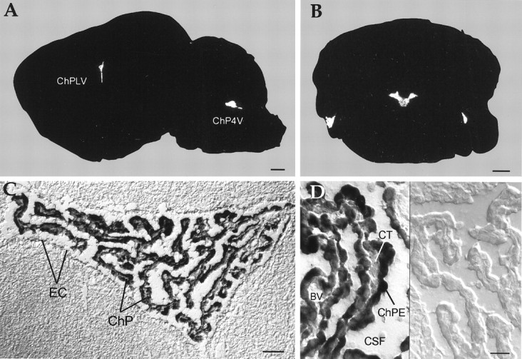
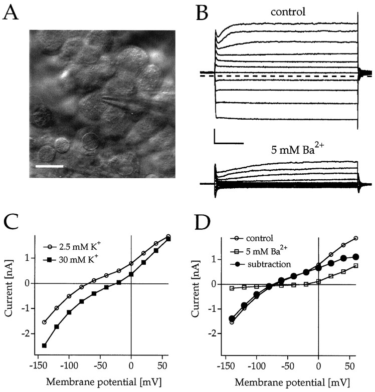
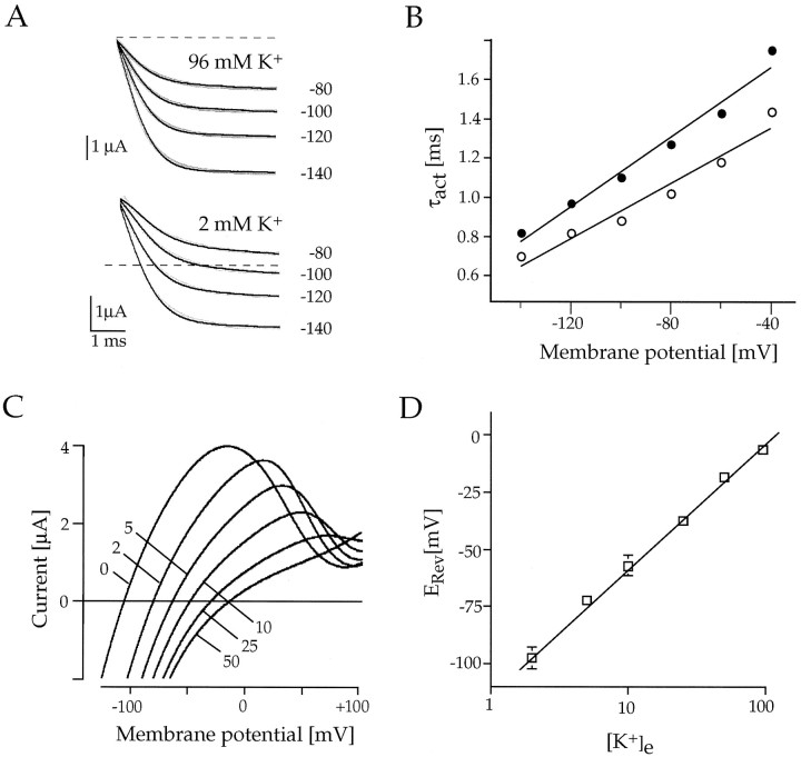
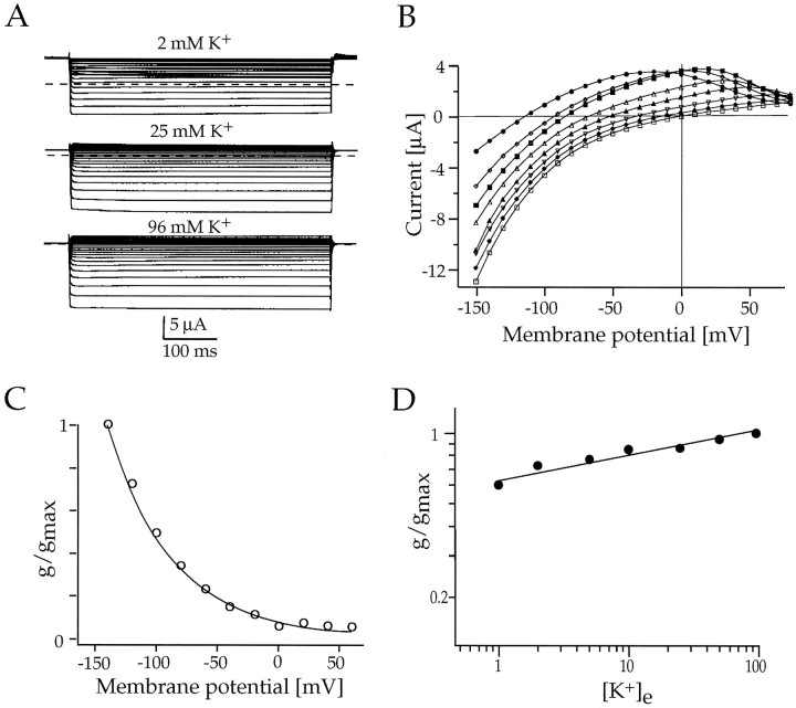
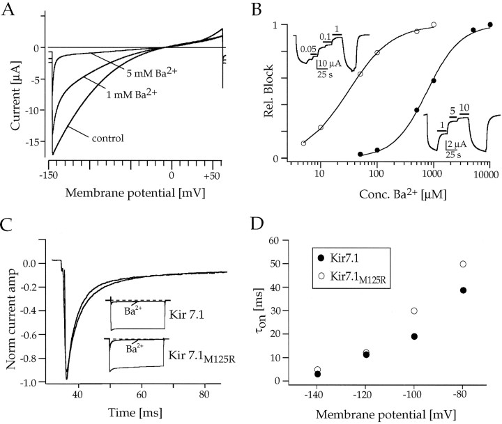
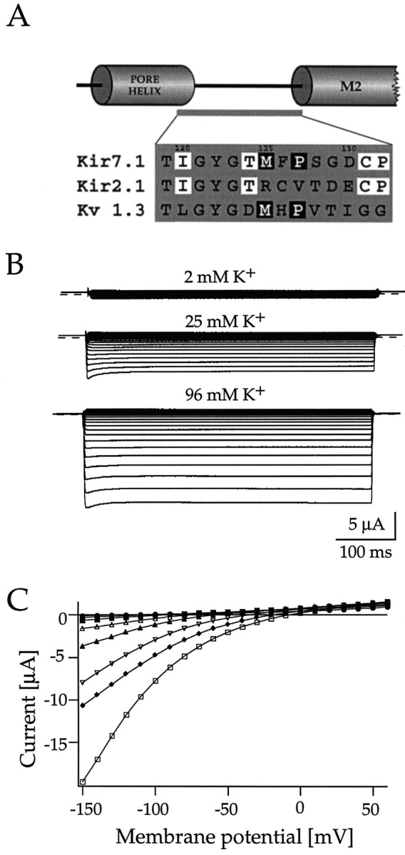
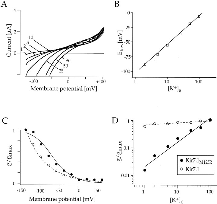
Rat and human cDNAs were isolated that both encoded a 360 amino acid polypeptide with a tertiary structure typical of inwardly rectifying K+ channel (Kir) subunits. The new proteins, termed Kir7.1, were <37% identical to other Kir subunits and showed various unique residues at conserved sites, particularly near the pore region. High levels of Kir7.1 transcripts were detected in rat brain, lung, kidney, and testis. In situ hybridization of rat brain sections demonstrated that Kir7.1 mRNA was absent from neurons and glia but strongly expressed in the secretory epithelial cells of the choroid plexus (as confirmed by in situ patch-clamp measurements). In cRNA-injected Xenopus oocytes Kir7.1 generated macroscopic Kir currents that showed a very shallow dependence on external K+ ([K+]e), which is in marked contrast to all other Kir channels. At a holding potential of -100 mV, the inward current through Kir7.1 averaged -3.8 +/- 1.04 microA with 2 mM [K+]e and -4.82 +/- 1.87 microA with 96 mM [K+]e. Kir7.1 has a methionine at position 125 in the pore region where other Kir channels have an arginine. When this residue was replaced by the conserved arginine in mutant Kir7.1 channels, the pronounced dependence of K+ permeability on [K+]e, characteristic for other Kir channels, was restored and the Ba2+ sensitivity was increased by a factor of approximately 25 (Ki = 27 microM). These findings support the important role of this site in the regulation of K+ permeability in Kir channels by extracellular cations.


Figures

Fig. 1.
Fig. 1.
Comparison of the amino acid sequences of Kir7.1 with representative subunits of other Kir subfamilies. The predicted 360 amino acids of human Kir7.1 (single-letter code) are shown aligned with sequences of human Kir1.2 (GenBank accession numberU73192), human Kir2.1 (U12507), human Kir3.3 (U52152), rat Kir5.1 (X83581), and human Kir6.2 (D50582). Residues are shaded inblack in instances in which other subunits are identical to Kir 7.1; asterisks denote residues conserved in all known Kir channels, and boxed residues (arrowheads) indicate where Kir7.1 is different from the consensus of all other Kir channels. Outlined also are the transmembrane segments M1 and M2 and the pore-forming P-region (H5). Amino acid gaps within the alignment are indicated by short bars. The GenBank accession number for the human and rat Kir7.1 sequences are AJ006128and AJ006129, respectively.
Fig. 2.
Fig. 2.
Northern blot analysis showing the distribution of Kir7.1 mRNA in rat heart, brain, spleen, lung, liver, skeletal muscle, kidney, and testis. Blots containing 2 μg of poly(A+) RNA from each tissue were hybridized with32P-labeled cDNA probes specific for rat Kir7.1. The positions of RNA size markers (in kb) are indicated (left).
Fig. 3.
Fig. 3.
mRNA localization of Kir7.1 in the rat brain as revealed by in situ hybridization. Brain sections were hybridized with 35S-labeled oligonucleotides (A, B) or digoxigenin-labeled cRNA probes (C, D), as described in Materials and Methods. X-ray film autoradiographs of sagittal (A) and coronal (B) sections show mRNA expression only in the choroid plexus of the fourth (A, B) and lateral (A) ventricles. Exposure time, 19 d.C, Bright-field photomicrographs with Nomarski optics show an overview of the choroid plexus (C) and epithelial cells (D) at higher magnification. Transcripts were found only by using antisense cRNA probes (left side), but not sense cRNA probes (right side). Note that epithelial cells surrounding the plexus vasculature are not labeled. BV, Blood vessel; ChP4V, choroid plexus of the fourth ventricle; ChPE, epithelial cells of the choroid plexus; ChPLV, choroid plexus of the lateral ventricle; CSF, cerebrospinal fluid;CT, connective tissue; EC, ependyma cells lining the ventricle; Scale bars: 1 mm in A, B; 50 μm in C; 20 μm in D.
Fig. 4.
Fig. 4.
Demonstration of intrinsic inwardly rectifying K+ conductances inhibited by Ba2+in rat choroid plexus epithelial cells in situ.A, IR-DIC image of an epithelial cell recorded with a patch-clamp pipette. Scale bar, 10 μm. B, Current responses of an epithelial cell to brief voltage steps between +60 and −140 mV from a holding potential of Vh = −60 mV in 2.5 mm [K+]e in the absence (control) and presence of 5 mm Ba2+. Calibration: 0.5 nA, 20 msec.C, Current–voltage (I–V) relationships of the endogenous current in 2.5 (○) and 30 mm [K+]e (▪) measured at the end of the voltage pulse. D, I–Vrelationship of the currents shown in B. The • represents subtraction currents before and after the application of 5 mm Ba2+.
Fig. 5.
Fig. 5.
Characterization of macroscopic Kir7.1 inwardly rectifying currents in Xenopus oocytes.A, Current responses of an oocyte expressing Kir7.1 to brief steps between −80 and −140 mV from a holding potential ofVh = 0 mV in 2 and 96 mm[K+]e, as indicated.B, Time constants of activation (τact) are plotted against the membrane potential for [K+]e = 2 mm (•) and for [K+]e = 96 mm (○).C, Kir7.1 currents in response to fast voltage ramps show outward currents and a shift of reversal potentials with altering [K+]e, as indicated.D, Zero currents (reversal potentials,ERev) of Kir7.1 currents that are in close agreement with EK are plotted versus the extracellular concentration of K+([K+]e) on a semi-logarithmic scale. Solid lines are linear regression fits to the data.
Fig. 6.
Fig. 6.
Whole-cell voltage-clamp responses ofXenopus oocytes expressing Kir7.1 channels.A, Macroscopic currents in response to 500 msec voltage steps between −80 and −140 mV from a holding potential ofVh = 0 mV in 2, 25, and 96 mm[K+]e, as indicated.B, Current–voltage (I–V) relationship of Kir7.1 currents measured at the end of the 500 msec voltage pulse in 0 (•), 1 (⋄), 2 (▪), 5 (▵), 10 (▴), 50 (♦), and 96 mm (■) extracellular K+.C, Normalized chord conductances of Kir7.1 (G/Gmax) in 96 mm K+ are plotted versus the membrane voltage. The G–V relationship was fit by a single exponential. D, Double-logarithmic plot of the Kir7.1 chord conductance as a function of [K+]e. Conductances were measured atGmax, and the data were fit toG =m([K+]e)n, where m and n are variables.
Fig. 7.
Fig. 7.
Analysis of the block by extracellular Ba2+ of wild-type Kir7.1 and mutant Kir7.1M125R inXenopus oocytes. A, Ramp current responses to voltage ramps of 2 sec duration between −150 and +60 mV show the voltage dependence of IKir7.1 block by 1 and 5 mm Ba2+. B, Current inhibition relative to a maximum block by a saturating concentration of Ba2+ is plotted versus the concentration of the blocking cation at a holding potential ofVh = −80 mV. Curves are least-squares fits of data points to a Hill equation (1/1 + [A/Ki]n, giving a Ki for Ba2+ of 670 μm for Kir7.1 (•) and aKi for Ba2+ of 27 μm for Kir7.1M125R (○). Kiis the concentration of cation producing 50% block; Aand n are variables. Insets, Shown are continuous recordings of wild-type and mutant Kir7.1 currents at −80 mV under Ba2+ block. The application of the blocker is indicated by black bars. C, Time- and voltage-dependent block of wild-type and mutant Kir7.1M125R channels by Ba2+. Shown are single-exponential fits to the time course of the current block by a saturating concentration of Ba2+ (10 mm for Kir7.1 and 500 μm for Kir7.1M125R). Insets, Shown are the complete current responses of Kir7.1 and Kir7.1M125R channels to 500 msec voltage steps between 0 and −120 mV. D, Relationship of time constants of activation (τon) and the membrane potential for wild-type Kir7.1 (•) and mutant Kir7.1M125R (○) channels at 96 mm external K+.
Fig. 8.
Fig. 8.
Macroscopic currents through mutant Kir7.1M125R channels in Xenopus oocytes. A, Sequence alignment of the core region between the pore helix and the M2 (inner) helix of Kir7.1, Kir2.1, and Kv1.3 channels, as suggested by Doyle et al. (1998), show the residues mutated in this study.Boxed in white are residues conserved in all Kir channels; boxed in black are residues identical between Kir7.1 and mostly present in Kv channels.B, Whole-cell voltage-clamp responses of oocytes expressing mutant Kir7.1M125R channels to 500 msec voltage steps between −80 and −140 mV from a holding potential ofVh = 0 mV in 2, 25, and 96 mm[K+]e, as indicated.C, Current–voltage (I–V) relationship of Kir7.1M125R currents measured at the end of the 500 msec voltage pulse in 0 (•), 1 (⋄), 2 (▪), 5 (▵), 10 (▴), 50 (♦), and 96 mm (■) extracellular K+.
Fig. 9.
Fig. 9.
Functional characteristics of mutant Kir7.1M125R channels in Xenopus oocytes. A, Kir7.1M125R currents in response to fast voltage ramps show a shift of reversal potentials with altering [K+]e, as indicated.B, Zero currents (reversal potentials,ERev) of Kir7.1M125R currents, which are in close agreement with EK, are plotted versus the extracellular concentration of K+([K+]e) on a semi-logarithmic scale. The solid line is a linear regression fit to the data. C, Normalized chord conductances of Kir7.1M125R (G/Gmax) in 96 mm K+ are plotted versus the membrane voltage (•). The ○ indicates the values for the wild-type Kir7.1. The G–V relationship was fit by a single Boltzmann function [G = 1/1 + exp(VoV1/2/k)], with a midpointV1/2 = −68.6 mV and a slope factor k of 23 mV. D, Double-logarithmic plot of the Kir7.1M125R chord conductance (•) as a function of [K+]e. The ○ indicates values for the wild-type Kir7.1). Conductances were measured atGmax, and data were fit toG =m([K+]e)n, where m and n are variables.

Similar articles

Cited by

References

    1. Altschul SF, Madden TL, Shaffer AA, Zhang J, Miller W, Lipman DJ. Gapped BLAST and PSI-BLAST: a new generation of protein database search programs. Nucleic Acids Res. 1997;25:3389–3402. - PMC - PubMed
    1. Armstrong C. The vision of the pore. Science. 1998;280:56–57. - PubMed
    1. Baumann A, Krah-Jentgens I, Müller R, Müller-Holtkamp F, Seidel R, Kecskemethy N, Casal J, Ferrus A, Pongs O. Molecular organization of the maternal effect region of the Shaker complex of Drosophila: characterization of an IA channel transcript with homology to vertebrate Na+ channel. EMBO J. 1987;6:3419–3429. - PMC - PubMed
    1. Cohen NA, Brenman JE, Snyder SH, Bredt DS. Binding of the inward rectifier K+ channel Kir2.3 to PSD-95 is regulated by protein kinase A phosphorylation. Neuron. 1996;17:759–767. - PubMed
    1. Doyle DA, Morais Cabral J, Pfuetzner RA, Kuo A, Gulbis JM, Cohen SL, Chait BT, Mackinnon R. The structure of the potassium channel: molecular basis of K+ conduction and selectivity. Science. 1998;280:69–77. - PubMed

Publication types

MeSH terms

Associated data