. 1998 Nov 1;18(21):8648-59.
doi: 10.1523/JNEUROSCI.18-21-08648.1998.

The localization of the brain-specific inorganic phosphate transporter suggests a specific presynaptic role in glutamatergic transmission

Affiliations

The localization of the brain-specific inorganic phosphate transporter suggests a specific presynaptic role in glutamatergic transmission

E E Bellocchio et al. J Neurosci. .

Abstract

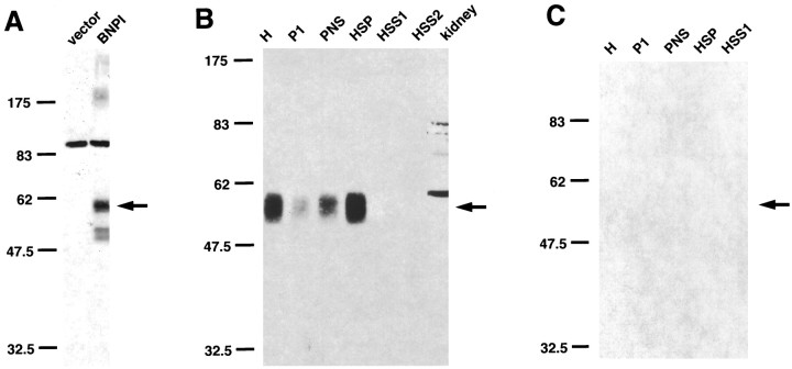
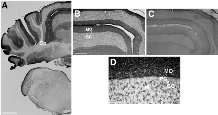
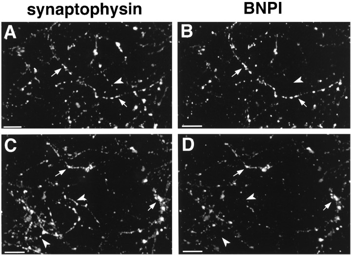
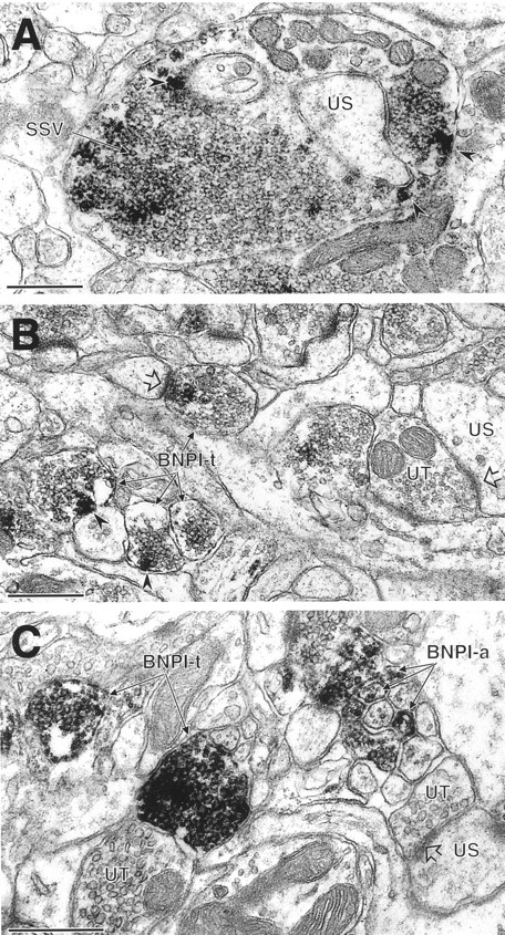
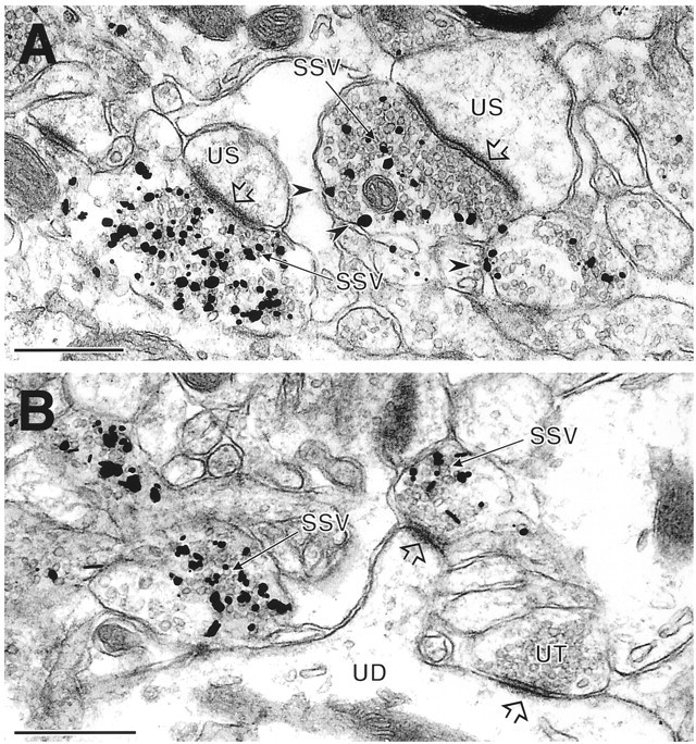
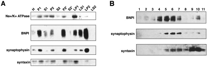
Molecular cloning has recently identified a vertebrate brain-specific Na+-dependent inorganic phosphate transporter (BNPI). BNPI has strong sequence similarity to EAT-4, a Caenorhabditis elegans protein implicated in glutamatergic transmission. To characterize the physiological role of BNPI, we have generated an antibody to the protein. Immunocytochemistry of rat brain sections shows a light microscopic pattern that is suggestive of reactivity in nerve terminals. Excitatory projections are labeled prominently, and ultrastructural analysis confirms that BNPI localizes almost exclusively to terminals forming asymmetric excitatory-type synapses. Although BNPI depends on a Na+ gradient and presumably functions at the plasma membrane, both electron microscopy and biochemical fractionation show that BNPI associates preferentially with the membranes of small synaptic vesicles. The results provide anatomic evidence of a specific presynaptic role for BNPI in glutamatergic neurotransmission, consistent with the phenotype of eat-4 mutants. Because an enzyme known as the phosphate-activated glutaminase produces glutamate for release as a neurotransmitter, BNPI may augment excitatory transmission by increasing cytoplasmic phosphate concentrations within the nerve terminal and hence increasing glutamate synthesis. Expression of BNPI on synaptic vesicles suggests a mechanism for neural activity to regulate the function of BNPI.


Figures

Fig. 1.
Fig. 1.
BNPI antiserum specifically recognizes a 60 kDa protein in transfected COS cells and rat brain. A, COS cells were transfected with rat BNPI cDNA or with vector alone. Equal amounts of protein from each postnuclear supernatant were separated by electrophoresis via 10% SDS-polyacrylamide, transferred to nitrocellulose, and immunoblotted with antiserum generated against the C terminus of BNPI. The antiserum recognizes a single broad ∼60 kDa band in COS cells expressing BNPI (arrow), but not in control cells. Note that the ∼85 kDa background species detected in both BNPI-transfected and control cells, as well as the faint ∼175, 52, and 50 kDa species present only in BNPI-transfected cells, does not occur in the brain. B, BNPI antiserum recognizes a single ∼60 kDa species in rat brain (arrow). Differential centrifugation of rat brain extracts was performed as described in Materials and Methods, and a Western blot containing equal amounts of protein from each fraction was immunolabeled by using the BNPI C-terminal antiserum preadsorbed with a control GST fusion protein. The homogenate (H) contains an ∼60 kDa immunoreactive species. Low-speed centrifugation (1075 × g for 20 min) to remove cell debris (P1) results in the sedimentation of some immunoreactive material, but the majority occurs in the postnuclear supernatant (PNS). Centrifugation of the PNS at 152,000 × gmax for 1 hr sediments BNPI in the high-speed pellet (HSP), whereas the high-speed supernatants (HSS1 and HSS2) contain little immunoreactive material. Postnuclear supernatant from the rat kidney (40 μg of protein) does not express the ∼60 kDa species, consistent with the brain-specific expression of BNPI. The molecular weights of standards (in kilodaltons) are shown to theleft. C, Preadsorption of the BNPI antiserum with the GST fusion protein used as an immunogen prevents detection of the ∼60 kDa species (arrow) in the same rat brain fractions that were used in B.
Fig. 2.
Fig. 2.
BNPI immunohistochemistry at the level of the basal ganglia. Representative 40 μm coronal sections from rat brain were immunolabeled for BNPI by using the antiserum preadsorbed with LAP and either the control GST fusion protein GST-VGAT (A–D) or GST-BNPI as a control (E). A, B, Sections through the basal ganglia show immunoreactivity distributed diffusely throughout the cortex (Cx) and caudate putamen (CPu). The cortex lacks a laminar pattern of immunoreactivity. White matter such as the corpus callosum (cc) shows little labeling. C, D, At high magnification, punctate immunoreactivity occurs in nerve fibers within caudate putamen (C) and cortex (D). Cell bodies show no labeling.E, Adsorption of the antibody with GST-BNPI abolishes immunolabeling (shown here for cortex), confirming the specificity of the reaction. Scale bars: A, 1 mm; B, 100 μm; C–E, 50 μm.
Fig. 3.
Fig. 3.
BNPI immunohistochemistry at the level of the hippocampus. Representative 40 μm coronal sections from rat brain were immunolabeled for BNPI by using the antiserum preadsorbed with LAP and either the control GST fusion protein GST-VGAT (A, B, D, E) or GST-BNPI as a control (C).A, A section through the hippocampus (Hp) shows a distinctive pattern of immunoreactivity, particularly within the molecular and polymorphic layers of the hippocampus. White matter such as the internal capsule (ic) shows little labeling. B, Under higher magnification the hippocampus shows dense immunoreactivity in stratum oriens (O) and stratum radiatum (R), with a marked reduction in labeling in stratum lacunosum moleculare (LM). In the dentate gyrus the outer two-thirds of the molecular layer (M) label more strongly for BNPI than the inner one-third, suggesting preferential localization to excitatory perforant path inputs from the entorhinal cortex. Strikingly, the granule cell body layer of the dentate gyrus (G) and the pyramidal cell body layer (P) of the hippocampus proper both lack substantial immunoreactivity.C, Adsorption of the antibody with GST-BNPI abolishes the immunolabeling, confirming the specificity of the reaction. D, E, A high-magnification view of CA3 (D) shows coarse granular labeling in stratum lucidum (L) at the periphery of the pyramidal cell layer (P), strongly suggestive of mossy fiber synapses. Stratum oriens (O) and stratum radiatum (R) of both CA3 (D) and CA1 (E) show weaker, more diffuse BNPI immunoreactivity. Scale bars: A, 1 mm; B,C, 500 μm; D, 100 μm;E, 50 μm.
Fig. 4.
Fig. 4.
BNPI immunohistochemistry at the level of the caudal midbrain. Representative 40 μm coronal sections from rat brain were immunolabeled for BNPI by using the antiserum preadsorbed with LAP and either the control GST fusion protein GST-VGAT (A, B, D) or GST-BNPI as a control (C, E).A, A section through the rostral pons shows immunoreactivity in gray matter of the periaqueductal gray (PAG) and particularly strong labeling in the pontine nuclei (Pn). White matter such as the corticospinal tracts in the cerebral peduncle (cp), the decussation of the superior cerebellar peduncle (xscp), the lateral lemnisci (ll), and the lateral tegmental tracts (ltg) shows little labeling. B, D,Sections through the midbrain observed under high magnification show uniform punctate immunoreactivity in the neuropil of the substantia nigra (B) and tectum (D). Cell bodies in both regions show no labeling. C, E,Adsorption of the antibody with GST-BNPI abolishes the majority of punctate immunoreactivity in the substantia nigra (C) and tectum (E), confirming the specificity of the reaction. Scale bars:A, 1 mm; B–E, 50 μm.
Fig. 5.
Fig. 5.
BNPI immunohistochemistry at the level of the medulla and cerebellum. Representative 40 μm coronal sections from rat brain were immunolabeled for BNPI by using the antiserum preadsorbed with LAP and either the control GST fusion protein GST-VGAT (A, B, D) or GST-BNPI as a control (C). A, A section through the cerebellum (Cb) shows prominent immunoreactivity in cerebellar cortex. B, A high-magnification view of the cerebellar cortex reveals strong dense labeling in the molecular layer (MO) and lighter labeling in the granule cell layer (GC). C, Adsorption of the antibody with GST-BNPI abolishes the immunolabeling, confirming the specificity of the reaction. D, At high magnification the granule cell layer (GC) shows large punctate structures characteristic of mossy fiber synapses onto granule cells. Within the molecular layer (MO) the immunoreactivity produces a dense coarse labeling pattern suggestive of climbing or parallel fiber synapses onto Purkinje cell dendrites. Cell bodies, including Purkinje cells (PC), granule and Golgi cells, and basket and stellate cells, show no immunoreactivity. Scale bars:A, 1 mm; B, C, 500 μm;D, 50 μm.
Fig. 6.
Fig. 6.
BNPI colocalizes with synaptophysin at a subset of varicosities in primary hippocampal cultures. After 14 d in vitro, primary hippocampal cultures from E19 rats were double-labeled for the synaptic vesicle marker synaptophysin (A, C) and BNPI (B, D). Synaptophysin was detected with a mouse monoclonal antibody and BNPI with the rabbit polyclonal antibody. The primary antibodies were recognized with appropriate secondary antibodies conjugated to rhodamine (A, C) and fluorescein (B, D). Examination of two fields (A, B and C, D) shows that synaptophysin distributes in a punctate manner along neuronal processes (arrows). Essentially all BNPI immunoreactivity distributes in a similar manner, colocalizing with synaptophysin at synaptic structures (arrows). However, many synaptophysin-immunoreactive synapses do not label for BNPI (arrowheads), indicating that BNPI localizes to a subset of terminals. Scale bars, 20 μm.
Fig. 7.
Fig. 7.
BNPI localizes to excitatory-type terminals in the rat hippocampal formation and caudate putamen nucleus.A, In stratum lucidum of the CA3 region of the hippocampus, BNPI peroxidase labeling is seen in a large, complex mossy fiber terminal that contacts an unlabeled spine (US). Although this section does not demonstrate unequivocally the asymmetric nature of the synapse, all mossy fiber terminals form exclusively asymmetric synapses onto dendritic spines (Amaral and Dent, 1981). Within the mossy fiber terminal the peroxidase reaction product is distributed diffusely around the membranes of numerous small synaptic vesicles (SSV). Intense labeling also occurs near the plasma membrane (arrowheads), where it appears to overlie large dense core vesicles. B, In the hilar layer of the dentate gyrus, several small axon terminals (BNPI-t) contain peroxidase labeling for BNPI that is associated with putative dense core vesicles near the plasma membrane (arrowheads). One of these labeled terminals forms an asymmetric synapse (open arrow) with an unlabeled spine. Many unlabeled spines and unlabeled axon terminals are seen in the neuropil. One of the unlabeled terminals (UT) forms an asymmetric synapse (open arrow) with an unlabeled spine (US). C, Intense peroxidase reaction product surrounds the membranes of small vesicles in selected unmyelinated axons (BNPI-a) and two axon terminals (BNPI-t) in the dorsal caudate putamen nucleus. Numerous other unlabeled axons and nerve terminals (UT) are present in the neuropil. The unlabeled terminal (UT) on the right forms an asymmetric synapse (open arrowhead) with an unlabeled dendritic spine (US). Scale bars, 0.5 μm.
Fig. 8.
Fig. 8.
BNPI localizes to synaptic vesicles at asymmetric synapses by immunogold-silver electron microscopy. Immunogold-silver electron microscopy localizes BNPI in axon terminals that form asymmetric excitatory-type synapses (open arrowheads) with unlabeled dendritic shafts (UD) or spines (US) in the rat caudate putamen nucleus.A, Immunogold-silver deposits are seen in direct contact with many small synaptic vesicles (SSV) within the BNPI-labeled terminals. Several gold particles also directly contact the plasma membrane (arrowheads) but only in the vicinity of synaptic vesicles. B, Immunogold-silver labeling for BNPI is associated with SSVs in two axon terminals, one of which forms an asymmetric synapse (open arrowhead) with a spine from the unlabeled dendrite (UD). An adjacent unlabeled terminal (UT) also forms an asymmetric synaptic contact (open arrowhead) with the shaft of the same dendrite. Scale bars, 0.5 μm.
Fig. 9.
Fig. 9.
BNPI resides on synaptic vesicles by differential centrifugation and velocity sedimentation. A,Synaptosomes and synaptic vesicles were prepared from rat brain. Equal amounts of protein from each fraction were loaded into lanes and analyzed by Western analysis. BNPI appears in both the insoluble debris (P1) and postnuclear supernatant (S1). Further, BNPI sediments with the plasma membrane marker Na+/K+-ATPase, the synaptic vesicle marker synaptophysin, and the presynaptic plasma membrane marker syntaxin in the crude synaptosomal fractions P2and P2′ rather than with the high-speed supernatantsS2 and S2′. After hypo-osmotic lysis of the synaptosomes, the Na+/K+-ATPase and syntaxin occur principally in LP1, strongly suggesting the localization of plasma membrane fragments to this fraction. In contrast, the first supernatant (LS1) contains more BNPI and synaptophysin than the first pellet (LP1), suggesting localization of BNPI to synaptic vesicles. Further, high-speed sedimentation ofLS1 shows localization of both BNPI and synaptophysin toLP2 rather than to LS2. Thus, BNPI cofractionates with synaptophysin rather than with the plasma membrane markers, suggesting localization to a population of synaptic vesicles. B, Fractions 1–11 were collected from the top of a 5–25% glycerol velocity gradient of LS1. Western analysis of equal volumes of each fraction shows that BNPI cofractionates with synaptophysin in the middle of the gradient. In contrast, the synaptic plasma membrane marker syntaxin occurs predominantly at the bottom of the gradient. Thus, BNPI occurs on synaptic vesicles rather than on the presynaptic plasma membrane. The small amount of syntaxin cofractionating with synaptophysin presumably reflects the low levels of syntaxin known to occur on synaptic vesicles.

Similar articles

Cited by

References

    1. Amaral DG, Dent JA. Development of the mossy fibers of the dentate gyrus. I. A light and electron microscopic study of the mossy fibers and their expansions. J Comp Neurol. 1981;195:51–86. - PubMed
    1. Aoki C, Kaneko T, Starr A, Pickel VM. Identification of mitochondrial and non-mitochondrial glutaminase within select neurons and glia of rat forebrain by electron microscopic immunocytochemistry. J Neurosci Res. 1991;28:531–548. - PubMed
    1. Arriza JL, Fairman WA, Wadiche JI, Murdoch GH, Kavanaugh MP, Amara SG. Functional comparison of three glutamate transporter subtypes cloned from human motor cortex. J Neurosci. 1994;14:5559–5569. - PMC - PubMed
    1. Arriza JL, Eliasof S, Kavanaugh MP, Amara SG. Excitatory amino acid transporter 5, a retinal glutamate transporter coupled to a chloride conductance. Proc Natl Acad Sci USA. 1997;94:4155–4160. - PMC - PubMed
    1. Avery L. The genetics of feeding in Caenorhabditis elegans. Genetics. 1993;133:897–917. - PMC - PubMed

Publication types

MeSH terms

LinkOut - more resources