. 1998 Nov 1;18(21):8740-50.
doi: 10.1523/JNEUROSCI.18-21-08740.1998.

Differences in Ca2+ channels governing generation of miniature and evoked excitatory synaptic currents in spinal laminae I and II

Affiliations

Differences in Ca2+ channels governing generation of miniature and evoked excitatory synaptic currents in spinal laminae I and II

J Bao et al. J Neurosci. .

Abstract

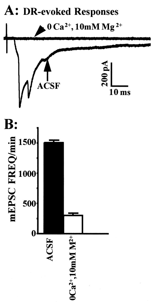
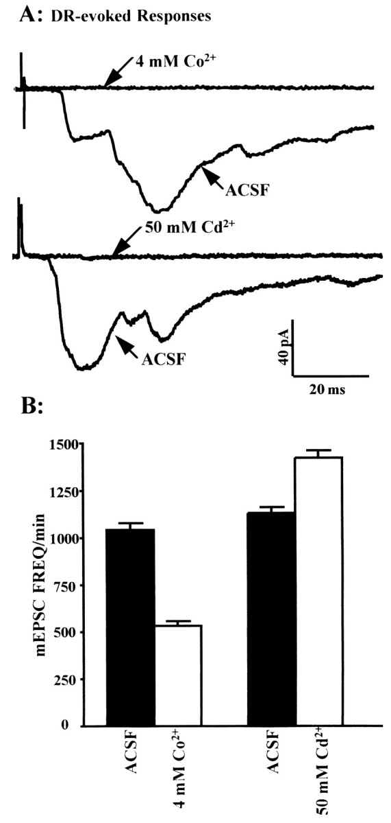
Many neurons of spinal laminae I and II, a region concerned with pain and other somatosensory mechanisms, display frequent miniature "spontaneous" EPSCs (mEPSCs). In a number of instances, mEPSCs occur often enough to influence neuronal excitability. To compare generation of mEPSCs to EPSCs evoked by dorsal root stimulation (DR-EPSCs), various agents affecting neuronal activity and Ca2+ channels were applied to in vitro slice preparations of rodent spinal cord during tight-seal, whole-cell, voltage-clamp recordings from laminae I and II neurons. The AMPA/kainate glutamate receptor antagonist CNQX (10-20 microM) regularly abolished DR-EPSCs. In many neurons CNQX also eliminated mEPSCs; however, in a number of cases a proportion of the mEPSCs were resistant to CNQX suggesting that in these instances different mediators or receptors were also involved. Cd2+ (10-50 microM) blocked evoked EPSCs without suppressing mEPSC occurrence. In contrast, Ni2+ (</=100 microM), a low-threshold Ca2+ channel antagonist, markedly decreased mEPSC frequency while leaving evoked monosynaptic EPSCs little changed. Selective organic antagonists of high-threshold (HVA) Ca2+ channels, nimodipine, omega-Conotoxin GVIA, and Agatoxin IVA partially suppressed DR-EPSCs, however, they had little or no effect on mEPSC frequency. La3+ and mibefradil, agents interfering with low-threshold Ca2+ channels, regularly decreased mEPSC frequency with little effect on fast-evoked EPSCs. Increased [K+]o (5-10 mM) in the superfusion, producing modest depolarizations, consistently increased mEPSC frequency; an increase suppressed by mibefradil but not by HVA Ca2+ channel antagonists. Together these observations indicate that different Ca2+ channels are important for evoked EPSCs and mEPSCs in spinal laminae I and II and implicate a low-threshold type of Ca2+ channel in generation of mEPSCs.


Figures

Fig. 1.
Fig. 1.
Effects of TTX on evoked and spontaneous mEPSCs. Tight-seal, whole-cell recording from a lamina IIo neuron in a transverse spinal cord slice before and during exposure to 1 μm TTX. Voltage-clamp mode; inward current is indicated by downward deflection; standard K+ internal pipette solution. A, Average of 10 consecutive responses evoked by electrical pulse stimulation of the ipsilateral, segmental dorsal root repeated at 5 sec intervals (DR-evoked). Initial component of inward current judged to be monosynaptic based on the stability of its latency to near threshold stimuli (data not shown). Illustrated responses were initiated by superthreshold stimuli; inward and outward current components could be dissociated by adjusting the intensity of the DR stimulus. B, Sample analog records of mEPSC activity under control conditions (ACSF) and during superfusion with 1 μm TTX (bottom). C, Number of mEPSCs per minute for the second through the fifth minute (± SEM) of a 5 min continuous recording with standard superfusion solution (ACSF) and with ACSF containing 1 μm TTX. Error bars indicate SEM. See Materials and Methods for additional details.
Fig. 2.
Fig. 2.
Reversible suppression of mEPSCs by an AMPA/kainate glutamate receptor antagonist (CNQX). Tight-seal, whole-cell, voltage-clamp recording from a lamina I-IIoneuron. Each vertical column displays a continuous record (from top down) of ongoing miniature currents recorded during an 8 sec period.Control, Superfusion with standard ACSF.CNQX, Recording toward the end of a 4 min superfusion of ACSF containing CNQX (10 mm). Wash, Recording after 5 min of superfusion with standard ACSF after the exposure to CNQX, showing partial recovery of mEPSC occurrence. See Materials and Methods for additional details.
Fig. 3.
Fig. 3.
Effects of zero Ca2+ and 10 mm Mg2+ in superfusing ACSF on DR-evoked and background mEPSCs recorded from a lamina II neuron.A, EPSCs evoked by stimulation of the segmental dorsal root (DR-evoked). Average of 10 consecutive DR-evoked responses under control conditions (ACSF) and after 5 min superfusion with ACSF containing zero Ca2+ and 10 mmMg2+. B, Average mEPSC frequency during superfusion with standard ACSF and with ACSF containing zero Ca2+ and 10 mm Mg2+. See Figure 1 legend and Materials and Methods for additional details.
Fig. 4.
Fig. 4.
Examples of the effects of Co2+and Cd2+ on evoked EPSCs and mEPSCs recorded from laminae I and II neurons. Tight-seal, whole-cell recordings in voltage-clamp mode. Only one agent was used on a given neuron and a spinal slice. A, Average EPSCs evoked by 10 successive stimuli at 5 sec intervals to the segmental dorsal root. The evoked responses were obtained at the end of a 5 min sample of ongoing activity for the data in B. ACSF, Control conditions using standard superfusion. 4 mmCo2+, Records obtained at end of a 5 min exposure to the Co2+ superfusate. 50 μmCd2+, Records obtained at the end of a 5 min exposure to the Cd2+superfusate. B, Average mEPSC frequency during control ACSF superfusion (solid bars) and during exposure to ACSF containing the indicated divalent ion (open bars); same neurons and manipulations as in A. The difference between the mean mEPSC frequency for the standard ACSF and for the exposure to divalent cation in both cases had a chance probability ofp < 0.001 (Student’s t test). See Figure 1 legend for additional details.
Fig. 5.
Fig. 5.
Effects of Ni2+ on evoked EPSCs and mEPSCs recorded from a lamina II neuron. A, Average of 10 consecutive DR-evoked responses under control (ACSF) and after 5 min superfusion with ACSF containing 100 μmNi2+. The initial inward current phase had the constant latency attributable to a monosynaptic connection. The later phases varied more in latency suggesting a polysynaptic linkage.B, Sample analog records from neuron of Ashowing background mEPSC activity under control conditions (ACSF) and during superfusion with ACSF containing 100 μmNi2+ (bottom). C, Average mEPSC frequency from neuron of A during control ACSF superfusion (solid bar) and during superfusion with ACSF containing 100 μm Ni2+(open bar). Difference between mean values inC had a chance probability of p < 0.001 (Student’s t test). See legends for Figures 1 and4 for other details.
Fig. 6.
Fig. 6.
Effects of La3+ and mibefradil (Ro 40–5967) on evoked EPSCs and mEPSCs of laminae I and II neurons.A, B, Tight-seal, whole-cell recordings in voltage clamp mode from different slices. (Note separate calibrations). EPSCs evoked by stimulation of the segmental dorsal root. La3+ (5 μm) and Ro 40–5967 (2.5 μm) were added to standard ACSF. See legends for Figures1 and 3 for additional information. C, Same neurons as in A and B. Bars showing mean mEPSC frequency (± SEM) for second to fifth minute of a 5 min sample of background activity during superfusion with the standard ACSF (solid bars) and ACSF containing the indicated agent (open bars). Differences between control and test mEPSC frequencies in both instances had chance probability ofp < 0.01 (Student’s t test).
Fig. 7.
Fig. 7.
Effects of mibefradil (Ro 40–5967) on background mEPSCs. Tight-seal, whole-cell recording from a neuron in lamina I-IIo. A, Average amplitude of mEPSCs during the 4 consecutive min of recording used for the graph ofB. B, Average mEPSC frequency (± SEM) during second to fifth minute of 5 min superfusion with the indicated solution. C, Amplitude distribution of mEPSCs during 1 min of control superfusion and during 1 min of superfusion with ACSF containing 5 μm Ro 40–5967. Same recording asA and B.
Fig. 8.
Fig. 8.
Effects of increased [K+]o on evoked EPSCs and mEPSCs and its block by mibefradil (Ro 40–5967). Each barindicates the average percentage change from the control level (standard ACSF superfusion). Filled bars, mEPSC frequency per minute. Open bars, Peak amplitude of the DR-evoked EPSC. [K+]o was increased from 2.5 to either 5 or to 10 mm. Right, [K+]o in the superfusion was increased to indicated level 10 min after exposure to 5 μm Ro 40–5967. The n above each pair of bars indicates the number of neurons tested.
Fig. 9.
Fig. 9.
Action of selective antagonists of HVA Ca2+ channels on EPSCs in laminae I-II neurons.A, Average DR-evoked EPSCs; i,ii, iii are from different spinal cord slices. Calibration in Ai applies to all.B, Each set of three bars represents observations made on a neuron whose responses are shown in A and indicates mEPSC frequency per minute (± SEM) for the last 4 min of 5 min observation periods. Solid filled bars, Control superfusion (ACSF). Open bars, Mean frequency after 10 min application of the indicated agent. Patterned bars, Effect of increasing [K+]o to 10 mm in the presence of the indicated Ca2+channel antagonist. Note: Increase in mEPSC frequency after nimodipine shown was greater than average (see Table 1C for additional information).

References

    1. Bao J, Li J, Perl ER. Different presynaptic Ca2+ channels influence evoked EPSCs and spontaneous, miniature EPSCs in rodent spinal I and II neurones. J Physiol (Lond) 1997;504.P:175P. - PubMed
    1. Bao J, Li J, Wall B, Perl ER. Low threshold (T type) Ca2+ channels control miniature EPSC frequency in the spinal substantia gelatinosa. Soc Neurosci Abstr. 1995;21:1572.
    1. Bardoni R, Goldstein PA, Lee CJ, Gu JG, MacDermott AB. ATP P2X receptors mediate fast synaptic transmission in the dorsal horn of the rat spinal cord. J Neurosci. 1997;17:5297–5304. - PMC - PubMed
    1. Bessou P, Perl ER. Response of cutaneous sensory units with unmyelinated fibers to noxious stimuli. J Neurophysiol. 1969;32:1025–1043. - PubMed
    1. Bezprozvanny I, Tsien RW. Voltage-dependent blockade of diverse types of voltage-gated Ca2+ channels expressed in Xenopus oocytes by the Ca2+ channel antagonist mibefradil (Ro 40–5967). Mol Pharmacol. 1995;48:540–549. - PubMed

Publication types

MeSH terms

LinkOut - more resources