. 1998 Nov 1;18(21):8770-9.
doi: 10.1523/JNEUROSCI.18-21-08770.1998.

Presynaptic induction and expression of homosynaptic depression at Aplysia sensorimotor neuron synapses

Affiliations

Presynaptic induction and expression of homosynaptic depression at Aplysia sensorimotor neuron synapses

B A Armitage et al. J Neurosci. .

Abstract

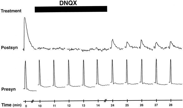
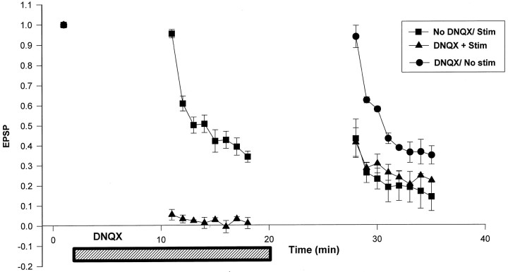
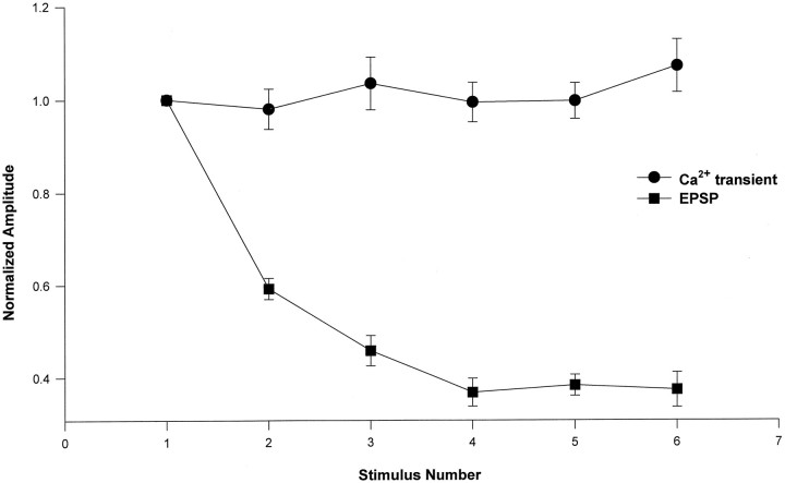
The cellular mechanisms underlying the induction and expression of homosynaptic depression at the glutamatergic synapse between Aplysia sensory and motor neurons were studied in dissociated cell culture. Intracellular microelectrodes were used to stimulate action potentials in the presynaptic sensory neuron and record the depolarizing EPSP from the motor neuron. Homosynaptic depression (HSD) was induced by repeatedly stimulating the sensory neuron at rates as low as one action potential per minute. Activation of postsynaptic Glu receptors was neither sufficient nor necessary to induce HSD. Thus, repeated applications of exogenous Glu did not depress the synaptically evoked EPSP. Moreover, normal HSD was observed when the sensory neuron was stimulated during a period when the Glu receptors were blocked with the antagonist DNQX. The induction of HSD is thus likely to occur within the presynaptic terminal. We explored the role of presynaptic calcium in the induction of HSD by injecting the sensory neuron with EGTA, a relatively slow calcium chelator that does not alter rapid release but effectively buffers the slow residual calcium transient thought to be important for plasticity. EGTA had little effect on HSD, indicating that residual Cai is not involved. HSD does not appear to involve a decrease in presynaptic calcium influx, because there was no change in the presynaptic calcium transient, measured by calcium indicator dyes, during HSD. We conclude that HSD is induced and expressed in the presynaptic terminal, possibly by a mechanism directly coupled to the release process.


Figures

Fig. 1.
Fig. 1.
Glu receptor activation is not sufficient to induce HSD. A, Effect of Glu at a representative sensory to motor neuron synapse. A1, HSD in response to low-frequency presynaptic stimulation. EPSPs were recorded from a postsynaptic motor neuron in response to intracellular stimulation of presynaptic action potentials in a sensory neuron. Responses were elicited at 1 min intervals.A2, The effect of exogenous Glu application on synaptically evoked EPSP. The first trace(bold) shows the first EPSP recorded from a motor neuron in response to firing an action potential in the presynaptic sensory neuron. Five brief Glu pulses (100 msec) were then applied from a puffer pipette to elicit postsynaptic depolarizations (thin traces) of approximately equal amplitude to the synaptically evoked EPSP. The final trace(bold) is a second synaptically evoked EPSP. All responses were elicited at 1 min intervals. Calibration:A1, 10 mV, 100 msec;A2, 10 mV, 250 msec.B, Summary of mean data for Glu applications. Protocol is identical to that shown in A. For each synapse analyzed using presynaptic stimulation, the amplitudes of the EPSPs evoked during successive presynaptic stimuli were normalized to that of the first EPSP (triangles,Control). The amplitudes of depolarizing responses to exogenous Glu were normalized to the first response to Glu (small squares, +Glu). In the same experiments, the EPSP evoked by presynaptic stimulation after the Glu pulses was normalized to the first evoked EPSP before the Glu pulses (large squares). The normalized responses were then averaged among the different cells. Triangles,n = 4; squares,n = 5. Error bars indicate SEM.
Fig. 2.
Fig. 2.
Effect of blockade of postsynaptic Glu receptors on induction of HSD. EPSPs were recorded from a postsynaptic motor neuron (top traces) in response to intracellular stimulation of action potentials in a presynaptic sensory neuron once per minute (bottom traces). After the first stimulus, 10 μm DNQX (filled bar) was applied by microperfusion. After the fifth stimulus, presynaptic stimulation was halted, and DNQX was washed out for a period of 10 min. Presynaptic stimulation was then resumed.
Fig. 3.
Fig. 3.
Activation of ionotropic Glu receptors is not required for induction of HSD. Mean data shown for induction of HSD during three experimental protocols to test the effects of DNQX. The first protocol is similar to that shown in Figure2 (triangles). The presynaptic cell was first stimulated to evoke an EPSP (left point, 0 min). Cells were then exposed to 10 μm DNQX for 10 min (bottom bar). In the continued presence of DNQX, eight additional presynaptic stimuli were applied at a rate of one per minute (middle points, 11–18 min). The DNQX was then washed from the bath for 10 min, and eight more presynaptic stimuli were applied (right points, 28–35 min). The second protocol was identical to the first, except that the cells were not exposed to DNQX (squares). In the third protocol, cells were exposed to DNQX as in the first protocol, except that the first group of eight presynaptic stimuli during DNQX were omitted (circles). For each cell, EPSP amplitudes were normalized to the first EPSP, and then the normalized values in each group were averaged. n = 5 for each protocol.
Fig. 4.
Fig. 4.
Presynaptic injection of EGTA does not inhibit induction of HSD. Peak EPSP amplitudes from four motor neurons are plotted in response to successive stimulation of the sensory neurons. EPSP amplitudes are normalized to that of the first EPSP. In two cells, the presynaptic microelectrode contained 100 mm EGTA (diamonds), and in a third cell, the microelectrode contained 1 m EGTA (triangles). In a control cell, the presynaptic electrode did not contain EGTA (squares). The presynaptic cell was stimulated at 20 sec intervals. Before these measurements were obtained, solution was ejected from the presynaptic electrode using 500 msec pressure pulses.
Fig. 5.
Fig. 5.
Ca influx in a single presynaptic varicosity does not change during induction of HSD. Cai transients, plotted as ΔF/F, were imaged from a single presynaptic varicosity in response to stimulation of six presynaptic action potentials at 20 sec intervals. The peak EPSP amplitude recorded from the postsynaptic motor neuron in response to the same presynaptic stimulus is given below each calcium transient. Time is shown in 1 sec intervals.
Fig. 6.
Fig. 6.
Spatial distribution of Cai influx does not change during HSD. Pseudocolor images of the peak calcium response (ΔF/F) in a presynaptic sensory neuron elicited by a single action potential.Red indicates high calcium concentration;blue indicates low calcium concentration.A shows the Cai response to the first stimulus, that in B to the sixth stimulus, during a series of six action potentials, elicited at 20 sec intervals.
Fig. 7.
Fig. 7.
Mean data for Cai transient amplitude during HSD. The mean peak amplitude of the presynaptic Caitransient (ΔF/F;circles) and the associated mean peak EPSP amplitude (squares) are plotted during successive presynaptic stimuli. A presynaptic action potential was elicited once every 20 sec. The average calcium transient in response to a given stimulus was calculated by first measuring the calcium transient in four to five individual varicosities per experiment. For each varicosity, the amplitude of the transient was then normalized to that of the first stimulus. These normalized values were then averaged for all the varicosities (n = 38) from all cells (n = 9). For each postsynaptic cell, the EPSP amplitudes were normalized to that of the first EPSP, and then the normalized values from all cells (n = 9) were averaged. Error bars indicate SEM.
Fig. 8.
Fig. 8.
Multiple stimuli activate a constant number of varicosities. Calcium responses in individual varicosities from presynaptic sensory neurons were measured in response to successive action potentials used to induce HSD (n = 38). For each varicosity, the amplitude of the calcium transient in response to each successive action potential, ΔF(i)/F, was normalized to that of the first action potential, ΔF(1)/F, yielding the ratio f(i)/f(1). Normalized Cai transients during six successive action potentials are plotted as a function of action potential number (i). Each varicosity is represented by a separateline. Action potentials were stimulated once every 20 sec.

References

    1. Adler EM, Augustine GJ, Duffy SN, Charlton MP. Alien intracellular calcium chelators attenuate neurotransmitter release at the squid giant synapse. J Neurosci. 1991;11:1496–1507. - PMC - PubMed
    1. Bailey CH, Chen M. Morphological basis of short-term habituation in Aplysia. J Neurosci. 1988;8:2452–2459. - PMC - PubMed
    1. Bao JX, Kandel ER, Hawkins RD. Involvement of pre- and postsynaptic mechanisms in posttetanic potentiation at Aplysia synapses. Science. 1997;275:969–973. - PubMed
    1. Bear MF, Malenka RC. Synaptic plasticity: LTP and LTD. Curr Opin Neurobiol. 1994;3:389–399. - PubMed
    1. Bliss TV, Lomo T. Long-lasting potentiation of synaptic transmission in the dentate area of the anaesthetized rabbit following stimulation of the perforant path. J Physiol (Lond) 1973;232:331–356. - PMC - PubMed

Publication types

MeSH terms

LinkOut - more resources