. 1998 Nov 1;18(21):8814-25.
doi: 10.1523/JNEUROSCI.18-21-08814.1998.

Extracellular signal-regulated kinase (ERK) controls immediate early gene induction on corticostriatal stimulation

Affiliations

Extracellular signal-regulated kinase (ERK) controls immediate early gene induction on corticostriatal stimulation

V Sgambato et al. J Neurosci. .

Abstract

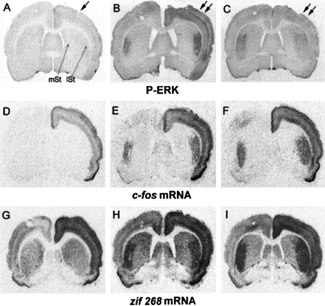
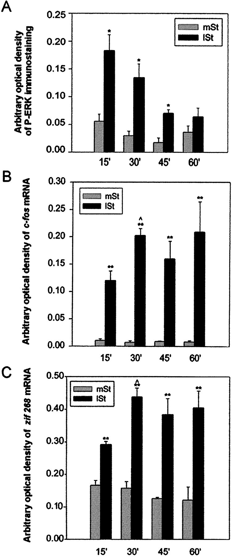
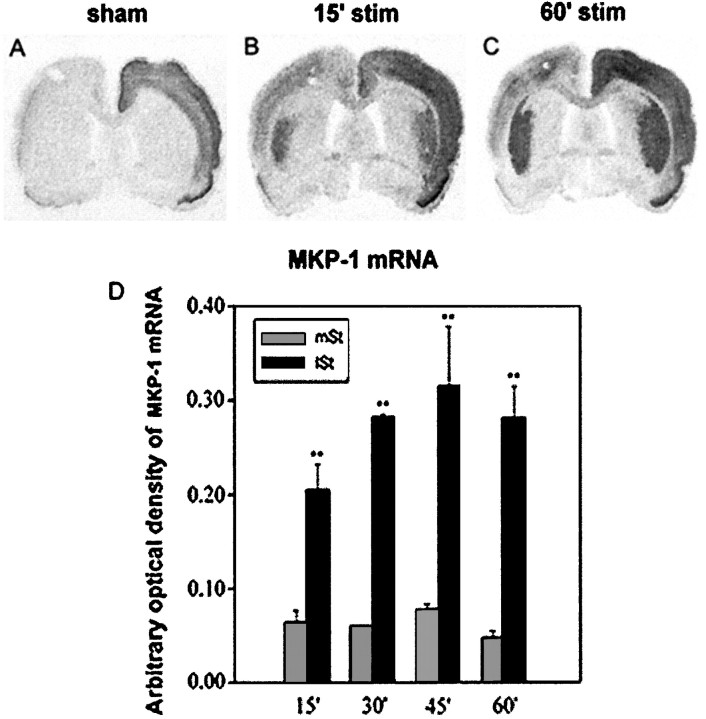
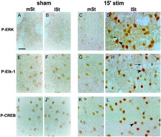
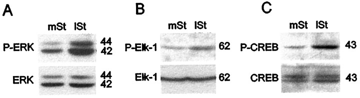
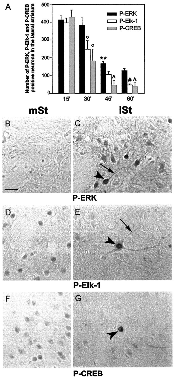
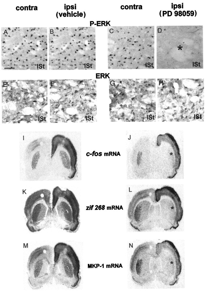
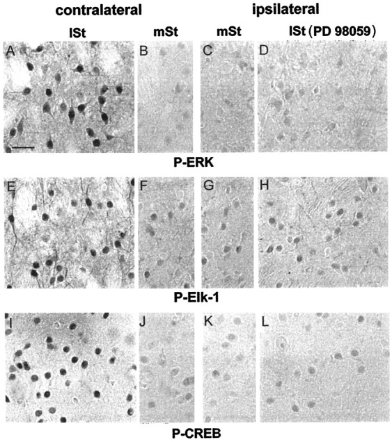
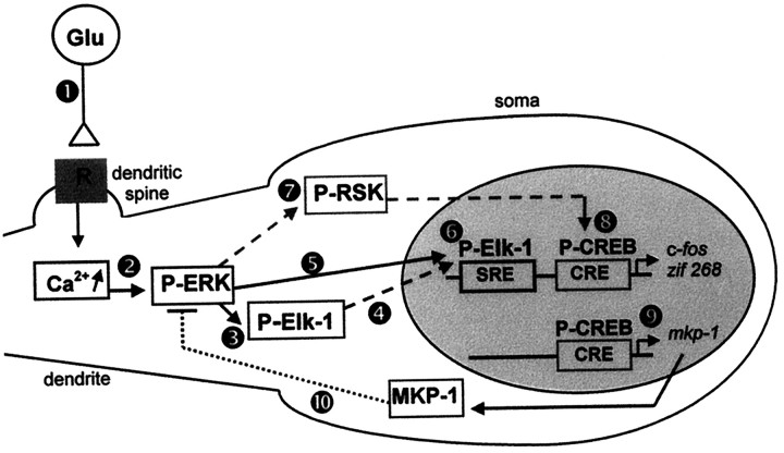
Activity-dependent changes in neuronal structure and synaptic remodeling depend critically on gene regulation. In an attempt to understand how glutamate receptor stimulation at the membrane leads to gene regulation in the nucleus, we traced intracellular signaling pathways targeting DNA regulatory elements of immediate early genes (IEGs). For this purpose we used an in vivo electrical stimulation of the glutamatergic corticostriatal pathway. We show that a transient activation of extracellular signal-regulated kinase (ERK) proteins (detected by immunocytochemistry with an anti-active antibody) is spatially coincident with the onset of IEG induction [c-fos, zif 268, and map kinase phosphatase-1 (MKP-1) detected by in situ hybridization] in the striatum, bilaterally. Both Elk-1 and CREB transcription factors (targeting SRE and CRE DNA regulatory elements, respectively) were hyperphosphorylated in register with ERK activation and IEG mRNA induction. However, their hyperphosphorylation occurred in different subcellular compartments: the cytoplasm and the nucleus for Elk-1 and the nucleus for CREB. The role of the ERK signaling cascade in gene regulation was confirmed after intrastriatal and unilateral injection of the specific ERK inhibitor PD 98059, which completely abolished c-fos, zif 268, and MKP-1 mRNA induction in the injected side. Of interest, both Elk-1 and CREB hyperphosphorylation also was impaired after PD 98059 injection. Thus two different ERK modules, one depending on the cytoplasmic activation of Elk-1 and the other one depending on the nuclear activation of CREB, control IEG transcriptional regulation in our model. Our findings provide significant insights into intracellular mechanisms underlying synaptic plasticity in the striatum.


Figures

Fig. 1.
Fig. 1.
ERK activation occurs in a tight spatial register with c-fos and zif 268 mRNA induction after unilateral stimulation of the corticostriatal pathway. Brain sections were processed for P-ERK immunoreactivity (A–C) and in situ hybridization of c-fos (D–F) and zif 268 (G–I) mRNAs. Sham-stimulated rats (sham; A, D, G) were electrode-implanted in the right cerebral cortex (arrow in A). Although P-ERK immunostaining (A) is not detectable, c-fos (D) and zif 268 (G) mRNAs are induced slightly in the implanted cortex, with no apparent upregulated signal in the striatum of sham-stimulated rats. Stimulated rats were electrode-implanted in the right cerebral cortex and received electrical cortical stimulation (double arrows in B, C) for either 15 min (15′ stim; B, E,H) or 60 min (60′ stim;C, F, I). At 15 min of stimulation a strong activation of P-ERK (B) occurred bilaterally in the lateral striatum (lSt), but not in the medial striatum (mSt). Note that this activation occurs in a spatial register with both c-fos(E) and zif 268(H) mRNA induction. At 60 min of cortical stimulation, although ERK activation (C) decreased in the lateral striatum, c-fos(F) and zif 268(I) mRNAs remained strongly induced bilaterally.
Fig. 2.
Fig. 2.
Kinetics of ERK activation and c-fos and zif 268 transcriptional regulation on corticostriatal stimulation. Statistical comparisons were performed from three independent animals for each time point, using an image analyzer (IMSTAR, Paris, France). A, Densitometric measurements of P-ERK immunostaining. Signals were significantly higher in the lateral striatum (lSt) than in the medial striatum (mSt) after 15, 30, and 45 min of stimulation (*p < 0.05, paired Student’s ttest), but not at 60 min. P-ERK immunoreactivity was maximal at 15 min and then progressively decreased to return to basal levels.B, C, Autoradiographic signals from c-fos (B) and zif 268 (C) mRNA detection. Both mRNA levels were significantly higher in the lSt than in themSt, regardless of the stimulation period used (**p < 0.01, paired Student’s ttest). Moreover, messenger levels were significantly higher at 30 than at 15 min of stimulation ( p < 0.05, unpaired Student’s t test) and then remained high. Note that the transient activation of ERK correlated with a sustained transcriptional regulation of both messengers.
Fig. 3.
Fig. 3.
MKP-1 mRNA induction on corticostriatal stimulation. Brain sections of sham-stimulated and stimulated rats were processed for in situ hybridization of MKP-1 mRNAs (A–C). In sham-stimulated rats (A) MKP-1 mRNA levels were detected in the implanted cortex, with no apparent signal in the striatum. By contrast, at 15 min of stimulation (B), although no signals were detected in the medial striatum, MKP-1 mRNA was strongly induced bilaterally in the lateral striatum. This induction persisted at 60 min of stimulation (C). Note that MKP-1 induction spatiotemporally correlated with the ERK activation and IEG induction shown in Figure 1. B, Statistical comparisons were performed from three independent animals with an image analyzer (IMSTAR) after densitometric measurements of mRNA signals on autoradiograms. MKP-1 mRNA levels were significantly higher in the lateral striatum (lSt) than in the medial striatum (mSt), regardless of the stimulation period used (**p < 0.01, paired Student’s ttest).
Fig. 4.
Fig. 4.
Hyperphosphorylation of ERK, Elk-1, and CREB after 15 min of corticostriatal stimulation. Adjacent brain sections of sham (left panels) or 15-min-stimulated (15′ stim; right panels) rats were processed in parallel for P-ERK (A–D), P-Elk-1 (E–H), and P-CREB (I–L) immunohistochemistry. In the medial striatum (mSt) and lateral striatum (lSt) of sham animals, although no constitutive expression of P-ERK was detectable (A, B), a slight constitutive nuclear expression was visible for P-Elk-1 (E, F) and P-CREB (I, J). In the mSt of 15′-stimulated rats, immunolabeling for P-ERK (C), P-Elk-1 (G), and P-CREB (K) was similar to that in sham. By contrast, a clear increase of P-ERK (D), P-Elk-1 (H), and P-CREB (L) was detectable in the lSt. The activation of ERK (D) occurred in both nuclear (arrowhead) and cytoplasmic (thin arrow) neuronal compartments. Similarly, Elk-1 hyperphosphorylation (H) occurred in the nucleus (arrowhead) as well as in cytoplasmic compartments (thin arrow). Finally, CREB activation (L) was restricted to the nucleus (arrowhead) of striatal cells. Data are representative of three independent rats for each group. Scale bar, 1 cm = 15 μm.
Fig. 5.
Fig. 5.
Western blot analysis of P-ERK, P-Elk-1, and P-CREB immunoreactivity in medial and lateral parts of the striatum after 15 min of stimulation. Extracts of medial (mSt) and lateral (lSt) striatum were dissected rapidly and processed for Western blots. Detection of anti-active proteins was analyzed first, and then total proteins were visualized on the same blot after a stripping procedure. Note the increase of P-ERK (A), P-Elk-1 (B), and P-CREB (C) immunoreactivities in thelSt when compared with the mSt and the same level of total ERKs (A), Elk-1 (B), and CREB (C) proteins in both striatal regions. Molecular weights (in kDa) are indicated to the right of each blot.
Fig. 6.
Fig. 6.
Inactivation of ERK, Elk-1, and CREB after 60 min of corticostriatal stimulation. A, Quantification of P-ERK, P-Elk-1, and P-CREB immunoreactive striatal cells at 15, 30, 45, and 60 min of corticostriatal stimulation (n = 3 rats for each stimulation period). Cells counts were performed with an image analyzer (Biocom) in the lateral striatum ipsilaterally to the stimulation (total surface area examined, 2.7 mm2). Statistical comparisons were performed with a one-way ANOVA. **p < 0.01 when comparing P-ERK immunostaining between 45 and 30 min; °p < 0.05 when comparing P-Elk-1 and P-CREB immunostaining between 15 and 30 min;  p < 0.05 when comparing P-CREB with P-ERK at 45 and 60 min; #p < 0.05 when comparing P-Elk-1 with P-ERK at 60 min. Adjacent brain sections of 60-min-stimulated rats were processed in parallel for P-ERK (B, C), P-Elk-1 (D, E), and P-CREB (F, G) immunohistochemistry. In the lateral striatum (lSt) few P-ERK immunoreactivecells were found (C) when compared with those in Figure 4D. These cells presented both nuclear (arrowhead) and cytoplasmic (thin arrow) staining. E, Very few large neurons remained immunolabeled for P-Elk-1 in the lSt when compared with those in Figure 4H. Here again, the labeling was detectable in the nucleus (arrowhead) as well as in the cytoplasmic compartments (thin arrow). Similarly, CREB hyperphosphorylation (G) occurred in the nucleus of very few cells (arrowhead). Note that the constitutive immunolabeling observed in the medial striatum (mSt) for P-Elk-1 (D) and P-CREB (F) is no longer detectable in thelSt (E and G, respectively). Data are representative of three independent 60-min-stimulated rats. Scale bar, 1 cm = 15 μm.
Fig. 7.
Fig. 7.
Parke Davis 98059 abolishes IEG induction after 15 min of corticostriatal stimulation. The MEK inhibitor PD 98059 (right columns) or the vehicle (left columns) was injected in the lateral striatum (lSt) unilaterally and ipsilaterally to the electrode implantation. After 15 min of stimulation, brains were processed for P-ERK (A–D) and ERK (E–H) immunoreactivity and in situ hybridization of c-fos (I, J),zif 268 (K, L), and MKP-1 (M, N) mRNAs. In vehicle-injected rats the stimulation induced a strong bilateral activation of ERK in the lSt (A, B). By contrast, in PD 98059-injected rats this stimulation produced ERK activation contralaterally (C), but not ipsilaterally (D; asterisk), to the inhibitor injection site. ERK immunostaining remained unchanged in vehicle-injected (E, F) and PD 98059-injected (G, H) rats. In vehicle-injected rats the cortical stimulation led to a bilateral induction of c-fos (I),zif 268 (K), and MKP-1 (M) mRNAs. In PD 98059-injected rats, although IEG mRNAs were still induced contralaterally to the inhibitor injection site, an impairment of induction for c-fos(J) zif 268(L), and MKP-1 (N) was observed ipsilaterally (asterisk) to the PD 98059 injection. These data are representative of three independent rats for each group. Scale bar, 1 cm = 50 μm.
Fig. 8.
Fig. 8.
ERK proteins dually control Elk-1 and CREB hyperphosphorylation. Brains of PD 98059-injected rats, stimulated for 15 min, were processed for P-ERK (A–D), P-Elk-1 (E–H), and P-CREB (I–L) immunoreactivity. The injection of PD 98059 completely prevented ERK activation in the lateral striatum (lSt) ipsilaterally (D), but not contralaterally (A), to the inhibitor injection site. In the medial striatum (mSt) P-ERK immunoreactivity remained low in both sites (B, C). On sections adjacent to those used below, Elk-1 and CREB hyperphosphorylation were impaired in the lSt ipsilaterally (H andL, respectively), but not contralaterally (E and I, respectively), to the PD 98059 injection. The constitutive P-Elk-1 (F,G) and P-CREB (J,K) immunostaining remained low in both medial striata and was comparable to the immunolabeling observed inlSt ipsilaterally to the PD 98059 injection (H, L). Scale bar, 1 cm = 25 μm.
Fig. 9.
Fig. 9.
Proposed model for ERK-dependent IEG induction on corticostriatal pathway stimulation. Cortical fibers impinge specifically on dendritic spines of striatal neurons (Smith and Bolam, 1990). The electrical stimulation of these afferents leads to glutamate release at corticostriatal synapses (Reubi and Cuenod, 1979), which in turn act via glutamatergic receptors (1). The subsequent and local elevation of intracellular calcium levels, via NMDA receptors, activates ERK machinery (2) located in the dendrites (Fiore et al., 1993a; Ortiz et al., 1995). Elk-1 proteins that are present in dendrites (Sgambato et al., 1998) are phosphorylated locally by ERK proteins (3). Because both ERK and Elk-1 hyperphosphorylated proteins also are found in the nucleus, we suggest that they are translocated into the nucleus on activation (4, 5). In the nucleus Elk-1 can mediate the transcriptional regulation of SRE-containing IEGs like c-fos and zif 268(6). In parallel, we show that activated ERK proteins lead to a hyperphosphorylation of the nuclear CREB protein. The activation of the CREB kinase RSK, a cytoplasmic substrate of ERK proteins (Strugill et al., 1988) (7), and its subsequent translocation into the nucleus (Chen et al., 1992) could explain the ERK-dependent CREB activation in our model. In the nucleus CREB mediates the transcriptional regulation of CRE-containing IEGs like c-fos, zif 268(8), and MKP-1 (9). The protein MKP-1 may act in a negative feedback loop (10) to downregulate ERK activation. Thus, two different ERK modules are critically involved in IEG induction: ERK/Elk-1/SRE and ERK/?/CREB/CRE.

Similar articles

Cited by

References

    1. Abo V, Deniau JM, Rogard M, Besson MJ. Involvement of the NMDA receptors in the induction of Fos expression in basal ganglia following electrical stimulation of the motor cortex. Eur J Neurosci [suppl] 1994;7:101.
    1. Alessi DR, Smythe C, Keyse SM. The human CL100 gene encodes a Tyr/Thr-protein phosphatase which potently and specifically inactivates MAP kinase and suppresses its activation by oncogenic ras in Xenopus oocyte extracts. Oncogene. 1993;8:2015–2020. - PubMed
    1. Alessi DR, Cuenda A, Cohen P, Dudley DT, Saltiel AR. PD 098059 is a specific inhibitor of the activation of mitogen-activated protein kinase in vitro and in vivo. J Biol Chem. 1995;270:27489–27494. - PubMed
    1. Bading H, Greenberg ME. Stimulation of protein tyrosine phosphorylation by NMDA receptor activation. Science. 1991;253:912–914. - PubMed
    1. Bading H, Ginty DD, Greenberg ME. Regulation of gene expression in hippocampal neurons by distinct signaling pathways. Science. 1993;260:181–186. - PubMed

Publication types

MeSH terms

Substances

LinkOut - more resources