. 1998 Nov 1;18(21):9038-54.
doi: 10.1523/JNEUROSCI.18-21-09038.1998.

Partial inactivation of the primary motor cortex hand area: effects on individuated finger movements

Affiliations

Partial inactivation of the primary motor cortex hand area: effects on individuated finger movements

M H Schieber et al. J Neurosci. .

Abstract

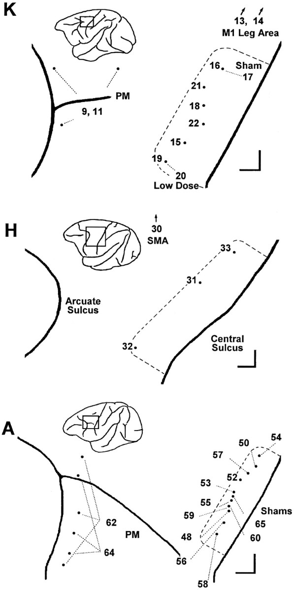
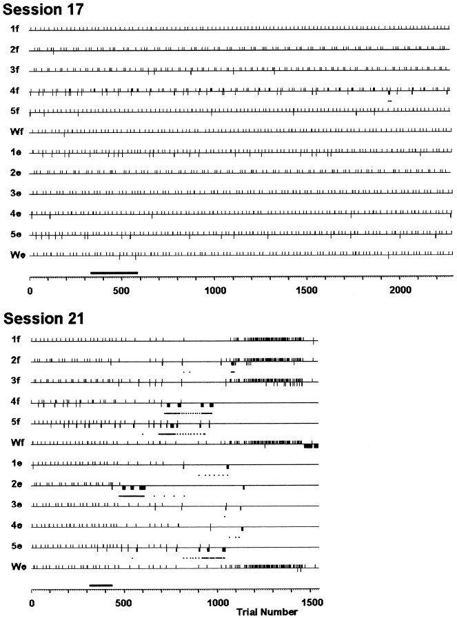
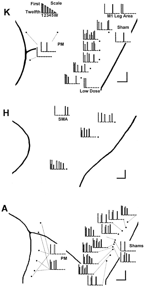
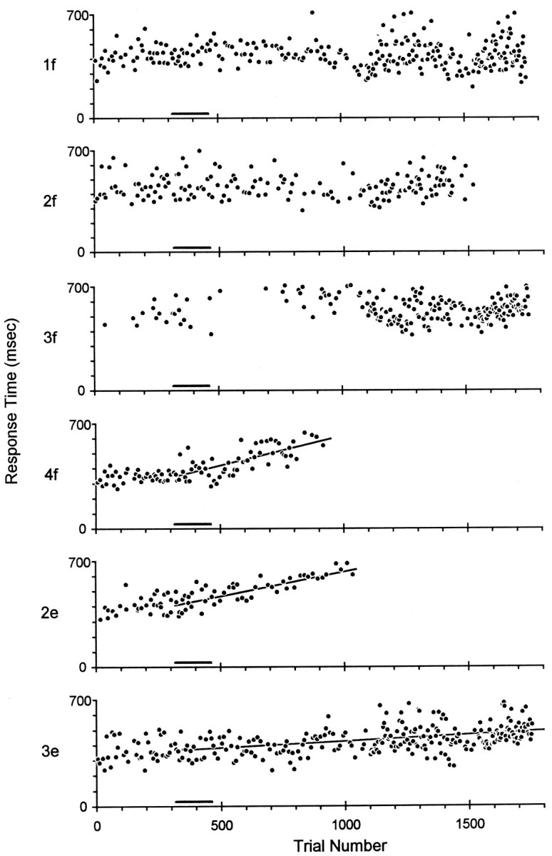
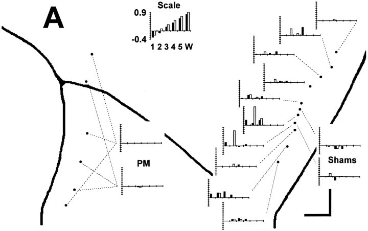
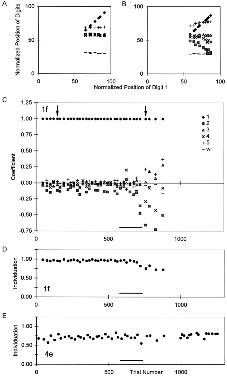
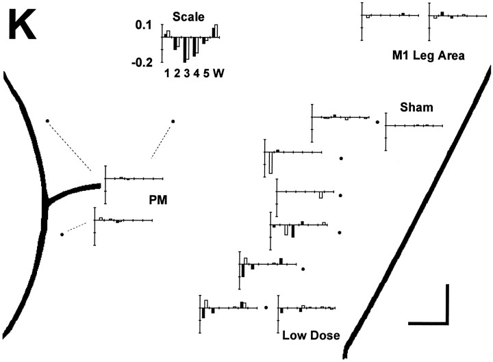
After large lesions of the primary motor cortex (M1), voluntary movements of affected body parts are weak and slow. In addition, the relative independence of moving one body part without others is lost; attempts at individuated movements of a given body part are accompanied by excessive, unintended motion of contiguous body parts. The effects of partial inactivation of the M1 hand area are comparatively unknown, however. If the M1 hand area contains the somatotopically ordered finger representations implied by the classic homunculus or simiusculus, then partial inactivation might produce weakness, slowness, and loss of independence of one or two adjacent digits without affecting other digits. But if control of each finger movement is distributed in the M1 hand area as many studies suggest, then partial inactivation might produce dissociation of weakness, slowness, and relative independence of movement, and which fingers movements are impaired might be unrelated to the location of the inactivation along the central sulcus. To investigate the motoric deficits resulting from partial inactivation of the M1 hand area, we therefore made single intracortical injections of muscimol as trained monkeys performed visually cued, individuated flexion-extension movements of the fingers and wrist. We found little if any evidence that which finger movements were impaired after each injection was related to the injection location along the central sulcus. Unimpaired fingers could be flanked on both sides by impaired fingers, and the flexion movements of a given finger could be unaffected even though the extension movements were impaired, or vice versa. Partial inactivation also could produce dissociated weakness and slowness versus loss of independence in a given finger movement. These findings suggest that control of each individuated finger movement is distributed widely in the M1 hand area.


Figures

Fig. 1.
Fig. 1.
Location of injections in each session. For each monkey, K, H, and A, the sites of muscimol injections are shown aspoints on an enlarged view of the left hemisphere, with the central sulcus to the viewer’s right and the arcuate sulcus to the left. A rectanglein the inset of each monkey’s left hemisphere shows the region enlarged. Session numbers are shown either next to the point representing the injection site or else connected to the point with a fine dashed line. The extent of the M1 hand area in each monkey, as assessed with single-neuron recording and ICMS, is indicated by a heavy dashed line. Scale bars at thebottom right of each enlargement indicate 1 mm in the rostral and medial directions. In addition to muscimol injections in the M1 hand area, comparison control injections were made in the in the PM just posterior to the arcuate sulcus, in the M1 leg area, and in the SMA. M1 leg area sites and SMA sites were medial to the region shown enlarged (arrows). In PM, multiple sites were injected in the same session (see Table 1).
Fig. 2.
Fig. 2.
Success and failure of different instructed movements in two sessions. Horizontal lines for each of the 12 instructed movements are marked with upward ticksfor each correctly performed trial and with downward ticks for each incorrectly performed trial of that instructed movement. Dots beneath these lines indicate the mean trial number of 10 consecutive trials of that instructed movement whenever 8 of the 10 were failures. Note that sequential ticks or dots occurring in the same row appear merged into a thickened tick or line segment, respectively, as minor divisions of the horizontal axis represent 10 sequential trials. A sham injection was performed in session 17, and a single muscimol injection was performed in session 21, during the trials indicated by thebar just above the horizontal trial number axis for each session. The sham injection in session 17 did not alter the monkey’s performance, whereas the muscimol injection in session 21 was followed by an increased failure rate for several instructed movements but not others. Session 17 spanned 93 min (injection, 13 min); session 21 lasted 102 min (injection, 5 min).
Fig. 3.
Fig. 3.
Failure on 8 of 10 consecutive trials. A bar graph for each session is used to show which instructed movements met the 8 of 10 consecutive failure criterion. Each bar graph has been positioned on the enlarged map of each monkey’s left hemispheric surface close to the point at which the injection was made or else connected to it with a fine dashed line. (Three dashed linesindicate the three different locations injected during each PM session.) Each instructed movement that failed (met the 8 of 10 criterion) is shown as a bar positioned along theabscissa to indicate the instructed digit andshaded to indicate the instructed direction:filled, flexion; open, extension. The height of each bar indicates the serial order in which different instructed movements failed within each session from 1st (tallest) to 12th (shortest). The scale at top thus would illustrate an idealized result in which an injection placed laterally in the hand area impaired instructed movements starting with those of the thumb and spreading somatotopically to those of the little finger and wrist, with instructed flexion of each digit failing before instructed extension. Such a result was not obtained, however. Although in each monkey (K, A, and H), muscimol injections in the M1 hand area caused more instructed movement failures than controls, which finger movements failed showed no systematic relationship to the location of the injection along the central sulcus.
Fig. 4.
Fig. 4.
Prolongation of response times. The response times, from cue onset to switch closure, on successful trials have been sorted according to instructed movement (1f, 2f, 3f, 4f, 2e, and 3e) and plotted as a function of sequential trial number in session 50. Muscimol was injected as monkey A performed trials 317–462 (black bar beneath each plot). Subsequently, response times became progressively longer in trials of 4f, 2e, and 3e, and linear regression showed slopes significantly different from zero, whereas response times in trials of 1f, 2f, and 3f remained stable. Shortly before 1000 trials, monkey A became unable to perform 4f and 2e, and these instructed movements were removed from the rotation, making subsequent trials of 1f, 2f, 3f, and 3e relatively more frequent. The total elapsed time in session 50 was 72 min, and the injection was performed over 5 min.
Fig. 5.
Fig. 5.
Response time changes in each session in Monkey A. A bar graph for each session in monkey A is used to show which instructed movements had progressive changes in response time with slope significantly different from zero. Bar graphs are positioned and formatted as in Figure 3, but here the height of each bar indicates the slope of the best-fit line of response time versus trial number in units of milliseconds per trial. The scale at top thus would illustrate an idealized result in which an injection placed medially in the hand area prolonged response times most dramatically for wrist and little finger movements and less for ring and middle finger movements, whereas response times of thumb movements shortened. Such a result was not obtained, however. Rather, different muscimol injections prolonged the response times of some instructed movements but not others in any given session. Which movements were affected showed little relation to the location of the injection along the central sulcus.
Fig. 6.
Fig. 6.
Changes in individuation in a single session.A, B, Plots of normalized finger positions (0, position at extension switch closure;100, position at flexion switch closure) from two successful trials of instructed movement 1f, one performed before (A) and the other after (B) the muscimol injection in session 15. In these two panels, the normalized position of each digit and of the wrist has been plotted as a function of the simultaneous normalized position of the instructed digit, digit 1. Whereas the noninstructed digits did not move as the monkey flexed his thumb before the muscimol injection (A), digits 2 and 4 extended as the monkey flexed his thumb after the injection (B).C, Relative motion coefficients (ordinate) derived from the slopes of such plots for each digit in each correctly performed trial of 1f in session 15. The single trials shown in A and B are indicated here by arrowheads. After the muscimol injection (bar at bottom), coefficients for noninstructed digits diverged from their near-zero baselines.D, Individuation Index calculated from these coefficients for each successful 1f trial. E, Individuation Index for trials of 4e in the same session. The Individuation Index of 1f trials decreased progressively after the muscimol injection, whereas the Individuation Index for 4e trials remained stable. The total elapsed time in session 15 was 100 min (injection, 8 min).
Fig. 7.
Fig. 7.
Individuation Index changes in each session in monkey K. A bar graph for each session in monkey K is used to show which instructed movements had progressive changes in Individuation Index with slope significantly different from zero. Bar graphs are positioned and formatted as in Figure 3, but here the height of each bar indicates the slope of the best-fit line of Individuation Index versus trial number, in Individuation Index units per 1000 trials. The scale at top thus would illustrate an idealized result in which an injection placed centrally in the hand area caused the greatest Individuation Index decrease for movements of the middle finger and less for index and ring finger movements, whereas Individuation Indexes of thumb and wrist movements increased. Such a result was not obtained, however. Rather, different muscimol injections impaired individuation of some instructed movements but not others in any given session. Which movements were affected showed little relation to the location of the injection along the central sulcus.

Similar articles

Cited by

References

    1. Andersen P, Hagan PJ, Phillips CG, Powell TP. Mapping by microstimulation of overlapping projections from area 4 to motor units of the baboon’s hand. Proc R Soc Lond B Biol Sci. 1975;188:31–36. - PubMed
    1. Ashe J. Force and the motor cortex. Behav Brain Res. 1997;86:1–15. - PubMed
    1. Beck CH, Chambers WW. Speed, accuracy, and strength of forelimb movement after unilateral pyramidotomy in rhesus monkeys. J Comp Physiol Psychol. 1970;70:1–22. - PubMed
    1. Beevor C. The Croonian Lectures on muscular movements. MacMillan; New York: 1903a. - PubMed
    1. Beevor C. Remarks on paralysis of the movements of the trunk in hemiplegia. MacMillan; New York: 1903b. - PMC - PubMed

Publication types

LinkOut - more resources