. 1998 Nov 1;18(21):9069-77.
doi: 10.1523/JNEUROSCI.18-21-09069.1998.

Functional consequences of central serotonin depletion produced by repeated fenfluramine administration in rats

Affiliations

Functional consequences of central serotonin depletion produced by repeated fenfluramine administration in rats

M H Baumann et al. J Neurosci. .

Abstract

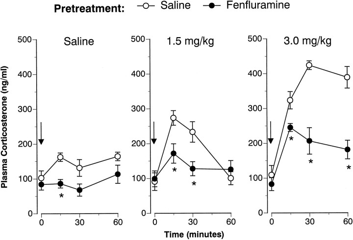
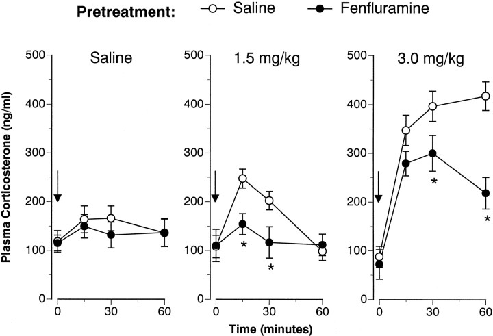
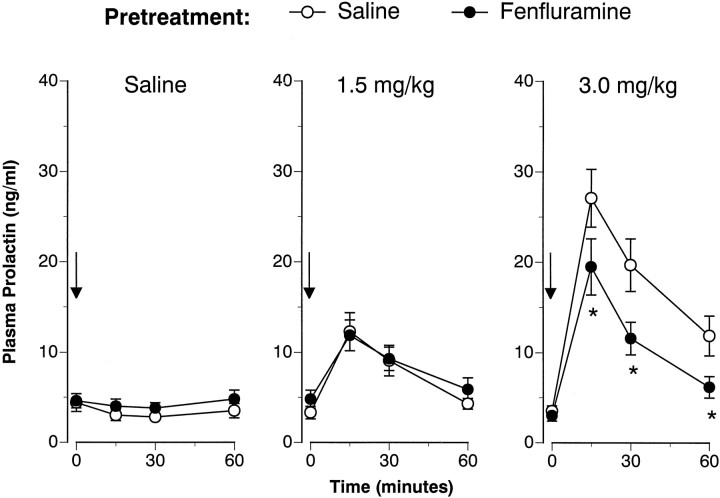
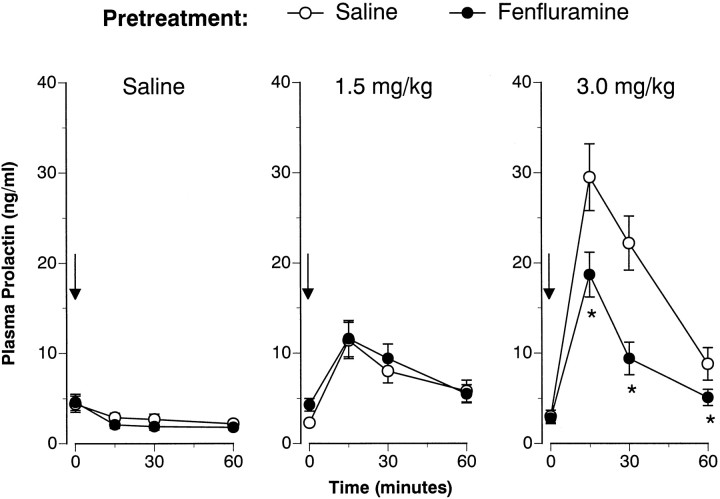
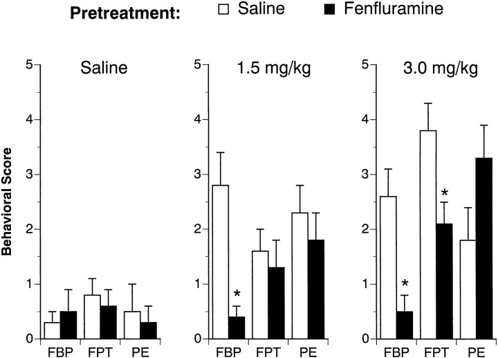
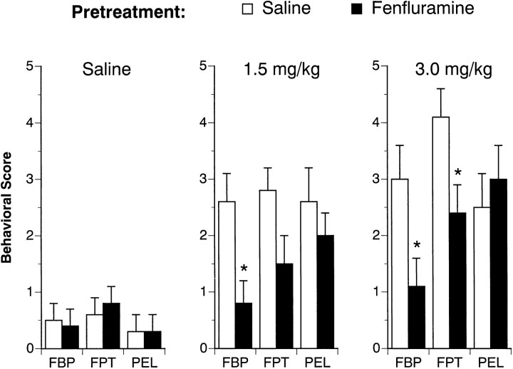
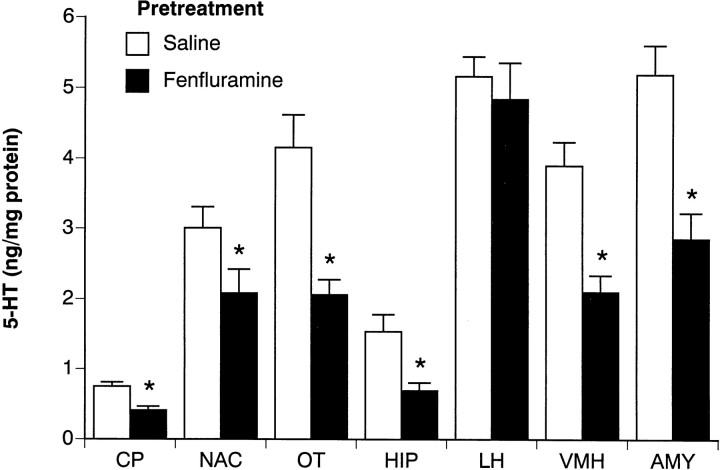
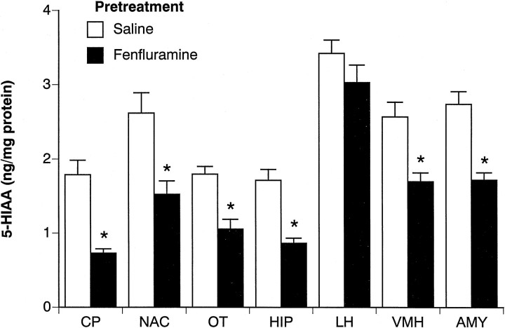
Repeated administration of D,L-fenfluramine (FEN) is known to cause prolonged depletion of forebrain serotonin (5-HT) in animals. Ironically, few studies have evaluated functional consequences of such FEN-induced 5-HT loss. In the present work, we examined neuroendocrine and behavioral responses evoked by acute FEN injection in rats that had previously received a 4 d FEN-dosing regimen known to deplete forebrain 5-HT (D,L-FEN, 20 mg/kg, s.c., b. i.d.). Rats were fitted with indwelling jugular catheters before the study to allow for repeated intravenous challenge injections and stress-free blood sampling. At 1 and 2 weeks after the 4 d dosing regimen, acute FEN (1.5 or 3.0 mg/kg, i.v.) produced dose-related elevations in plasma corticosterone and prolactin; these hormonal responses were markedly attenuated in FEN-pretreated rats. Behavioral effects of acute FEN, namely flat body posture and forepaw treading, were also blunted in FEN-pretreated rats. Interestingly, rats exposed to repeated FEN did not display overt abnormalities in hormonal or behavioral parameters under basal (i.e., unprovoked) conditions, despite dramatic decreases in postmortem tissue levels of 5-HT in numerous brain areas. Our results suggest that FEN-induced 5-HT depletion is accompanied by multiple impairments in 5-HT function. Although the clinical relevance of our data are debatable, the findings clearly show the utility of the FEN challenge test for uncovering in vivo functional deficits that might otherwise go undetected. FEN should remain an important pharmacological tool for determining the role of 5-HT neurons in mediating diverse physiological and behavioral processes.


Figures

Fig. 1.
Fig. 1.
FEN-induced corticosterone release determined 1 week after cessation of the 4 d saline (1 ml/kg, s.c., b.i.d.) or FEN (20 mg/kg, s.c., b.i.d.) dosing regimen. Rats from each pretreatment group were challenged with intravenous saline or FEN (1.5 or 3.0 mg/kg). Serial blood samples were withdrawn immediately before (time 0) and at 15, 30, and 60 min after intravenous injection. Corticosterone values are mean ± SEM for n = 8 rats per group. *p < 0.05 with respect to corresponding saline pretreatment group at the specified time point.
Fig. 2.
Fig. 2.
FEN-induced corticosterone release assessed 2 weeks after the 4 d saline or FEN-dosing regimen. The data depicted here are obtained from the same subjects described in Figure1. Refer to the legend of Figure 1 for further details.n = 7 or 8 rats per group. *p< 0.05 with respect to corresponding saline pretreatment group.
Fig. 3.
Fig. 3.
FEN-induced prolactin secretion determined 1 week after cessation of the 4 d saline (1 ml/kg, s.c., b.i.d.) or FEN (20 mg/kg, s.c., b.i.d.) dosing regimen. Rats from each pretreatment group were challenged with intravenous saline or FEN (1.5 or 3.0 mg/kg). Serial blood samples were withdrawn immediately before (time 0) and at 15, 30, and 60 min after intravenous injection. Prolactin levels are mean ± SEM expressed as nanogram per milliliter equivalents of rPRL-RP-3 for n = 8 rats per group. *p < 0.05 with respect to corresponding saline pretreatment group at the specified time point.
Fig. 4.
Fig. 4.
FEN-induced prolactin secretion assessed 2 weeks after the 4 d saline or FEN-dosing regimen. The data depicted here are obtained from the same subjects described in Figure 3. Refer to the legend of Figure 3 for further details. n = 7 or 8 rats per group. *p < 0.05 with respect to corresponding saline pretreatment group.
Fig. 5.
Fig. 5.
FEN-induced behavioral responses determined 1 week after cessation of the 4 d saline (1 ml/kg, s.c., b.i.d.) or FEN (20 mg/kg, s.c., b.i.d.) dosing regimen. Rats from each pretreatment group were challenged with intravenous saline or FEN (1.5 or 3.0 mg/kg). The occurrence of flat body posture (FBP), forepaw treading (FPT), and penile erections (PE) was assessed at 2, 10, 20, and 30 min after injection. Numerical scores were recorded for each behavior at each time point according to a graded rating scale (see Materials and Methods). Rats received a final “behavioral score” for each behavior, which consisted of the summed scores for that behavior over all time points. Data are expressed as mean ± SEM forn = 8 rats per group. *p < 0.05 with respect to corresponding saline pretreatment group.
Fig. 6.
Fig. 6.
FEN-induced behavioral responses evaluated 2 weeks after the 4 d saline or FEN-dosing regimen. The data depicted here are obtained from the same subjects described in Figure 5. Refer to the legend of Figure 5 for further details. n = 8 rats per group. *p < 0.05 with respect to corresponding saline pretreatment group.
Fig. 7.
Fig. 7.
Postmortem tissue levels of 5-HT in microdissected brain regions from rats challenged with acute saline. Rats had previously received repeated saline or FEN for 4 d and were decapitated 2 weeks later after the final challenge test session. Micropunches of tissue were assayed for 5-HT by HPLC-EC techniques (see Materials and Methods). Regions examined were caudate putamen (CP), nucleus accumbens (NAC), olfactory tubercle (OT), hippocampus CA3 (HIP), lateral hypothalamus (LH), ventromedial hypothalamus (VMH), and basolateral amygdala (AMY). Data are mean ± SEM expressed as nanograms per milligrams of protein forn = 8 rats per group. *p < 0.05 compared with saline pretreatment group.
Fig. 8.
Fig. 8.
Postmortem tissue levels of 5-HIAA in microdissected brain regions from rats challenged with acute saline. Rats had previously received repeated saline or FEN for 4 d and were decapitated 2 weeks later after the final challenge test session. Micropunches of tissue were assayed for 5-HIAA by HPLC-EC techniques (see Materials and Methods). Data are mean ± SEM expressed as nanograms per milligram of protein for n = 8 rats per group. *p < 0.05 compared with saline pretreatment group.

References

    1. Appel NM, Contrera JF, De Souza EB. Fenfluramine selectively and differentially decreases the density of serotonergic nerve terminals in rat brain: evidence from immunocytochemical studies. J Pharmacol Exp Ther. 1989;249:928–943. - PubMed
    1. Appel NM, Mitchell WM, Contrera JF, De Souza EB. Effects of high-dose fenfluramine treatment on monoamine uptake sites in rat brain: assessment using quantitative autoradiography. Synapse. 1990;6:33–44. - PubMed
    1. Appel NM, Owens MJ, Culp S, Zaczek R, Contrera JF, Bissette G, Nemeroff CB, De Souza EB. Role for brain corticotropin-releasing factor in the weight-reducing effects of chronic fenfluramine treatment in rats. Endocrinology. 1991;128:3237–3246. - PubMed
    1. Auerbach SB, Minzenberg MJ, Wilkinson LO. Extracellular serotonin and 5-hydroxyindoleacetic acid in hypothalamus of the unanesthetized rat measured by in vivo dialysis coupled to high-performance liquid chromatography with electrochemical detection. Brain Res. 1989;499:281–290. - PubMed
    1. Baumann MH, Rothman RB. Combined phentermine/fenfluramine administration and central serotonin neurons. Synapse. 1998;28:339–342. - PubMed

Publication types

LinkOut - more resources