. 1998 Oct 27;95(22):12860-5.
doi: 10.1073/pnas.95.22.12860.

Folding in vivo of a newly translated yeast cytosolic enzyme is mediated by the SSA class of cytosolic yeast Hsp70 proteins

Affiliations

Folding in vivo of a newly translated yeast cytosolic enzyme is mediated by the SSA class of cytosolic yeast Hsp70 proteins

S Kim et al. Proc Natl Acad Sci U S A. .

Abstract

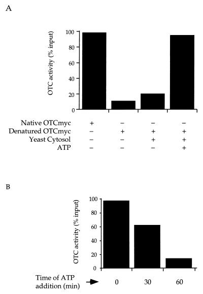
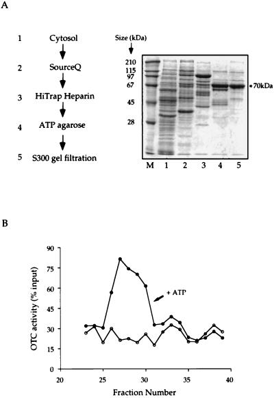
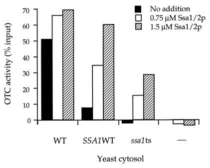
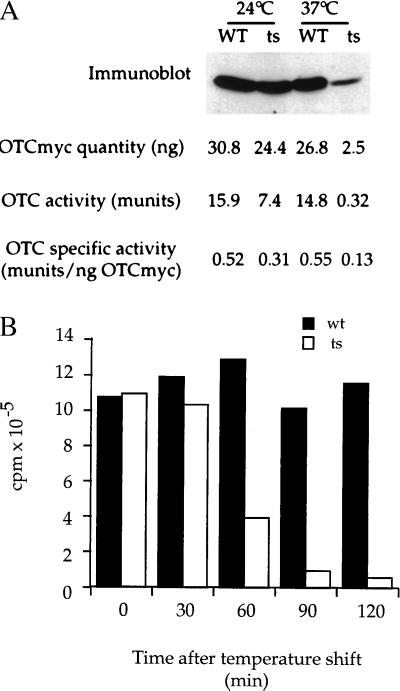
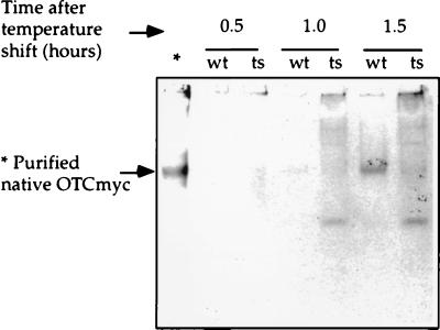
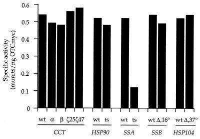
The nature of chaperone action in the eukaryotic cytosol that assists newly translated cytosolic proteins to reach the native state has remained poorly defined. Actin, tubulin, and Galpha transducin are assisted by the cytosolic chaperonin, CCT, but many other proteins, for example, ornithine transcarbamoylase (OTC), a cytosolic homotrimeric enzyme of yeast, do not require CCT action. Here, we observe that yeast cytosolic OTC is assisted to its native state by the SSA class of yeast cytosolic Hsp70 proteins. In vitro, refolding of OTC diluted from denaturant was assisted by crude yeast cytosol and ATP and found to be directed by SSA1/2. In vivo, when OTC was induced in a temperature-sensitive SSA-deficient strain, it exhibited reduced specific activity, and nonnative subunits were detected in the soluble fraction. These findings indicate that, in vivo, the Hsp70 system assists in folding at least some newly translated cytosolic enzymes, most likely functioning in a posttranslational manner.


Figures

Figure 1
Figure 1
(A) Crude yeast cytosol mediates ATP-dependent renaturation of OTC or a COOH-terminal myc-tagged derivative. (B) Extent of renaturation is reduced when ATP is added at later times. OTC or OTC-myc was diluted 100-fold from 6 M guanidine⋅HCl into buffer or yeast cytosolic extract prepared as described in Methods, with or without ATP. OTC enzymatic activity was assayed after 1 h. Activity was expressed as a percentage of the activity of input material that had not been subjected to denaturation.
Figure 2
Figure 2
Fractionation of yeast cytosolic extract to enrich the OTC-myc renaturing activity. (A) Steps of fractionation and SDS/PAGE analysis of fractions with peak renaturing activity from the various steps. (B) Typical profile of renaturing activity of fractions eluted from Q-Sepharose Fast Flow chromatography. Crude yeast cytosol was prepared and subjected to the sequence of fractionation steps shown (see Methods for details). A fraction from the peak of renaturing activity from each of the various steps was displayed in SDS/PAGE. In B the peak of ATP-dependent renaturing activity from Q-Sepharose FastFlow fractionation is shown, expressed as recovery of input denatured OTC in the assay of each fraction. The peak elutes at fractions 26–30 and corresponds to elution at 280–320 mM NaCl.
Figure 3
Figure 3
Extent of OTC-myc renaturation by yeast cytosolic extract is related to the level of SSA (Hsp70) protein, influenced either in vivo, by the SSA genotype of the strain from which extract is prepared, or in vitro, by addition of purified SSA1/2 protein. Assay for renaturation of OTC-myc was carried out as in Fig. 1 for extracts prepared from three different yeast strains: wild-type strain (WT), with all four SSA genes intact; SSA1WT strain, with only SSA1 intact and SSA2–4 disrupted; or ssa1ts strain, with a ssa1–45 ts version of SSA1 and SSA2–4 disrupted. A control was also carried out with buffer only (–). The extracts were tested for renaturing activity unsupplemented (black bars), supplemented with the mixture of purified SSA1 and SSA2 proteins obtained from wild-type yeast grown at 30°C to levels of either 0.75 μM (open bars), or 1.5 μM (hatched bars). Activity is expressed by subtracting activity measured in the absence of ATP from that in its presence and expressing this difference as a percentage of the total input enzymatic activity. Note that provision of SSA1 and SSA2 proteins to a buffer extract is insufficient to promote OTC-myc renaturation (right-hand bars), consistent with requirement for other components that are present in cytosolic extract.
Figure 4
Figure 4
Specific activity of newly translated OTC-myc is reduced in SSA-deficient cells as compared with wild-type (SSA1+ SSA2–4-deleted) cells at both permissive (24°C) and nonpermissive (37°C) growth temperatures. (A) Cells grown at 24°C were placed in galactose-containing medium at either 24°C or 37°C. After 90 min, equal amounts of cells were harvested and extracted as described in Methods (note that ts SSA cells arrest growth but not protein synthesis immediately after shift to 37°C). Extracts were analyzed both for amount of induced OTC-myc protein by immunoblotting and for amount of induced OTC enzymatic activity by assay as described in Methods. The immunoblot signal for OTC-myc was scanned densitometrically to compare the relative amounts of OTC-myc in wild-type and mutant cells against standards applied to the same gels (see Methods), and specific activity was calculated by dividing the amount of OTC enzymatic activity by the relative amount of OTC-myc protein. (B) To measure total translation, cells were shifted to 37°C (t0) and pulse-radiolabeled with [35S]methionine for 5 min at each time point. Radiolabeled proteins were quantitated by trichloroacetic acid precipitation and scintillation counting. Wild-type cells (wt), solid bars; ts SSA-deficient cells (ts), open bars. Note that OTC enzymatic activity is reduced by ≈50% in ts mutant cells at 24°C as compared with wild-type cells, whereas synthesis of OTC-myc protein is reduced by only ≈20%. At 37°C, both translation and activity are greatly reduced relative to wild-type cells, with activity affected to a greater extent (45-fold vs. 10-fold effect on OTC-myc synthesis). Reduced translation of OTC-myc at 37°C in mutant cells reflects a general defect of translation (B). To establish that the measurements of reduced levels of OTC-myc protein in the ts mutant cells at 37°C determined by densitometric gel scanning were accurate, reduced amounts of wild-type cell extract were loaded side-by-side with mutant cell extract, so as to produce identical signals (not shown). The level of OTC-myc protein produced with a 90-min induction at 37°C in mutant cells is ≈10% that of wild-type cells. To measure the amounts of OTC-myc protein produced, known amounts of purified OTC-myc were immunoblotted from the same gels (see Methods).
Figure 5
Figure 5
Nondenaturing gel analysis of soluble fractions from wild-type (wt) and SSA-deficient (ts) cells after galactose induction of OTC-myc at 37°C. At each time point, equal amounts of wild-type and mutant cells were harvested, and lysates were prepared. After ultracentrifugation at 340,000 × g for 10 min, supernatants were fractionated in an 8% polyacrylamide gel, pH 8.8, at 4°C and immunoblotting was then carried out with anti-myc monoclonal antibody 9E10. ∗ Indicates control lane displaying OTC-myc purified from E. coli.
Figure 6
Figure 6
Specific activity of OTC-myc induced in a variety of chaperone-deficient mutant strains under nonpermissive growth conditions. OTC specific activity is expressed as nanomoles of citrulline produced per minute (munits) per nanogram of OTC-myc protein. Wild-type or mutant strains for the indicated cytosolic chaperones were placed at the temperature nonpermissive for growth of the mutant for 2 h (excepting Hsp104, where 37°C does not significantly affect growth) in media containing galactose to induce OTC-myc. Extracts were prepared from equal amounts of wild-type and mutant cells, as in Fig. 4 and were analyzed as in Fig. 4 both for amount of induced OTC-myc protein by immunoblotting and for amount of induced OTC enzymatic activity. For each strain, the specific activity of the induced OTC-myc protein is displayed, measured as the amount of OTC enzymatic activity per nanogram of OTC-myc protein.

References

    1. Fayet O, Ziegelhoffer T, Georgopoulos C. J Bacteriol. 1989;171:1379–1385. - PMC - PubMed
    1. Horwich A L, Low K B, Fenton W A, Hirshfield I N, Furtak K. Cell. 1993;74:909–917. - PubMed
    1. Ewalt K L, Hendrick J P, Houry W A, Hartl F-U. Cell. 1997;90:491–500. - PubMed
    1. Bukau B, Horwich A L. Cell. 1998;92:351–366. - PubMed
    1. Johnson J L, Craig E A. Cell. 1997;90:201–204. - PubMed

Publication types

MeSH terms

Substances