. 1998 Oct 27;95(22):12967-72.
doi: 10.1073/pnas.95.22.12967.

A conventional myosin motor drives neurite outgrowth

Affiliations

A conventional myosin motor drives neurite outgrowth

S R Wylie et al. Proc Natl Acad Sci U S A. .

Abstract

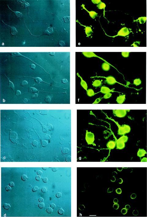
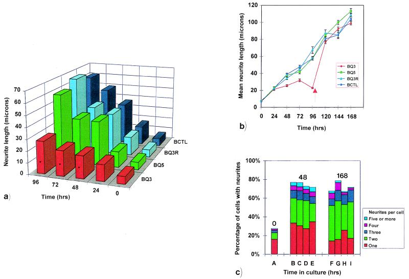
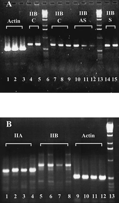
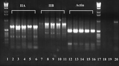
Neuritic outgrowth is a striking example of directed motility, powered through the actions of molecular motors. Members of the myosin superfamily of actin-associated motors have been implicated in this complex process. Although conventional myosin II is known to be present in neurons, where it is localized at the leading edge of growth cones and in the cell cortex close to the plasma membrane, its functional involvement in growth cone motility has remained unproven. Here, we show that antisense oligodeoxyribonucleotides, complementary to a specific isoform of conventional myosin (myosin IIB), attenuate filopodial extension whereas sense and scrambled control oligodeoxyribonucleotides have no effect. Attenuation is shown to be reversible, neurite outgrowth being restored after cessation of the antisense regimen. Myosin IIB mRNA was present during active neurite extension, but levels were minimal in phenotypically rounded cells before neurite outgrowth and message levels decreased during antisense treatment. By contrast, the myosin IIA isoform is shown to be expressed constitutively both before and during neurite outgrowth and throughout exposure to myosin IIB antisense oligodeoxyribonucleotides. These results provide direct evidence that a conventional two-headed myosin is required for growth cone motility and is responsible, at least in part, for driving neuritic process outgrowth.


Figures

Figure 1
Figure 1
The effect of antisense or control oligonucleotides, derived from myosin IIB sequence, on the phenotype of cultured mouse Neuro-2A cells. Cells shown were observed at 96 hours by both DIC (ad) and indirect immunofluorescence (eh) microscopy and were either untreated (a and e) or treated with scrambled (b and f), sense (c and g), or antisense (d and h) oligonucleotides. Antibodies used were rabbit anti-nonmuscle myosin II and fluorescein-conjugated goat anti-rabbit IgG. Note that small processes still remain after antisense treatment, this being seen most easily in the DIC images (d). (Bar = 20 μm.)
Figure 2
Figure 2
The effect of antisense oligonucleotides, derived from myosin IIB sequence, on neurite outgrowth from cultured mouse Neuro-2A cells. Data was collected from 65 to 382 cells that were measured at every time point. The average number of cells measured in a, b, and c below were, respectively, 219, n = 17; 198, n = 29; 312, n = 9, where n is the number of data sets. (a) Histogram of representative experiment (one of four) demonstrating the effect of oligonucleotide treatments on median neurite length from bipolar cells over a 96-hour period. Application of the Mann–Whitney u test at each of the 48-, 72-, and 96-hour time points shows that BQ3 (antisense, red) treatment leads to neurite lengths that are significantly different (asterisks indicate P < 0.05) from those arising from BQ5 (sense, green), BQ3R (scrambled, blue), or BCTL (untreated, cyan) controls. A nonparametric test was chosen because both the raw data for all length measurements and their logarithmic transformations did not display a Gaussian distribution, as determined by an Anderson–Darling plot analysis (it may be noted that such discrimination was only possible because of the large number of cells measured at each time point in this study). (b) Effect of recovery from oligonucleotide treatment on mean neurite length representative of the entire cell population. After 96 hours, cells were incubated in oligonucleotide-free media (arrow). SEM is shown for every time point. (c) The effect of oligonucleotide treatment on the number of neurites arising from each cell at zero (column A), 48 (columns B–E), and 168 (columns F–I) hours. χ2 analysis indicates that, at any one time point, the number of neurites per cell is not affected by the form of oligonucleotide treatment. Cells were either untreated (BCTL, columns A, B, and F) or treated with scrambled (BQ3R, columns C and G), sense (BQ5, columns D and H), or antisense (BQ3, columns E and I) oligonucleotides.
Figure 3
Figure 3
(A) RT-PCR demonstration that myosin IIB mRNA does not accumulate during the regime of antisense treatment. RT-PCR was performed by using RNA extracted from Neuro-2A cells after 24, 48, and 72 hours in culture, cells being untreated (lanes 1, 7, 8, 9) or treated with BQ3 (antisense, lanes 3, 10, 11, and 12) or BQ5 (sense, lanes 2, 14, and 15) oligonucleotides. Lanes 1, 2 and 3 represent actin amplicons from the 72-hour time point of control, sense-, and antisense-treated cells, respectively. Lanes 7, 8 and 9 are myosin IIB amplicons from the 24-, 48-, and 72-hour time points of control (C) cells; lanes 4 and 5 are also 72-hour amplicons from control cells but from replicate experiments, demonstrating amplicon reproducibility between experiments. Lanes 10, 11 and 12 are myosin IIB amplicons from the 24-, 48-, and 72-hour time points of antisense-treated (AS) cells. Lanes 14 and 15 are myosin IIB amplicons from the 48- and 72-hour time points of sense-treated (S) cells. Markers, 1-kilobase DNA ladders (Life Technologies), are seen in Lanes 6 and 13. Densitometry indicates that myosin IIB amplicon levels after antisense treatment are present at 50 and 29% of the control amplicon levels for the 48- and 72-hour time points, respectively. Conditions and primers used are described in Materials and Methods. (B) RT-PCR demonstration that myosin IIA mRNA remains unaffected during the regime of myosin IIB antisense treatment. RT-PCR was performed by using RNA extracted from Neuro-2A cells after 96 hours in culture, cells being untreated (Lanes 1, 5, and 9) or treated with BQ5 (sense, lanes 2, 6, and 10), BQ3 (antisense, lanes 3, 7, and 11), or BQ3R (scrambled, lanes 4, 8, and 12) oligonucleotides. Amplicons shown use primers for myosin IIA (lanes 1–4), myosin IIB (lanes 5–8), and actin (lanes 9–12). Markers, 1-kilobase DNA ladders (Life Technologies), are seen in Lane 13. Conditions and primers used are described in Materials and Methods.
Figure 4
Figure 4
RT-PCR demonstration that myosin IIB expression is concurrent with neurite outgrowth whereas myosin IIA exhibits constitutive expression. Neuro-2A RNA samples were examined by RT-PCR at zero (lanes 2, 7, and 12), 24 (lanes 3, 8, and 13), 48 (lanes 4, 9, and 14), 72 (lanes 5, 10, and 15), and 96 (lanes 6, 11, and 16) hours after cell plating, generating amplicons from myosin IIA (lanes 2–6), myosin IIB (lanes 7–11), or actin (positive control, lanes 12–16) primers. Markers, 1-kilobase DNA ladders (Life Technologies), are seen in lanes 1 and 17. Conditions and primers used are described in Materials and Methods. These observations were repeated on templates derived from six different cDNA preparations. Two negative controls comprise an absence of template to test for systematic DNA contamination (lane 18) and an absence of reverse transcriptase to test for the presence of genomic DNA (lane 19). A positive control used G3PDH primers and human placental RNA (CLONTECH) and tested for the integrity of reverse transcriptase and Taq polymerase; it yielded the expected 963-bp fragment (lane 20).

References

    1. Letourneau P C, Kater S B, Macagno E R, editors. The Nerve Growth Cone. New York: Raven; 1991.
    1. Puszkin S, Berl S, Puszkin E, Clarke D. Science. 1968;161:170–171. - PubMed
    1. Puszkin S, Nicklas W J, Berl S. J Neurochem. 1972;19:1319–1333. - PubMed
    1. Wagner M C, Barylko B, Albanesi J P. J Cell Biol. 1992;119:163–170. - PMC - PubMed
    1. Lewis A K, Bridgman P C. Cell Motil Cytoskeleton. 1996;33:130–150. - PubMed

Publication types

LinkOut - more resources