. 1998 Oct 27;95(22):12983-8.
doi: 10.1073/pnas.95.22.12983.

BRCA1 is associated with the centrosome during mitosis

Affiliations

BRCA1 is associated with the centrosome during mitosis

L C Hsu et al. Proc Natl Acad Sci U S A. .

Abstract

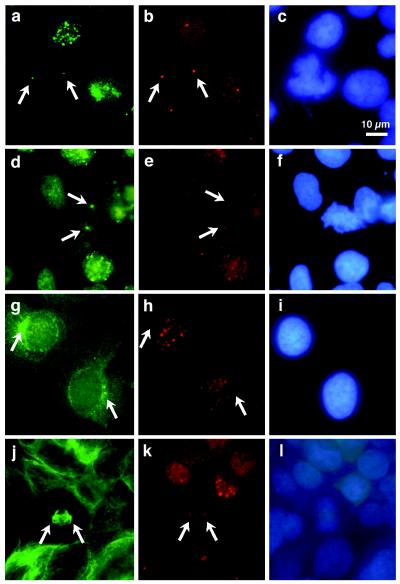
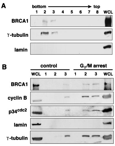
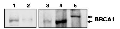
Centrosomes and their associated microtubules direct events during mitosis and control the organization of animal cell structures and movement during interphase. The centrosome replicates during the cell cycle, directs the assembly of bipolar mitotic spindles, and plays an important role in maintaining the fidelity of cell division. Recently, tumor suppressors such as p53 and retinoblastoma protein pRB have been localized to the centrosome in a cell cycle-dependent manner. Immunofluorescence microscopy and analysis of isolated centrosomes now provide evidence that BRCA1 protein, a suppressor of tumorigenesis in breast and ovary, also is associated with centrosomes during mitosis. Our results indicate that BRCA1 localizes with the centrosome during mitosis and coimmunoprecipitates with gamma-tubulin, a centrosomal component essential for nucleation of microtubules. Furthermore, gamma-tubulin associates preferentially with a hypophosphorylated form of BRCA1.


Figures

Figure 1
Figure 1
Coimmunostaining of COS-7 cells with mouse monoclonal γ-tubulin antibody and rabbit polyclonal BRCA1 antibody C-20. (A) A prometaphase to metaphase cell; (B) an early anaphase cell, and (C) a late anaphase cell. The colocalized signals of BRCA1 and γ-tubulin are yellow. DAPI counterstained DNA. Arrows indicate the positions of centrosomes.
Figure 2
Figure 2
Immunostaining of COS-7 and E6/BE46 cells. Cells were costained with mouse monoclonal BRCA1 antibody MS110 (a) and rabbit polyclonal pericentrin antibody 4B (b); with MS110 (d) and rabbit polyclonal BRCA1 antibody C-20 (e); with mouse monoclonal γ-tubulin antibody (g) and C-20 (h); or with mouse monoclonal α-tubulin antibody (j) and C-20 (k). c, f, i, and l include DAPI counterstain of DNA. af highlight mitotic COS-7 cells; gi are interphase COS-7 cells; jl illustrate a mitotic E6/BE46 cell. Arrows indicate the positions of centrosomes.
Figure 3
Figure 3
BRCA1 protein is detected in centrosome fractions purified by a discontinuous sucrose gradient. (A) COS-7 cells (≈1 × 10⁷) were harvested in exponential growth phase. The proteins in each sucrose gradient fraction were separated by 4–15% gradient SDS/PAGE and subjected to Western blot analysis. (B) Approximately 5 × 10⁶ of control COS-7 cells (cell cycle distribution: G0/G1, 37.21%; S, 45.15%; G2/M, 17.64%; CV, 6.25%) and cells arrested in G2/M by sequential treatment with thymidine and nocodazole (G0/G1, 5.51%; S, 36.60%; G2/M, 57.89%; CV, 8.69%) were used for centrosome preparations. Western blot analyses of fractions 1–3 of each sample are shown here. Whole-cell lysates were prepared from ≈2 × 10⁵ cells. MS110 was used to detect BRCA1.
Figure 4
Figure 4
Coimmunoprecipitation of BRCA1 and γ-tubulin. Immunoprecipitation was performed by using rabbit polyclonal BRCA1 antibody C-20 (lane 2) or mouse monoclonal γ-tubulin antibody (lane 4), from 400 μg of COS-7 cell lysate. Normal rabbit IgG (lane 1) and mouse IgG (lane 3) were negative controls. Samples were separated by SDS/6% polyacrylamide gels and immunoblotted by BRCA1 MS110 and γ-tubulin antibody.
Figure 5
Figure 5
γ-tubulin associates preferentially with hypophosphorylated BRCA1. Samples were separated by 4–12% SDS/PAGE, and immunoblotted by BRCA1 MS110. Lane 1, isolated centrosomes; lanes 2 and 3, in vitro transcription/translation product of full-length BRCA1; lane 4, immunoprecipitation by γ-tubulin antibody; and lane 5, immunoprecipitation by C-20.

References

    1. Newman B, Mu H, Butler L M, Millikan R C, Moorman P G, King M-C. J Am Med Assn. 1998;279:915–921. - PubMed
    1. Szabo C I, King M-C. Hum Mol Genet. 1995;4:1811–1817. - PubMed
    1. Couch F J, Weber B L the Breast Cancer Information Core. Hum Mutat. 1996;8:8–18. - PubMed
    1. Miki Y, Swensen J, Shattuck-Eidens D, Futreal P A, Harshman K, Tavtigian S, Liu Q, Cochran C, Bennett L M, Ding W, et al. Science. 1994;266:66–71. - PubMed
    1. Chen Y, Chen C-F, Riley D J, Allred D C, Chen P-L, Von Hoff D, Osborne C K, Lee W-H. Science. 1995;270:789–791. - PubMed

Publication types

MeSH terms