. 1998 Oct 27;95(22):13012-7.
doi: 10.1073/pnas.95.22.13012.

Ionizing radiation and short wavelength UV activate NF-kappaB through two distinct mechanisms

Affiliations

Ionizing radiation and short wavelength UV activate NF-kappaB through two distinct mechanisms

N Li et al. Proc Natl Acad Sci U S A. .

Abstract

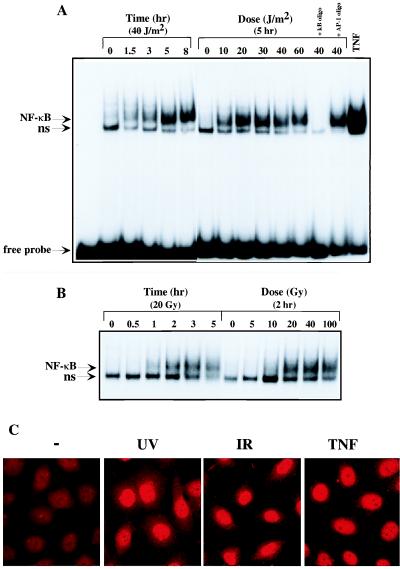
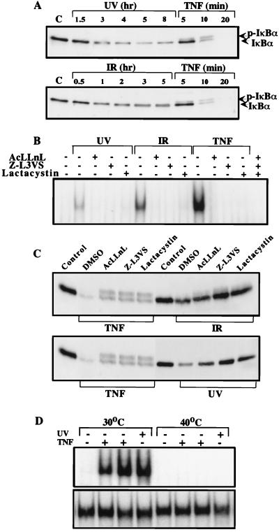
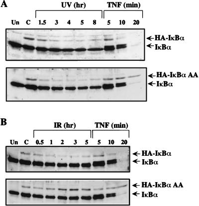
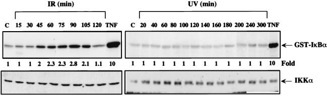
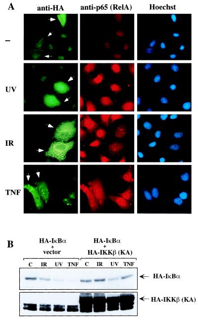
We examined the mechanisms by which two different types of photonic radiation, short wavelength UV (UV-C) and gamma radiation, activate transcription factor NF-kappaB. Exposure of mammalian cells to either form of radiation resulted in induction with similar kinetics of NF-kappaB DNA binding activity, nuclear translocation of its p65(RelA) subunit, and degradation of the major NF-kappaB inhibitor IkappaBalpha. In both cases, induction of NF-kappaB activity was attenuated by proteasome inhibitors and a mutation in ubiquitin-activating enzyme, suggesting that both UV-C and gamma radiation induce degradation of IkappaBs by means of the ubiquitin/proteasome pathway. However, although the induction of IkappaBalpha degradation by gamma rays was dependent on its phosphorylation at Ser-32 and Ser-36, UV-C-induced IkappaBalpha degradation was not dependent on phosphorylation of these residues. Even the "super repressor" IkappaBalpha mutant, which contains alanines at positions 32 and 36, was still susceptible to UV-C-induced degradation. Correspondingly, we found that gamma radiation led to activation of IKK, the protein kinase that phosphorylates IkappaBalpha at Ser-32 and Ser-36, whereas UV-C radiation did not. Furthermore, expression of a catalytically inactive IKKbeta mutant prevented NF-kappaB activation by gamma radiation, but not by UV-C. These results indicate that gamma radiation and UV-C activate NF-kappaB through two distinct mechanisms.


Figures

Figure 1
Figure 1
UV-C and IR activate NF-κB. Serum-starved HeLa cells were exposed to the indicated doses of UV-C (A) or IR (B). At the indicated times nuclear extracts were prepared and NF-κB DNA binding activity determined by EMSA. Specificity of binding was determined by competition with 100-fold excess unlabeled κB oligonucleotide or an AP-1 site oligonucleotide. As a positive control, EMSA was also performed with the nuclear extract of cells exposed to TNF (10 ng/ml) for 30 min. Migration positions of the NF-κB and nonspecific (ns) protein–DNA complexes and free probe are indicated. (C) UV-C and IR induce NF-κB nuclear translocation. HeLa cells grown on coverslips were untreated (−) or exposed to UV-C (40 J/m2), IR (20 Gy), or TNF (10 ng/ml) and fixed after 5, 3, or 0.5 hr, respectively. Cells were then incubated with anti-p65(RelA), stained with rhodamine-conjugated goat anti-rabbit IgG, and photographed through an epifluorescence-equipped microscope with a 63× oil objective and an automatic camera. Exposure times were considerably longer for the UV and IR irradiated samples to obtain exposure similar to the TNF treated samples.
Figure 2
Figure 2
UV-C and IR induce IκBα degradation. (A) HeLa cells were exposed to UV-C (40 J/m2), IR (20 Gy), or TNF (10 ng/ml) and whole cell extracts were prepared at the indicated times. Proteins (10 μg) were separated by electrophoresis on a 12.5% SDS/polyacrylamide gel and immunoblotted with anti-IκBα. Lane C, untreated cells. Positions of IκBα and phosphorylated IκBα (p-IκBα) are indicated. (B and C) Proteasome inhibitors block UV- and IR-induced NF-κB activation and IκBα degradation. HeLa cells were either untreated (control) or incubated with 100 μM AcLLnL, 50 μM Z-L3vs, or 10 μM clasto-lactacystin β-lactone (Lactacystin) for 1 hr before exposure to UV-C (40 J/m2), IR (20 Gy), or TNF (10 ng/ml). Cell extracts were prepared after 4 hr, 2 hr, and 15 min, respectively. NF-κB DNA binding activity was measured by EMSA (B) and IκBα degradation examined by immunoblotting with anti-IκBα (C). (D) Ubiquitination is required for NF-κB activation. ts20 cells were cultured at either the permissive (30°C) or restrictive (40°C) temperature for 6 hr to allow inactivation of the temperature-sensitive E1. Cells were then treated with TNF for 15 or 30 min or exposed to UV. Cell extracts were prepared and NF-κB (Upper) and NF-1 (Lower) DNA binding activities were measured by EMSA.
Figure 3
Figure 3
Ser-32 and Ser-36 are required for IκBα degradation in response to IR, but not by UV-C. Pools of stably transfected HeLa cells expressing HA-tagged wild-type IκBα or the IκBα AA mutant were either untreated or exposed to UV-C (40 J/m2) (A), IR (20 Gy) (B), and TNF (10 ng/ml). At the indicated times, lysates were prepared, separated by SDS/PAGE, and immunoblotted with anti-IκBα. Lanes Un and C, untransfected and untreated transfected cells, respectively. The migration positions of endogenous IκBα and the transfected HA-IκBα are indicated. To visualize HA-tagged IκBα the film was overexposed.
Figure 4
Figure 4
IKK is activated by IR but not by UV-C irradiation. HeLa cells were either untreated (lane C) or exposed to UV-C (40 J/m2), IR (20 Gy), or TNF (10 ng/ml). At the indicated times after treatment (10 min for TNF), whole cell lysates were prepared. IKK activity was measured by the immunecomplex kinase assay (Upper). The amount of IKKα was determined by immunoblotting (Lower).
Figure 5
Figure 5
IKK is necessary for NF-κB nuclear translocation and IκBα degradation induced by IR but not by UV-C. (A) HeLa cells grown on coverslips were transfected with an expression vector encoding HA-IKKβ (KA). After 36 hr the cells were exposed to the indicated stimuli or left untreated (−) and fixed. Samples were then incubated with a mixture of anti-p65 (RelA) and anti-HA. After washing, samples were stained with rhodamine-conjugated goat anti-rabbit IgG, fluorescein isothiocyanate-conjugated goat anti-mouse IgG-IgM, and Hoechst dye and photographed through an epifluorescence-equipped microscope. Arrowheads indicate cells that express HA-IKKβ (KA). (B) HeLa cells were transfected with an expression vector encoding HA-IκBα together with an empty vector or with a vector encoding HA-IKKβ (KA). Twenty-four hours later, cells were left untreated (C) or treated with IR, UV, or TNF. After 2 hr, 4 hr, or 15 min, cells were harvested and lysates were analyzed by immunoblotting with anti-HA.

References

    1. Herrlich P, Ponta H, Rahmsdorf H J. Rev Physiol Biochem Pharmacol. 1992;119:187–223. - PubMed
    1. Canman C E, Kastan M B. Nature (London) 1996;384:213–214. - PubMed
    1. Ko L J, Prives C. Genes Dev. 1996;10:1054–1072. - PubMed
    1. Liu Z-G, Baskaran R, Lea-Chou E T, Wood L D, Chen Y, Karin M, Wang J Y J. Nature (London) 1996;384:273–276. - PubMed
    1. El-Deiry W S, Tokino T, Velculescu V E, Levy D B, Parsons R, Trent J M, Lin D, Mercer W E, Kinzler K W, Vogelstein B. Cell. 1993;75:817–825. - PubMed

Publication types

MeSH terms

LinkOut - more resources