. 1998 Oct 27;95(22):13018-23.
doi: 10.1073/pnas.95.22.13018.

Identification of a gene encoding an acyl CoA:diacylglycerol acyltransferase, a key enzyme in triacylglycerol synthesis

Affiliations

Identification of a gene encoding an acyl CoA:diacylglycerol acyltransferase, a key enzyme in triacylglycerol synthesis

S Cases et al. Proc Natl Acad Sci U S A. .

Abstract

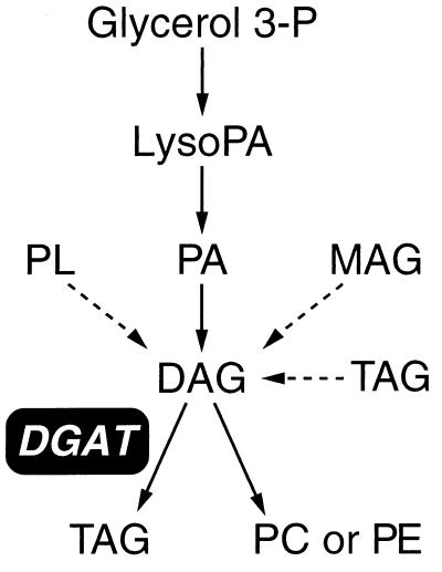
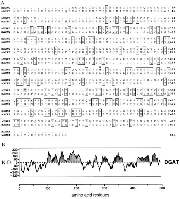
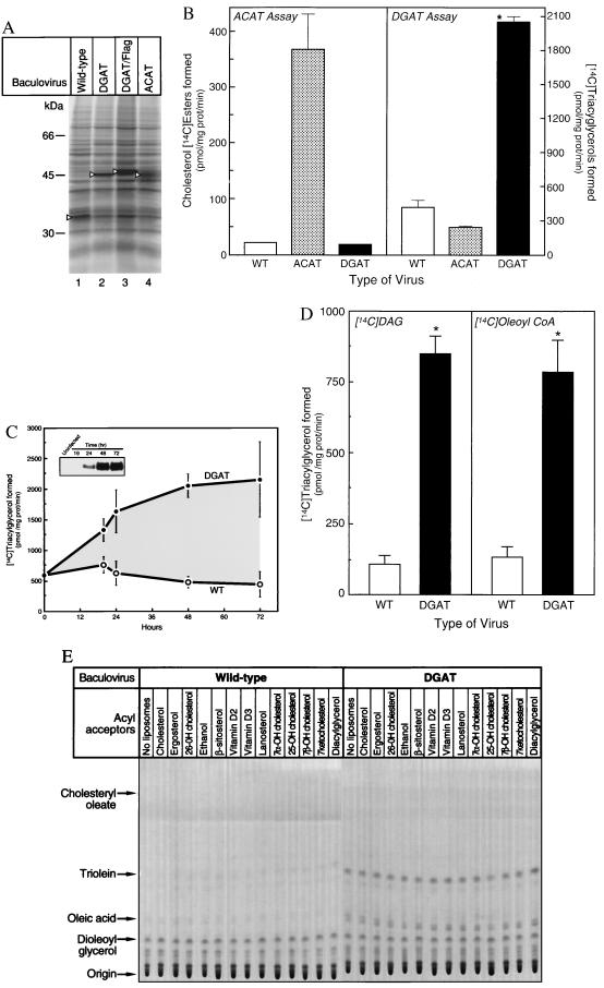
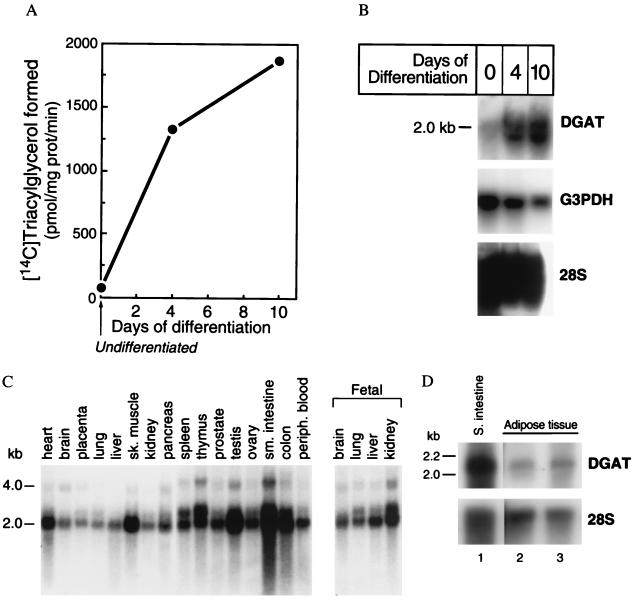
Triacylglycerols are quantitatively the most important storage form of energy for eukaryotic cells. Acyl CoA:diacylglycerol acyltransferase (DGAT, EC 2.3.1.20) catalyzes the terminal and only committed step in triacylglycerol synthesis, by using diacylglycerol and fatty acyl CoA as substrates. DGAT plays a fundamental role in the metabolism of cellular diacylglycerol and is important in higher eukaryotes for physiologic processes involving triacylglycerol metabolism such as intestinal fat absorption, lipoprotein assembly, adipose tissue formation, and lactation. DGAT is an integral membrane protein that has never been purified to homogeneity, nor has its gene been cloned. We identified an expressed sequence tag clone that shared regions of similarity with acyl CoA:cholesterol acyltransferase, an enzyme that also uses fatty acyl CoA as a substrate. Expression of a mouse cDNA for this expressed sequence tag in insect cells resulted in high levels of DGAT activity in cell membranes. No other acyltransferase activity was detected when a variety of substrates, including cholesterol, were used as acyl acceptors. The gene was expressed in all tissues examined; during differentiation of NIH 3T3-L1 cells into adipocytes, its expression increased markedly in parallel with increases in DGAT activity. The identification of this cDNA encoding a DGAT will greatly facilitate studies of cellular glycerolipid metabolism and its regulation.


Figures

Figure 1
Figure 1
Role of DGAT in glycerolipid metabolism. DAG used by DGAT potentially originates from hydrolysis of phosphatidic acid (PA), from the esterification of 2-monoacylglycerol (MAG), or from triacylglycerol (TAG) or phospholipid (PL) hydrolysis. The MAG pathway is thought to be especially important in enterocytes of the small intestine (3). P, phosphate; LysoPA, lysophosphatidic acid; PC, phosphatidyl choline; PE, phosphatidyl ethanolamine.
Figure 2
Figure 2
The mouse DGAT protein. (A) Predicted amino acid sequence of the mouse DGAT cDNA. The predicted amino acid sequence of mouse DGAT (mDGAT) is shown in alignment with mouse ACAT (mACAT) (32). The two sequences are ≈20% identical (identical residues are boxed). A potential N-linked glycosylation site (asterisk) and tyrosine phosphorylation site (shaded) are indicated. A serine residue in ACAT known to be necessary for catalytic function is also indicated (triangle). (B) Hydrophobicity plot of DGAT as assessed by Kyte–Doolittle (K–D) analysis (33). Hydrophobic regions are shaded.
Figure 3
Figure 3
Enzymatic activities in insect cell membranes expressing DGAT. Cells were infected with wild-type baculovirus (WT), mouse ACAT, or mouse DGAT recombinant baculoviruses, and membranes were assayed for enzymatic activity. (A) Metabolic labeling. Cell proteins, 48 h after infection, were metabolically labeled with [35S]methionine and [35S]cysteine, and whole-cell lysates were analyzed by SDS/PAGE and autoradiography. The expression of viral polyhedrin protein (lane 1), mouse DGAT (lane 2), FLAG-tagged mouse DGAT (lane 3), and mouse ACAT (lane 4) is indicated (triangles). (B) ACAT and DGAT activities. Data represent the mean (±SE) of five experiments. ∗, P < 0.001. vs. WT. (C) Time course of DGAT virus infection. Insect cell membranes were isolated at the indicated times after infection. Expression of the FLAG-tagged DGAT was detected by immunoblotting with an anti-FLAG antibody (Inset), and DGAT activity was measured. The doublet band observed in this experiment was not routinely observed, and its significance is unknown. Data represent the mean (±SE) of three experiments. (D) Comparison of the rate of triacylglycerol synthesis with either DAG or oleoyl CoA as the radiolabeled substrate. Assays contained the same amounts of oleoyl CoA (5 nmol) and DAG (2.5 μg) in all cases. The specific activity for DGAT virus-infected cells is less than that observed in A because of the reduction in DAG substrate concentration (i.e., this experiment was not performed at apparent VMAX). Data are the mean (±SE) of five experiments. ∗, P < 0.001 vs. WT. (E) Acyl acceptor specificity of DGAT. Reaction products from wild-type or DGAT virus-infected membranes assayed with [14C]oleoyl CoA and various acyl acceptor substrates were analyzed by TLC. Note that [14C]oleoyl CoA is incorporated specifically into triacylglycerols for all reactions containing membranes expressing DGAT. Hydrolysis of the labeled oleoyl CoA to oleic acid (as shown in this experiment) was observed in some, but not all, preparation of membranes expressing DGAT; this finding was associated with membranes expressing the highest levels of DGAT activity.
Figure 4
Figure 4
Analysis of DGAT mRNA expression. (A) DGAT activity and (B) DGAT mRNA expression during differentiation of 3T3-L1 cells into adipocytes. Mouse 3T3-L1 adipocyte differentiation was induced, and RNA and membranes were isolated from undifferentiated cells or cells harvested 4 and 10 days later. Results are shown for DGAT and two controls for RNA loading [glyceraldehyde-3-phosphate dehydrogenase (G3PDH) and 28S RNA]. Quantitation of DGAT mRNA in triplicate samples, by PhosphorImager analysis and correction for loading relative to 28S RNA (20) as an internal standard, demonstrated that DGAT levels were increased 5-fold by day 4 and 8-fold by day 10 of differentiation. The experiment was repeated three times with similar results. (C) DGAT expression in human tissues as assessed by Northern blot analysis. (D) DGAT expression in mouse small intestine and adipose tissue from two mice (lanes 2 and 3). The membrane was stripped and reprobed for 28S RNA (20).
Figure 5
Figure 5
Mouse chromosomal location of DGAT gene. Genetic mapping of the Dgat to mouse chromosome 15 was performed by linkage analysis performed with a panel of 67 progeny from an interspecific backcross [(C57BL/6J × Mus spretus)F1 × C57BL/6J] (23). A segment of the chromosome is drawn with the centromere toward the top. The ratios of the number of recombinants to the total number of informative mice and the recombination frequencies ±SE (in cM) for each pair of loci are indicated. For pairs of loci that cosegregate, the upper 95% confidence interval is shown in parentheses. No recombination was observed between Dgat, D15Ucla2, and Tef (0/65 mice). The DGAT gene has been assigned the name Dgat. The data have been deposited in the Mouse Genome Database under accession number MGD-J:44983.

References

    1. Bell R M, Coleman R A. Annu Rev Biochem. 1980;49:459–487. - PubMed
    1. Lehner R, Kuksis A. Prog Lipid Res. 1996;35:169–201. - PubMed
    1. Brindley D N. In: Biochemistry of Lipids, Lipoproteins and Membranes. Vance D E, Vance J E, editors. Amsterdam: Elsevier; 1991. pp. 171–203.
    1. Nishizuka Y. Science. 1992;258:607–614. - PubMed
    1. Haagsman H P, Van Golde L M G. Arch Biochem Biophys. 1981;208:395–402. - PubMed

Publication types

MeSH terms

Associated data