. 1998 Oct 27;95(22):13042-7.
doi: 10.1073/pnas.95.22.13042.

Detection of targeted GFP-Hox gene fusions during mouse embryogenesis

Affiliations

Detection of targeted GFP-Hox gene fusions during mouse embryogenesis

A R Godwin et al. Proc Natl Acad Sci U S A. .

Abstract

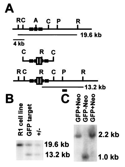
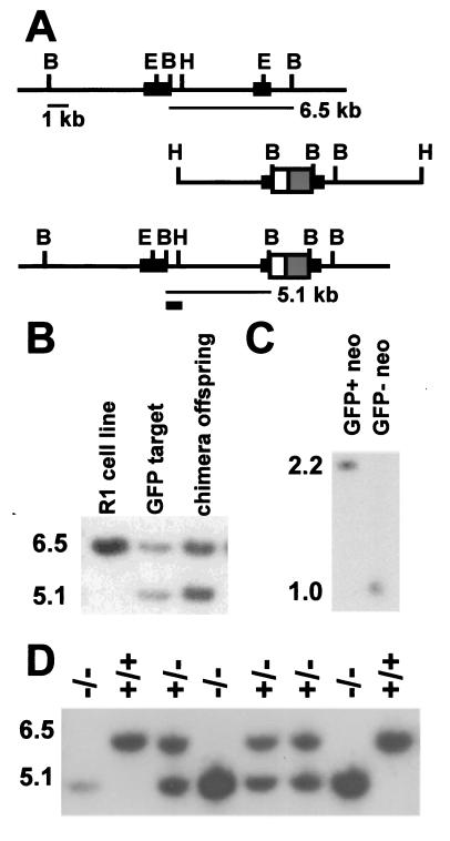
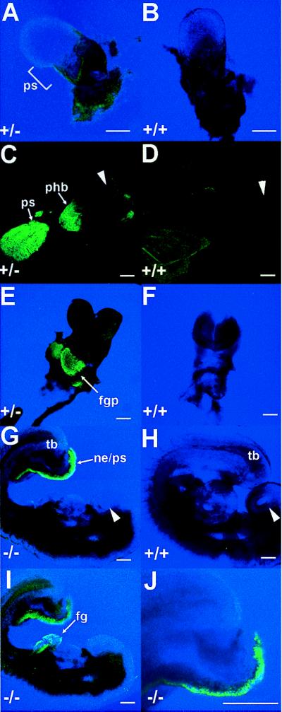
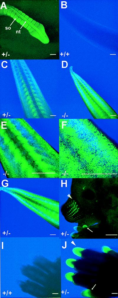
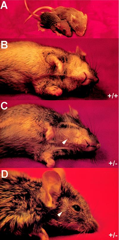
The ability to use a vital cell marker to study mouse embryogenesis will open new avenues of experimental research. Recently, the use of transgenic mice, containing multiple copies of the jellyfish gene encoding the green fluorescent protein (GFP), has begun to realize this potential. Here, we show that the fluorescent signals produced by single-copy, targeted GFP in-frame fusions with two different murine Hox genes, Hoxa1 and Hoxc13, are readily detectable by using confocal microscopy. Since Hoxa1 is expressed early and Hoxc13 is expressed late in mouse embryogenesis, this study shows that single-copy GFP gene fusions can be used through most of mouse embryogenesis. Previously, targeted lacZ gene fusions have been very useful for analyzing mouse mutants. Use of GFP gene fusions extends the benefits of targeted lacZ gene fusions by providing the additional utility of a vital marker. Our analysis of the Hoxc13(GFPneo) embryos reveals GFP expression in each of the sites expected from analysis of Hoxc13(lacZneo) embryos. Similarly, Hoxa1(GFPneo) expression was detected in all of the sites predicted from RNA in situ analysis. GFP expression in the foregut pocket of Hoxa1(GFPneo) embryos suggests a role for Hoxa1 in foregut-mediated differentiation of the cardiogenic mesoderm.


Figures

Figure 1
Figure 1
Hoxa1GFPneo-targeting vector and Southern blot analysis. (A) Large solid boxes represent Hoxa1 exons, white box the GFP gene, and gray box the loxP flanked MC1neo cassette, AatII (A), ClaI (C), and PacI (P), and EcoRV (R). The position of the 3′-flanking probe used for Southern transfer analysis is indicated by the small solid box. The first line shows the wild-type genomic structure, the second line the targeting vector, and the third line the genomic structure resulting from homologous recombination. (B) Southern blot analysis of targeted cell line and chimera progeny. Shown are EcoRV digests of the parental R1 cell line, a targeted ES cell line, and offspring from a chimera. The wild-type allele is detected by a 19.6-kb restriction fragment, whereas the mutant allele is detected by a 13.2-kb restriction fragment. (C) Analysis of progeny of a Hoxa1GFPneo heterozygote crossed to a Cre deleter mouse. Shown are Southern transfer analyses of BamHI-digested DNA probed with an NcoI–AflII pEGFP fragment. A 2.2-kb fragment is detected in the Hoxa1GFPneo mice that shifts to 1.0 kb upon Cre-mediated removal of the neomycin resistance gene.
Figure 2
Figure 2
Hoxc13GFPneo-targeting vector and Southern blot analysis. (A) Genomic structure and targeting vector. Symbols in A are as described in Fig. 1, except large solid boxes represent Hoxc13 exons, BamHI (B), BstEII (E), and HindIII (H). The position of the 5′-flanking probe used for Southern transfer analysis is indicated by the small solid box. (B) Southern blot analysis of targeted cell line and offspring from a chimera. Shown are BamHI digests of the parental R1 cell line, a targeted ES cell line, and an offspring from a chimera. (C) Analysis of progeny of a Hoxc13GFPneo heterozygote crossed to a Cre deleter mouse. Analysis is as described for Fig. 1C. (D) Genotypic analysis of a litter resulting from an intercross of Hoxc13GFPneo allele heterozygotes. Southern transfer analysis of BamHI-digested DNA was performed and shows two wild-type offspring carrying only the 6.5-kb fragment, three heterozygotes carrying both the 6.5- and 5.1-kb fragments, and three homozygous mutant offspring carrying only the 5.1-kb fragment.
Figure 3
Figure 3
Embryonic expression of the Hoxa1GFPneo allele. (A) Expression in Hoxa1GFPneo heterozygote at E 7.0 in the primitive streak (ps). (B) Autofluorescence of extra-embryonic tissues in a wild-type embryo at E 7.0. Note same level of autofluorescence in a heterozygote (A). (C) Expression at E 8.25 in Hoxa1GFPneo heterozygote in rhombomere 3/4 of the presumptive hindbrain (phb), the regressing primitive streak (ps), and tail bud. Arrowhead indicates dorsal headfold region. (D) Lack of autofluorescence in a wild-type embryo at E 8.25 by using the same microscopic conditions as in C. Arrowhead indicates dorsal headfold region. (E) Expression in the developing foregut pocket (fgp) at E 8.75 in a Hoxa1GFPneo heterozygote. (F) Lack of fluorescence in a wild-type embryo at E 8.75 by using the same microscopic conditions as in E. (G) Expression in a Hoxa1GFPneo homozygote at E 9.0 in the caudal most region of the embryo in the nephrogenic duct (ne), tail bud (tb), and primitive streak (ps). Arrowhead indicates nasal placode. (H) Lack of fluorescence in wild-type embryo at E 9.0 by using the same microscopic conditions as in G and I. Arrowhead indicates nasal placode. (I) Expression in a Hoxa1GFPneo homozygote at E 9.0 in the foregut (fg) and tail bud (tb). (J) Higher magnification image of caudal expression showing single cell resolution. (For AJ, scale bar is 20 microns.)
Figure 4
Figure 4
Expression of the Hoxc13GFPneo and Hoxc13GFPlox alleles. (A) Neural tube (nt) and somite (so) expression in the tail of a Hoxc13GFPneo heterozygote at E 11.5. (B) Tail expression in a wild-type embryo at E 13.5. The same microscopic conditions were used for B-D. (C) Tail expression in a Hoxc13GFPneo heterozygote at E 13.5. (D) Tail expression in a Hoxc13GFPneo homozygote at E 13.5. (E) Higher magnification of tail expression in Hoxc13GFPneo homozygote at E 13.5. (F) Higher magnification of tail expression in a Hoxc13GFPneo homozygote at E 13.5. (G) Tail expression in Hoxc13GFPlox heterozygote at E 13.5. (H) Vibrissae (arrowhead) and nail (arrow) expression in a Hoxc13GFPlox heterozygote at E 13.5. (I) Nail expression in a wild-type embryo at E 15.5. (J) Nail expression in a Hoxc13GFPneo heterozygote at E 15.5. The same microscopic conditions were used for I and J. Wild-type embryos showed no fluorescence under the same experimental conditions used for A, G, and H (data not shown). (Scale bar is 20 microns, except for H where it is 500 microns).
Figure 5
Figure 5
Hair phenotype of Hoxc13GFPneo and Hoxc13GFPlox mice. (A) Wild-type and Hoxc13GFPlox homozygous littermates at postnatal day 16. (B) Wild-type mouse ventral side. (C) Hoxc13GFPneo heterozygous littermate ventral side. Arrowhead indicates a region of fur that is patchy. (D) Hoxc13GFPneo heterozygous face. Arrowhead indicates a region of fur that is patchy.

References

    1. Chalfie M, Tu Y, Euskirchen G, Ward W W, Prasher D C. Science. 1994;263:802–805. - PubMed
    1. Heim R, Prasher D C, Tsien R Y. Proc Natl Acad Sci USA. 1994;91:12501–12504. - PMC - PubMed
    1. Prasher D C. Trends Genet. 1995;11:320–323. - PubMed
    1. Jacobson R H, Zhang X-J, DuBose R F, Matthews B W. Nature (London) 1994;369:761–766. - PubMed
    1. Kouhara H, Kurebayashi S, Hashimoto K, Kasayama S, Koga M, Kishimoto T, Sato B. Oncogene. 1995;10:2315–2322. - PubMed

Publication types

MeSH terms