. 1998 Oct 27;95(22):13079-84.
doi: 10.1073/pnas.95.22.13079.

A viral suppressor of gene silencing in plants

Affiliations

A viral suppressor of gene silencing in plants

R Anandalakshmi et al. Proc Natl Acad Sci U S A. .

Abstract

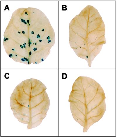
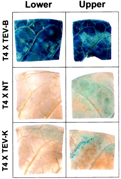
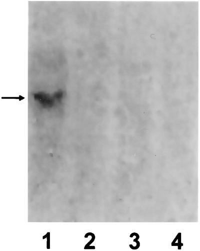
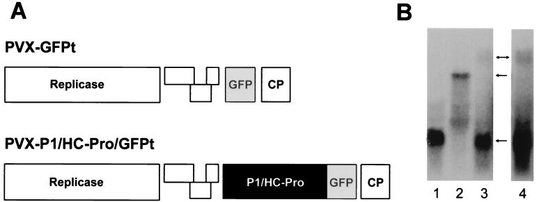
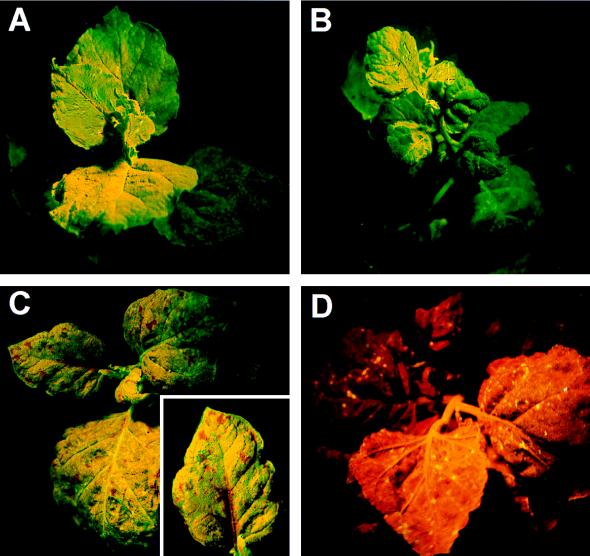
Gene silencing is an important but little understood regulatory mechanism in plants. Here we report that a viral sequence, initially identified as a mediator of synergistic viral disease, acts to suppress the establishment of both transgene-induced and virus-induced posttranscriptional gene silencing. The viral suppressor of silencing comprises the 5'-proximal region of the tobacco etch potyviral genomic RNA encoding P1, helper component-proteinase (HC-Pro) and a small part of P3, and is termed the P1/HC-Pro sequence. A reversal of silencing assay was used to assess the effect of the P1/HC-Pro sequence on transgenic tobacco plants (line T4) that are posttranscriptionally silenced for the uidA reporter gene. Silencing was lifted in offspring of T4 crosses with four independent transgenic lines expressing P1/HC-Pro, but not in offspring of control crosses. Viral vectors were used to assess the effect of P1/HC-Pro expression on virus-induced gene silencing (VIGS). The ability of a potato virus X vector expressing green fluorescent protein to induce silencing of a green fluorescent protein transgene was eliminated or greatly reduced when P1/HC-Pro was expressed from the same vector or from coinfecting potato virus X vectors. Expression of the HC-Pro coding sequence alone was sufficient to suppress virus-induced gene silencing, and the HC-Pro protein product was required for the suppression. This discovery points to the role of gene silencing as a natural antiviral defense system in plants and offers different approaches to elucidate the molecular basis of gene silencing.


Figures

Figure 1
Figure 1
PVX–GUS infection of offspring of GUS-silenced transgenic line T4. Three-week-old seedlings were inoculated with PVX–GUS (27), and the inoculated leaves were assayed for GUS activity by histochemical staining at 5 days postinoculation. Blue foci are indicative of viral replication. (A) T4 crossed with line TEV-B, which expresses the P1/HC-Pro sequence; (B) homozygous T4; (C) T4 crossed with line TEV-K, which contains a P1/HC-Pro sequence carrying a mutation that inactivates HC-Pro; and (D) T4 crossed with nontransformed N. tabacum cv xanthi nc.
Figure 2
Figure 2
Histochemical staining of GUS activity in leaves of offspring of GUS-silenced transgenic line T4 crossed with line TEV-B (Upper), nontransformed tobacco (Middle), or line TEV-K (Bottom). Leaves of 4- to 5-week-old seedlings were assayed as described.
Figure 3
Figure 3
Northern blot analysis of GUS mRNA levels in offspring of line T4 × TEV-B (lane 1), T4 × nontransformed tobacco (lane 3), or T4 × TEV-K (lane 4), or in homozygous T4 plants (lane 2). For each sample, total RNA was extracted from an entire 3-week-old seedling (excluding roots), quantitated, separated by denaturing gel electrophoresis (5 μg/lane), blotted to membrane, and probed with randomly primed cDNA specific for the GUS transgene. The arrow indicates the position of GUS mRNA.
Figure 4
Figure 4
VIGS of transgene encoded GFP in the absence or presence of P1/HC-Pro expression. (A) Schematic diagrams of the PVX vector constructs carrying the GFPt coding sequence (PVX–GFPt) or the TEV P1/HC-Pro sequence and GFPt. GFPt is the S65T version of GFP. (B) Northern blot analysis of GFP transgene mRNA levels in mock-inoculated GFP transgenic plants (lane 1) or plants infected with either PVX–GFPt (lane 2) or PVX–P1/HC-Pro/GFPt (lanes 3 and 4). Lane 4 shows a 3-fold longer exposure of lane 3. Total RNA was extracted from upper leaves of the plants at 20 days after inoculation and analyzed as described in the legend for Fig. 3, except that a GFP-specific probe was used. Arrows indicate the positions of PVX–P1/HC-Pro/GFPt genomic RNA, PVX–GFPt genomic RNA, and GFP transgene mRNA (from top to bottom, respectively).
Figure 5
Figure 5
VIGS of transgene encoded GFP in mixed virus infections. Transgenic GFP N. benthamiana plants coinoculated with (A) PVX–GFP and PVX–5′TEV showing complete suppression of VIGS of GFP, (B) PVX–GFP and PVX–HC showing an almost complete suppression of VIGS of GFP, (C) PVX–GFP and PVX–HC showing partial suppression of VIGS of GFP, and (D) PVX–GFP and PVX–noHC showing complete VIGS of GFP. The Inset in C is a closer view of a partially silenced leaf showing silencing in the vicinity of veins.

References

    1. Bingham P M. Cell. 1997;90:385–387. - PubMed
    1. Ingelbrecht I, Van Houdt H, Van Montagu M, Depicker A. Proc Natl Acad Sci USA. 1994;91:10502–1453. - PMC - PubMed
    1. Smith H A, Swaney S L, Parks T D, Wernsman E A, Dougherty W G. Plant Cell. 1994;6:1441–1443. - PMC - PubMed
    1. Park Y-D, Papp I, Moscone E A, Iglesias V A, Vaucheret H, Matzke M A, Matzke A J M. Plant J. 1996;9:183–194. - PubMed
    1. English J J, Mueller E, Baulcombe D C. Plant Cell. 1996;8:179–188. - PMC - PubMed

LinkOut - more resources