. 1998 Oct 27;95(22):13103-7.
doi: 10.1073/pnas.95.22.13103.

Pathogenic implications of mutations in the tau gene in pallido-ponto-nigral degeneration and related neurodegenerative disorders linked to chromosome 17

Affiliations

Pathogenic implications of mutations in the tau gene in pallido-ponto-nigral degeneration and related neurodegenerative disorders linked to chromosome 17

L N Clark et al. Proc Natl Acad Sci U S A. .

Abstract

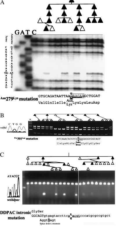
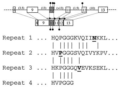
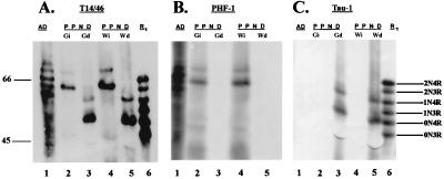
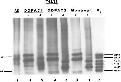
Pallido-ponto-nigral degeneration (PPND) is one of the most well characterized familial neurodegenerative disorders linked to chromosome 17q21-22. These hereditary disorders are known collectively as frontotemporal dementia (FTD) and parkinsonism linked to chromosome 17 (FTDP-17). Although the clinical features and associated regional variations in the neuronal loss observed in different FTDP-17 kindreds are diverse, the diagnostic lesions of FTDP-17 brains are tau-rich filaments in the cytoplasm of specific subpopulations of neurons and glial cells. The microtubule associated protein (tau) gene is located on chromosome 17q21-22. For these reasons, we investigated the possibility that PPND and other FTDP-17 syndromes might be caused by mutations in the tau gene. Two missense mutations in exon 10 of the tau gene that segregate with disease, Asn279(Lys) in the PPND kindred and Pro301(Leu) in four other FTDP-17 kindreds, were found. A third mutation was found in the intron adjacent to the 3' splice site of exon 10 in patients from another FTDP-17 family. Transcripts that contain exon 10 encode tau isoforms with four microtubule (MT)-binding repeats (4Rtau) as opposed to tau isoforms with three MT-binding repeats (3Rtau). The insoluble tau aggregates isolated from brains of patients with each mutation were analyzed by immunoblotting using tau-specific antibodies. For each of three mutations, abnormal tau with an apparent Mr of 64 and 69 was observed. The dephosphorylated material comigrated with tau isoforms containing exon 10 having four MT-binding repeats but not with 3Rtau. Thus, the brains of patients with both the missense mutations and the splice junction mutation contain aggregates of insoluble 4Rtau in filamentous inclusions, which may lead to neurodegeneration.


Figures

Figure 1
Figure 1
Mutation analysis in PPND and other FTDP-17 syndromes. Pedigree and mutation analyses for three kindreds are shown. Direct DNA sequence (A) or restriction endonuclease cleavage analyses (B and C) are shown for three mutations. A shows the analysis for the PPND kindred with a Asn279Lys mutation. B shows the Pro301Leu mutation segregating in the Montreal family. C shows the segregation of the intronic mutation in the DDPAC family. The affected DNA sequence is shown below each panel. The underlined sequence is the recognition sequence for a restriction endonuclease. The potential exon-splicing-enhancer sequence is enclosed in boxes in A. Sexes have been hidden, and birth orders have been modified to maintain patient confidentiality. Individuals with shaded symbols are younger than the average age of onset in Table 1.
Figure 2
Figure 2
Tau mutations in PPND and other FTDP-17 syndromes. Organization of the carboxy-terminus of the tau gene is shown. Exons are shown as boxes. The numbers in parentheses are the size of the intron, in kilobases. Exon 10, which is spliced alternatively to produce a transcript for 3Rtau and 4Rtau, is shaded. Black circles indicate the locations of the four known missense mutations. The intronic DDPAC mutation is indicated by a black diamond. The repeated microtubule binding domain’s amino acid sequence is shown below. Mutated amino acids are indicated in bold. Arrows in the diagram represent the repeats.
Figure 3
Figure 3
Western blots of native and dephosphorylated tau from a PPND brain. Insoluble fractions from gray and white matter were probed with the anti-tau mAb identified above each blot. Dephosphorylation reveals two predominant bands in tau from PPND brain that align with recombinant 4Rtau isoforms with an apparent molecular weight of 52 and 59. Molecular weight standards are shown in the left margin, and the positions of each of the nonphosphorylated recombinant human tau isoforms are labeled. Lane 1 shows PHFtau from AD brain. Lanes 2 and 3 show native and dephosphorylated tau, respectively, from gray matter of a PPND brain. Lanes 4 and 5 show native and dephosphorylated tau, respectively, from white matter of a PPND brain. Lane 6 shows all six recombinant tau isoforms (Rtau). Gi and Gd are native (i.e., insoluble) and dephosphorylated tau, respectively, from gray matter of a PPND brain. Wi and Wd are native and dephosphorylated tau, respectively, from white matter of a PPND brain. 0N3R, 1N3R, and 2N3R are three repeat tau isoforms with no vs. one or two amino terminal inserts, respectively. 0N4R, 1N4R, and 2N4R are four repeat tau isoforms with no vs. one or two amino terminal inserts, respectively.
Figure 4
Figure 4
Western blots of native and dephosphorylated tau from the DDPAC and Montreal kindreds. Insoluble fractions obtained from the frontal cortex of two different brains of a DDPAC kindred and a brain from the Montreal kindred were probed with the two phosphorylation independent mAbs T14 and T46. Dephosphorylation reveals two major and one minor band that align with all recombinant 4Rtau isoforms (lanes 3, 5, and 7). The major bands align with 0N4Rtau and 1N4Rtau isoforms, and the minor band aligns with the 2N4Rtau isoform. Molecular weight standards are shown in the left margin, and the positions of each of the nonphosphorylated recombinant tau isoforms are labeled. Lane 1 shows PHFtau from the brain of a patient with AD. Lanes 2 and 4 show native tau, and lanes 3 and 5 show dephosphorylated tau from two different DDPAC brains. Lanes 6 and 7 show native and dephosphorylated tau from a brain of the Montreal kindred. Abbreviations are as in Fig. 3.

References

    1. Foster N L, Wilhelmsen K C, Sima A A F, Jones M Z, D’Amato C, Gilman S. Ann Neurol. 1997;41:706–715. - PubMed
    1. Wilhelmsen K C, Lynch T, Nygaard T G. Am J Hum Genet. 1994;55:1159–1165. - PMC - PubMed
    1. Wszolek Z K, Pfeiffer R F, Bhatt M H, Schelper R L, Cordes M, Snow B J, Rodnitzky R L, Wolters E C, Arwert F, Calne D B. Ann Neurol. 1992;32:312–320. - PubMed
    1. Reed L A, Schmidt M L, Wszolek Z K, Balin B J, Soontornniyomkij V, Lee V M Y, Trojanowski J Q. J Neuropathol Exp Neurol. 1998;57:588–601. - PubMed
    1. Froelich S, Basun H, Forsell C, Lilius L, Axelman K, Andreadis A, Lannfelt L. Am J Med Genet. 1997;74:380–385. - PubMed

Publication types

MeSH terms